REVIEW 4 major objections 6 minor 81 references
Probing Stress and Magnetism at High Pressures with Two-Dimensional Quantum Sensors
T0 review · 4 major / 6 minor · reviewed 2026-08-10 · deepseek-v4-flash
Pith's one-line read A 2D layer of boron-vacancy centers inside a diamond anvil cell maps stress and magnetism up to 4 GPa, with three times the pressure response of NV centers.
desk verdict Solid methods paper: the VB- pressure calibration is credible and useful, but the magnetic phase-transition claim needs one more control before it carries weight. read the letter →
The pith
A machine-rendered reading of the paper's core claim, the machinery that carries it, and where it could break.
The reading
What carries the argument
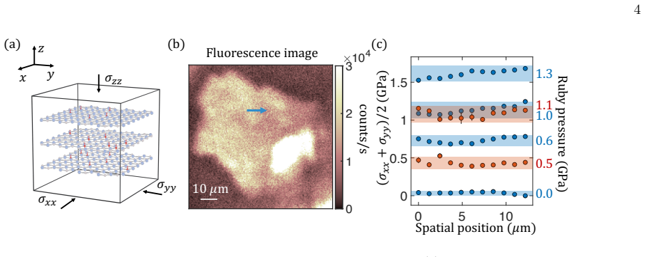
The central object is the negatively charged boron-vacancy center ($V_B^-$), a spin-1 defect in hexagonal boron nitride whose ground-state zero-field splitting $D_{gs}=(2\pi)\times 3.48$ GHz can be read out by ODMR at room temperature. The carrying mechanism is the linear stress coupling in the Hamiltonian $H_{gs}=D_{gs}S_z^2+\gamma B_{ext}S_z+h(\sigma_{xx}+\sigma_{yy})S_z^2/2+h'(\sigma_{xx}-\sigma_{yy})(S_x^2-S_y^2)/2$, which makes the ZFS respond to the lateral stress sum while remaining insensitive to out-of-plane stress; the measured coefficient $h=(2\pi)\times(43\pm 7)$ MHz/GPa converts ODMR frequency shifts directly into local stress values. This spin-stress transducer is combined with heterostructure assembly to place the sensor within nanometers of a target sample, and the Zeeman term $\gamma B_{ext}S_z$ supplies the dual magnetic-field readout.
What would settle it
Take the same hBN/Cr$_{1+\delta}$Te$_2$ stack and measure the stray field while rotating the external field direction at pressures around 0.5 GPa; if the signal reappears for some orientation, the transition is a spin reorientation, not a loss of moment. Alternatively, an independent magnetometry probe, such as a micro-SQUID or X-ray magnetic circular dichroism on the pressurized flake, that shows the magnetization amplitude still nonzero above 0.5 GPa would falsify the non-magnetic transition claim.
Extended reading notes
Core claim
The central discovery is that $V_B^-$ spin defects in hBN retain optically detected magnetic resonance (ODMR) up to roughly 4 GPa and that their spin resonance frequencies shift linearly with pressure at a rate of $(2\pi)\times(43\pm 7)$ MHz/GPa, in agreement with the first-principles value $(2\pi)\times 39.2$ MHz/GPa and about three times the NV-center susceptibility. The measured response is attributed to coupling of the spin triplet to the in-plane stress components $\sigma_{xx}$ and $\sigma_{yy}$, with the sensor placed directly on the anvil culet and calibrated against ruby fluorescence. Using this response the paper maps lateral stress profiles across a 15-micrometer region and shows that non-hydrostaticity produces marked gradients above 1 GPa. In a heterostructure with a Cr$_{1+\delta}$Te$_2$ flake, it images a stray-field pattern consistent with in-plane magnetization at 0 GPa and its disappearance above 0.5 GPa, interpreted as a ferro-to-nonmagnetic transition caused by pressure-reduced exchange.
Load-bearing premise
The load-bearing reading is that the disappearance of the stray field at the hBN sensor shows that Cr$_{1+\delta}$Te$_2$ loses its magnetization; this fails if the flake's magnetization reorients out of the sensing direction, if the ODMR contrast decays with pressure, or if the heterostructure delaminates, and the paper does not include an independent magnetization measurement to rule those out.
Editorial extensions
If this is right
- Stress and magnetic field can be imaged simultaneously inside the high-pressure chamber at sub-micrometer resolution, without relying on stress continuity assumptions between anvil and sample.
- The operating range of the sensor should extend beyond 4 GPa by replacing NaCl with a more hydrostatic medium such as argon or neon, following the contrast loss observed at 4 GPa.
- Integrating $V_B^-$ with other 2D materials enables probing interfacial magnetic and superconducting phenomena under pressure at nanoscale standoff.
- The measured pressure susceptibility of $(2\pi)\times(43\pm 7)$ MHz/GPa gives a direct material parameter for designing hBN-based stress sensors in other settings.
- The observed magnetic transition in Cr$_{1+\delta}$Te$_2$ near 0.5 GPa indicates pressure can control exchange interactions in self-intercalated van der Waals magnets at room temperature.
Reading between the lines
- The transition in Cr$_{1+\delta}$Te$_2$ could be a spin reorientation rather than a loss of moment, since the measurement tracks only the out-of-plane stray-field component; angle-dependent or vector-field ODMR would distinguish these.
- The same hBN sensor should be able to detect pressure-driven Meissner repulsion in thin-film superconductors if the stray-field sensitivity near 1 GPa is sufficient, since the platform's standoff is set by the hBN thickness.
- If the sensitivity can be raised, for instance by cavity-enhanced fluorescence, the platform may become a routine probe of mechanical deformation and paleomagnetism in geological samples.
- The near threefold improvement over NV centers suggests that 2D spin defects, not just diamond, are a viable route to quantitative pressure metrology in nanoscale environments.
Editorial analysis
A structured set of objections, weighed in public.
Referee Report
Summary. The manuscript reports a two-dimensional quantum sensing platform based on VB- centers in hBN integrated into a diamond anvil cell. The authors calibrate the pressure-induced shift of the VB- zero-field splitting as h = (2π) x (43 ± 7) MHz/GPa using ruby fluorescence as an external pressure standard, finding consistency with a previous DFT prediction and about three times the response of NV centers in diamond. They then use this calibration to infer a lateral stress profile along a linecut across the DAC and to track the stray magnetic field of a Cr1+δTe2 flake as a function of pressure. The stray field disappears above ~0.5 GPa, which they interpret as a pressure-driven ferro-to-nonmagnetic transition. The paper also reports the pressure dependence of the hyperfine coupling, sensitivity estimates, and DFT calculations of exchange coupling versus pressure.
Significance. The quantitative pressure susceptibility result is a solid and useful contribution: it is measured on two samples with pressurization/depressurization cycles, calibrated against an independent ruby standard, and compared with an ab initio prediction rather than fitted to it. If the magnetic transition were established by an independent magnetization measurement, the demonstration of a 2D sensor inside the DAC would be of high interest for high-pressure quantum sensing. As it stands, the stress-mapping demonstration is convincing only as a line-scan capability, and the magnetic phase-transition claim is not yet supported at the level claimed.
major comments (4)
- [Imaging pressure-driven magnetism in heterogeneous device (Fig. 4b/c)] The claim of a ferro-to-nonmagnetic transition near 0.5 GPa is not established because the only evidence is the disappearance of the VB- stray-field signal. The manuscript itself states that ODMR contrast decreases with pressure and that the thickness of the flake in the DAC was not directly measured (Methods, Fig. E2). A vanishing signal could equally result from pressure-induced contrast loss, flake delamination or cracking, or a reorientation of the magnetization into a configuration whose field at the sensor is suppressed; the depressurization pattern at 0.13 GPa, which differs qualitatively from the initial state, shows that the magnetic texture does not simply return. An independent magnetization measurement (e.g., SQUID or MOKE under pressure) or, at minimum, a control experiment with a nonmagnetic flake and an in-situ contrast/linewidth calibration at the sensor location is required before this transition can be claimed.
- [Methods, FIRST-PRINCIPLES CALCULATIONS (Fig. E3)] The supporting DFT calculations are performed for CrTe2, not for the self-intercalated Cr1+δTe2 studied in the experiment, and they yield a monotonic decrease of the exchange constant J with pressure rather than a computed transition at 0.5 GPa. The mean-field relation TC ∝ J does not predict a transition pressure unless a quantitative TC(P) crossing zero is computed and compared with the measured onset. Therefore the statement that the observed transition 'can be explained by ... agreeing with density functional theory predictions' overstates the level of agreement; the DFT shows a trend consistent in direction with a weakening of ferromagnetism, but does not independently predict the observed transition.
- [Abstract and Fig. 3c / Fig. 4b/c] The words 'mapping' and 'imaging' in the abstract overstate the spatial data shown. The stress result is a one-dimensional linecut across ~15 µm (Fig. 3c), and the magnetic data consist of a linecut plus a single 2D confocal scan at 0.13 GPa (Fig. 4b/c insets). No 2D stress map and no pressure-dependent 2D magnetic maps are presented. The authors should either provide true 2D maps at multiple pressures or qualify the claims as line-scan profiling.
- [Fig. 3c and Fig. 4c] The central quantitative plots lack error bars and detection-limit information. Without error bars, the emergence of the stress gradient above 1 GPa cannot be distinguished from noise, and the 'maximum stray field' versus pressure curve in Fig. 4c is the sole evidence for the transition at 0.5 GPa. The authors should report repeated measurements or propagated uncertainties, and they should state the minimum detectable stray field at each pressure, especially in light of the pressure-dependent ODMR contrast.
minor comments (6)
- [Introduction] The phrase 'by investigating the of pressure-induced magnetism' contains a missing word and should be revised.
- [Fig. 1 caption] The caption lists panels (a) and (c) but no (b), while the text refers to Fig. 1b; the panel labels should be made consistent.
- [Methods, DAC sample preparation] The phrase 'a anvil' should be 'an anvil'; in the hBN growth section, 'evcuated' and 'complimented' appear to be typos for 'evacuated' and 'complemented'.
- [Fig. E2 caption] The word 'Spacial' should be 'Spatial'.
- [Fig. 4c] The maximum stray field is defined differently for pressurization (difference between the two edges of the sample) and depressurization (difference between the center and a distant point); the main text should justify this non-uniform definition because it complicates comparison of the two branches.
- [Sensitivity estimates] The manuscript cites Supplementary Materials for the sensitivity values ηp and ηB, but the supplementary file is not included in the provided text; these numbers should either be derived in the main text or the supplement should be supplied.
Circularity Check
No significant circularity: the central pressure susceptibility is calibrated against an external ruby standard and cross-checked with an independent DFT calculation; stress and magnetic imaging reuse that calibration in a standard, non-bootstrapping way.
full rationale
The paper's central quantitative claim is a measured quantity, not a derived prediction: the V_B- zero-field splitting shift with pressure is fit against ruby R2 fluorescence (an external standard), yielding (2π)×(43±7) MHz/GPa. This is compared with an ab initio value of (2π)×39.2 MHz/GPa from ref. 38, whose author list does not overlap the present paper; agreement is therefore independent evidence, not a self-citation. The stress maps in Fig. 3 use the same calibrated h to convert local ODMR shifts to stress, which is ordinary sensor calibration rather than circular derivation. The magnetic-transition claim in Fig. 4 infers loss of magnetization from disappearance of the stray field at the hBN sensor; this is underdetermined (delamination, spin reorientation, or ODMR contrast loss are not excluded), but the inference is an experimental interpretation, not a quantity that is defined as its own input, and the proposed exchange-reduction explanation is supported by a separate DFT calculation. The only self-citation (ref. 53, 'our recent work') is used to discount a prior temperature-stress interpretation; even if it were wrong, the measured pressure susceptibility and its agreement with independent DFT would stand, so the self-citation is not load-bearing. No step in the derivation chain reduces by construction to its own input.
Assumptions & free parameters
free parameters (3)
- VB- pressure susceptibility h =
(2π) × (43 ± 7) MHz/GPa
- Hyperfine pressure dependence dAzz/dP =
(2π) × (0.5 ± 0.2) MHz/GPa
- DFT Hubbard U value =
5 eV and 6 eV (two values)
assumptions (6)
- domain assumption The VB- spin Hamiltonian in Eq. 1 couples only to in-plane stress components σxx and σyy, with out-of-plane stress having a negligible effect on the ZFS.
- domain assumption The NaCl pressure medium provides a quasi-hydrostatic environment, so σxx ≈ σyy ≈ σzz at the calibration pressures.
- domain assumption The ruby R2 fluorescence shift is a reliable, independent pressure calibrant.
- domain assumption The mean-field relation TC ∝ J connects the DFT-computed exchange constant to the observed magnetic transition.
- domain assumption DFT calculations on CrTe2, without self-intercalated chromium, adequately represent the magnetic behavior of Cr1+δTe2 with δ ≈ 0.5.
- domain assumption The stray field measured at the hBN sensor layer reflects the magnetization of the entire Cr1+δTe2 flake, with no significant screening, delamination, or stress decoupling between layers.
Cite this review
Pith. "Pith review of Probing Stress and Magnetism at High Pressures with Two-Dimensional Quantum Sensors." pith.science (2026). https://pith.science/paper/VGNYEZKM
@misc{pith2026250103319,
  author       = {Pith},
  title        = {Pith review of: Probing Stress and Magnetism at High Pressures with Two-Dimensional Quantum Sensors},
  year         = {2026},
  howpublished = {\url{https://pith.science/paper/VGNYEZKM}},
  note         = {Machine review of arXiv:2501.03319}
}
abstract
Pressure serves as a fundamental tuning parameter capable of drastically modifying all properties of matter. The advent of diamond anvil cells (DACs) has enabled a compact and tabletop platform for generating extreme pressure conditions in laboratory settings. However, the limited spatial dimensions and ultrahigh pressures within these environments present significant challenges for conventional spectroscopy techniques. In this work, we integrate optical spin defects within a thin layer of two-dimensional (2D) materials directly into the high-pressure chamber, enabling an in situ quantum sensing platform for mapping local stress and magnetic environments up to 4~GPa. Compared to nitrogen-vacancy (NV) centers embedded in diamond anvils, our 2D sensors exhibit around three times stronger response to local stress and provide nanoscale proximity to the target sample in heterogeneous devices. We showcase the versatility of our approach by imaging both stress gradients within the high-pressure chamber and a pressure-driven magnetic phase transition in a room-temperature self-intercalated van der Waals ferromagnet, Cr$_{1+\delta}$Te$_2$. Our work demonstrates an integrated quantum sensing device for high-pressure experiments, offering potential applications in probing pressure-induced phenomena such as superconductivity, magnetism, and mechanical deformation.
Figures


Figures from the paper (1 more)

Reference graph
Works this paper leans on
-
[80]
Liu, Y., Kwon, S., de Coster, G. J., Lake, R. K. & Ne- upane, M. R. Structural, electronic, and magnetic prop- erties of crte 2. Physical Review Materials 6, 084004 (2022)
work page 2022
-
[1]
The scale bars in (b) and (c) are 2 nm −1
and [112]. The scale bars in (b) and (c) are 2 nm −1. TEM studies were conducted using 120 kV JEOL JEM 1400plus. Fig. E1a is a low magnification TEM image of a chromium telluride nanoplate. Selected area electron diffraction (SAED) patterns were collected along [001] and [112] zone axes, which are shown in Fig. E1b and c, respectively. The main diffractio...
-
[2]
Doherty, M. W. et al. Electronic properties and metrol- ogy applications of the diamond NV- center under pres- sure. Physical review letters 112, 047601 (2014)
work page 2014
-
[3]
Hsieh, S. et al. Imaging stress and magnetism at high pressures using a nanoscale quantum sensor. Science 366, 1349–1354 (2019)
2019
-
[4]
Bhattacharyya, P. et al. Imaging the meissner effect in hydride superconductors using quantum sensors. Nature 627, 73–79 (2024)
work page 2024
-
[5]
Lesik, M. et al. Magnetic measurements on micrometer- sized samples under high pressure using designed nv cen- ters. Science 366, 1359–1362 (2019)
2019
-
[6]
Yip, K. Y. et al. Measuring magnetic field texture in cor- related electron systems under extreme conditions. Sci- ence 366, 1355–1359 (2019)
2019
-
[7]
Wang, M. et al. Imaging magnetic transition of magnetite to megabar pressures using quantum sensors in diamond anvil cell. Nature Communications 15, 8843 (2024)
work page 2024
Show all 81 references
-
[8]
Steele, L. et al. Optically detected magnetic resonance of nitrogen vacancies in a diamond anvil cell using designer diamond anvils. Applied Physics Letters 111 (2017)
2017
-
[9]
Shang, Y.-X. et al. Magnetic sensing inside a diamond anvil cell via nitrogen-vacancy center spins. Chinese Physics Letters 36, 086201 (2019)
2019
-
[10]
Hamlin, J. J. & Zhou, B. B. Extreme diamond-based quantum sensors. Science 366, 1312–1313 (2019)
2019
-
[11]
P., Cabriales, W
Shelton, D. P., Cabriales, W. & Salamat, A. Magne- tometry in a diamond anvil cell using nitrogen vacancy centers in a nanodiamond ensemble. Review of Scientific Instruments 95 (2024)
2024
-
[12]
Ho, K. O. et al. Probing local pressure environment in anvil cells with nitrogen-vacancy (n-v-) centers in dia- mond. Physical Review Applied 13, 024041 (2020)
2020
-
[13]
Dai, J.-H. et al. Optically detected magnetic resonance of diamond nitrogen-vacancy centers under megabar pres- sures. Chinese Physics Letters 39, 117601 (2022)
2022
-
[14]
Rovny, J. et al. Nanoscale diamond quantum sensors for many-body physics. Nature Reviews Physics 1–16 (2024)
2024
-
[15]
Hilberer, A. et al. Enabling quantum sensing under ex- treme pressure: Nitrogen-vacancy magnetometry up to 130 gpa. Physical Review B 107, L220102 (2023)
2023
-
[16]
Wen, J. et al. Probing the meissner effect in pressurized bilayer nickelate superconductors using diamond quan- tum sensors. arXiv preprint arXiv:2410.10275 (2024)
2024 arXiv
-
[17]
Vaidya, S., Gao, X., Dikshit, S., Aharonovich, I. & Li, T. Quantum sensing and imaging with spin defects in hexag- onal boron nitride. Advances in Physics: X 8, 2206049 (2023)
2023
-
[18]
I., Parto, K
Azzam, S. I., Parto, K. & Moody, G. Prospects and challenges of quantum emitters in 2d materials. Applied Physics Letters 118 (2021)
2021
-
[19]
& Zhang, J
Ren, S., Tan, Q. & Zhang, J. Review on the quantum emitters in two-dimensional materials. Journal of Semi- conductors 40, 071903 (2019)
2019
-
[20]
Su, C. et al. Tuning colour centres at a twisted hexagonal boron nitride interface. Nature materials 21, 896–902 (2022)
2022
-
[21]
Scholten, S. C. et al. Multi-species optically addressable spin defects in a van der waals material. Nature Commu- nications 15, 6727 (2024)
2024
-
[22]
Healey, A. et al. Quantum microscopy with van der waals heterostructures. Nature Physics 19, 87–91 (2023)
2023
-
[23]
& Toth, M
Aharonovich, I., Tetienne, J.-P. & Toth, M. Quantum emitters in hexagonal boron nitride. Nano Letters 22, 9227–9235 (2022)
2022
-
[24]
Gottscholl, A. et al. Initialization and read-out of in- trinsic spin defects in a van der waals crystal at room temperature. Nature materials 19, 540–545 (2020)
2020
-
[25]
Gottscholl, A. et al. Room temperature coherent con- trol of spin defects in hexagonal boron nitride. Science Advances 7, eabf3630 (2021)
2021
-
[26]
Gong, R. et al. Coherent dynamics of strongly interacting electronic spin defects in hexagonal boron nitride. Nature Communications 14, 3299 (2023)
2023
-
[27]
Naclerio, A. E. & Kidambi, P. R. A review of scal- able hexagonal boron nitride (h-bn) synthesis for present and future applications. Advanced Materials 35, 2207374 (2023)
2023
-
[28]
Durand, A. et al. Optically active spin defects in few- layer thick hexagonal boron nitride.Phys. Rev. Lett. 131, 116902 (2023)
2023
-
[29]
Stern, H. L. et al. Room-temperature optically detected 8 magnetic resonance of single defects in hexagonal boron nitride. Nature Communications 13, 618 (2022)
2022
-
[30]
& Gali, A
Li, S., Thiering, G., Udvarhelyi, P., Iv´ ady, V. & Gali, A. Carbon defect qubit in two-dimensional WS 2. Nature Communications 13, 1210 (2022)
2022
-
[31]
Gao, X. et al. Nanotube spin defects for omnidirectional magnetic field sensing. arXiv preprint arXiv:2310.02709 (2023)
2023 arXiv
-
[32]
Kumar, P. et al. Magnetic imaging with spin defects in hexagonal boron nitride. Physical Review Applied 18, L061002 (2022)
2022
-
[33]
Das, S. et al. Quantum sensing of spin dynamics using boron-vacancy centers in hexagonal boron nitride. Phys- ical Review Letters 133, 166704 (2024)
2024
-
[34]
Lyu, X. et al. Strain quantum sensing with spin defects in hexagonal boron nitride. Nano Letters 22, 6553–6559 (2022)
2022
-
[35]
Zabelotsky, T. et al. Creation of boron vacancies in hexagonal boron nitride exfoliated from bulk crystals for quantum sensing. ACS Applied Nano Materials 6, 21671– 21678 (2023)
2023
-
[36]
Gao, X. et al. High-contrast plasmonic-enhanced shal- low spin defects in hexagonal boron nitride for quantum sensing. Nano Letters 21, 7708–7714 (2021)
2021
-
[37]
L., Chen, W
Huang, Y. L., Chen, W. & Wee, A. T. Two-dimensional magnetic transition metal chalcogenides. SmartMat 2, 139–153 (2021)
2021
-
[38]
Zhou, J. et al. Sensing spin wave excitations by spin de- fects in few-layer-thick hexagonal boron nitride. Science Advances 10, eadk8495 (2024)
2024
-
[39]
Udvarhelyi, P. et al. A planar defect spin sensor in a two- dimensional material susceptible to strain and electric fields. npj Computational Materials 9, 150 (2023)
2023
-
[40]
Gottscholl, A. et al. Spin defects in hBN as promising temperature, pressure and magnetic field quantum sen- sors. Nature communications 12, 4480 (2021)
2021
-
[41]
& Capitani, F
Celeste, A., Borondics, F. & Capitani, F. Hydrostatic- ity of pressure-transmitting media for high pressure in- frared spectroscopy. High Pressure Research 39, 608–618 (2019)
2019
-
[42]
& Jin, C
You, S., Chen, L. & Jin, C. Hydrostaticity of pressure media in diamond anvil cells. Chinese Physics Letters 26, 204–206 (2009)
2009
-
[43]
& Haga, Y
Tateiwa, N. & Haga, Y. Appropriate pressure- transmitting media for cryogenic experiment in the dia- mond anvil cell up to 10 gpa. InJournal of Physics: Con- ference Series, vol. 215, 012178 (IOP Publishing, 2010)
2010
-
[44]
Gong, R. et al. Isotope engineering for spin defects in van der waals materials. Nature Communications 15, 104 (2024)
2024
-
[45]
Clua-Provost, T. et al. Isotopic control of the boron- vacancy spin defect in hexagonal boron nitride. Physical Review Letters 131, 126901 (2023)
2023
-
[46]
Janzen, E. et al. Boron and nitrogen isotope effects on hexagonal boron nitride properties. Advanced Materials 36, 2306033 (2024)
2024
-
[47]
& Kobayashi, K
Sasaki, K., Taniguchi, T. & Kobayashi, K. Nitrogen iso- tope effects on boron vacancy quantum sensors in hexag- onal boron nitride. Applied Physics Express 16, 095003 (2023)
2023
-
[48]
& Taylor, R
Sterer, E., Pasternak, M. & Taylor, R. A multipurpose miniature diamond anvil cell. Review of scientific instru- ments 61, 1117–1119 (1990)
1990
-
[49]
Plo, J. et al. Isotope substitution and polytype control for point defects identification: the case of the ultraviolet color center in hexagonal boron nitride. arXiv preprint arXiv:2405.20837 (2024)
2024 arXiv
-
[50]
& Le Marchand, G
Klotz, S., Chervin, J., Munsch, P. & Le Marchand, G. Hydrostatic limits of 11 pressure transmitting media. Journal of Physics D: Applied Physics 42, 075413 (2009)
2009
-
[51]
J., Bujak, M., Zhao, J., Gatta, G
Angel, R. J., Bujak, M., Zhao, J., Gatta, G. D. & Jacob- sen, S. D. Effective hydrostatic limits of pressure media for high-pressure crystallographic studies. Journal of Ap- plied Crystallography 40, 26–32 (2007)
2007
-
[52]
Hydrostaticity in high pressure experi- ments: some general observations and guidelines for high pressure experimenters
Takemura, K. Hydrostaticity in high pressure experi- ments: some general observations and guidelines for high pressure experimenters. High Pressure Research 41, 155– 174 (2021)
2021
-
[53]
Barson, M. S. et al. Nanomechanical sensing using spins in diamond. Nano letters 17, 1496–1503 (2017)
2017
-
[54]
Liu, Z. et al. Temperature dependent spin-phonon cou- pling of boron-vacancy centers in hexagonal boron ni- tride. arXiv preprint arXiv:2404.15493 (2024)
2024 arXiv
-
[55]
Yang, T. et al. Spin defects in hexagonal boron nitride for strain sensing on nanopillar arrays. Nanoscale 14, 5239–5244 (2022)
2022
-
[56]
Curie, D. et al. Correlative nanoscale imaging of strained hbn spin defects. ACS Applied Materials & Interfaces 14, 41361–41368 (2022)
2022
-
[57]
Lee, W. et al. Intrinsic high-fidelity spin polarization of charged vacancies in hexagonal boron nitride. arXiv preprint arXiv:2406.11953 (2024)
2024 arXiv
-
[58]
Clua-Provost, T. et al. Spin-dependent photodynamics of boron-vacancy centers in hexagonal boron nitride. Phys- ical Review B 110, 014104 (2024)
2024
-
[59]
Qian, C. et al. Unveiling the zero-phonon line of the boron vacancy center by cavity-enhanced emission. Nano Letters 22, 5137–5142 (2022)
2022
-
[60]
Fr¨ och, J. E.et al. Coupling spin defects in hexagonal boron nitride to monolithic bullseye cavities. Nano Let- ters 21, 6549–6555 (2021)
2021
-
[61]
Nonahal, M. et al. Coupling spin defects in hexago- nal boron nitride to titanium dioxide ring resonators. Nanoscale 14, 14950–14955 (2022)
2022
-
[62]
& Zhang, X
Gong, C. & Zhang, X. Two-dimensional magnetic crys- tals and emergent heterostructure devices. Science 363, eaav4450 (2019)
2019
-
[63]
Gibertini, M., Koperski, M., Morpurgo, A. F. & Novoselov, K. S. Magnetic 2d materials and heterostruc- tures. Nature nanotechnology 14, 408–419 (2019)
2019
-
[64]
Jiang, X. et al. Recent progress on 2d magnets: Funda- mental mechanism, structural design and modification. Applied Physics Reviews 8 (2021)
2021
-
[65]
Wang, Q. H. et al. The magnetic genome of two- dimensional van der waals materials. ACS nano 16, 6960–7079 (2022)
2022
-
[66]
S., Mandrus, D
Burch, K. S., Mandrus, D. & Park, J.-G. Magnetism in two-dimensional van der waals materials. Nature 563, 47–52 (2018)
2018
-
[67]
Conner, C. et al. Enhanced antiferromagnetic phase in metastable self-intercalated cr {1 +x} te 2 compounds. arXiv preprint arXiv:2411.13721 (2024)
2024 arXiv
-
[68]
Gong, C. et al. Discovery of intrinsic ferromagnetism in two-dimensional van der waals crystals. Nature 546, 265–269 (2017)
2017
-
[69]
Deng, Y. et al. Gate-tunable room-temperature ferro- magnetism in two-dimensional fe3gete2. Nature 563, 94– 99 (2018). 9
2018
-
[70]
Coughlin, A. L. et al. Near degeneracy of magnetic phases in two-dimensional chromium telluride with en- hanced perpendicular magnetic anisotropy. ACS nano 14, 15256–15266 (2020)
2020
-
[71]
Coughlin, A. L. et al. Van der waals superstructure and twisting in self-intercalated magnet with near room- temperature perpendicular ferromagnetism. Nano letters 21, 9517–9525 (2021)
2021
-
[72]
Lin, Z. et al. Pressure-induced spin reorientation tran- sition in layered ferromagnetic insulator cr 2 ge 2 te 6. Physical Review Materials 2, 051004 (2018)
2018
-
[73]
Sun, Y. et al. Effects of hydrostatic pressure on spin-lattice coupling in two-dimensional ferromagnetic cr2ge2te6. Applied Physics Letters 112 (2018)
2018
-
[74]
Coughlin, A. L. et al. Extreme air sensitivity and nonself- limited oxidation of two-dimensional magnetic tellurides. ACS Materials Letters 5, 1945–1953 (2023)
2023
-
[75]
Bl¨ ochl, P. E. Projector augmented-wave method.Physi- cal review B 50, 17953 (1994)
1994
-
[76]
& Joubert, D
Kresse, G. & Joubert, D. From ultrasoft pseudopoten- tials to the projector augmented-wave method. Physical review b 59, 1758 (1999)
1999
-
[77]
P., Burke, K
Perdew, J. P., Burke, K. & Ernzerhof, M. Generalized gradient approximation made simple. Physical review let- ters 77, 3865 (1996)
1996
-
[78]
I., Aryasetiawan, F
Anisimov, V. I., Aryasetiawan, F. & Lichtenstein, A. First-principles calculations of the electronic structure and spectra of strongly correlated systems: the lda+ u method. Journal of Physics: Condensed Matter 9, 767 (1997)
1997
-
[79]
L., Botton, G
Dudarev, S. L., Botton, G. A., Savrasov, S. Y., Humphreys, C. & Sutton, A. P. Electron-energy-loss spectra and the structural stability of nickel oxide: An lsda+ u study. Physical Review B 57, 1505 (1998)
1998
-
[81]
Mu, Z. et al. Magnetic imaging under high pressure with a spin-based quantum sensor integrated in a van der waals heterostructure. arXiv preprint (2025). To appear in the same arXiv posting
2025
Reviewed August 10, 2026 · model on record in the stance chip above.
Discussion (0). Continue with ORCID to comment.