REVIEW 4 major objections 6 minor 52 references
TFCDiff claims that ECG denoising works best when a conditional diffusion model runs in the DCT domain on raw 10-second multi-beat recordings, with a temporal-feature enhancement stream, rather than operating beat-by-beat in the time domain
Reviewed by Pith at T0; open to challenge. T0 means a machine referee read the full paper against a public rubric. the ladder, T0–T4 →
T0 review · deepseek-v4-flash
2026-08-03 21:06 UTC pith:G2GQP33E
load-bearing objection Serious, well-executed DCT-domain diffusion for multi-beat ECG denoising with a genuine external test, but the SimEMG advantage is likely inflated by an untested 50-Hz band limit. the 4 major comments →
TFCDiff: Robust ECG Denoising via Time-Frequency Complementary Diffusion
The pith
A machine-rendered reading of the paper's core claim, the machinery that carries it, and where it could break.
Core claim
The paper's finding is that a conditional diffusion model trained and sampled entirely in the truncated DCT domain—conditioned on the noisy signal's DCT coefficients, with a U-Net that intermittently converts feature maps to the time domain—outperforms time-domain diffusion, autoencoders, and filters on raw 10-second ECG segments. The authors report best results with an SNR-scaling factor of 150, a DC-derived scaling bound, and k-generation averaging. On the synthesized test set, TFCDiff-10 achieves SSD 21.5 au² and ImSNR 14.1 dB, surpassing all eight benchmarks; on an unseen real EMG-contaminated database, even one-generation TFCDiff beats the strongest non-diffusion baseline on all five me
What carries the argument
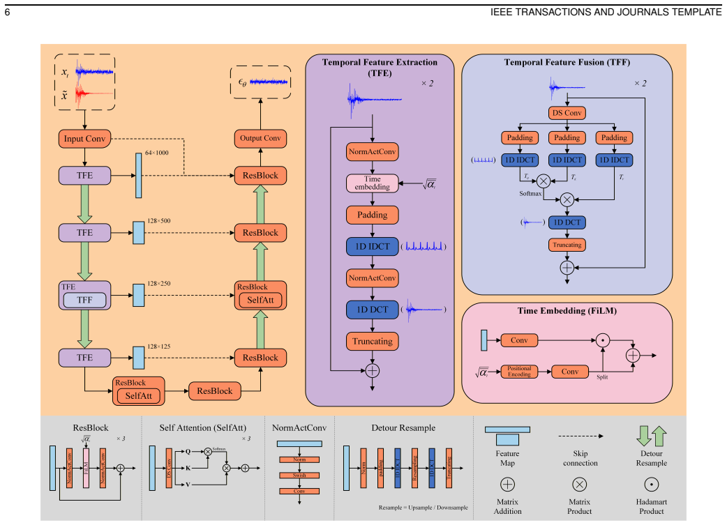
The central machinery is conditional diffusion in the DCT domain. A 10-second ECG is DCT-transformed, truncated to the first ~1000 coefficients (below 50 Hz), and both clean target and noisy conditioning input are scaled by a percentile-derived bound (η≈3) from the DC component. The noise predictor is a 1-D U-Net with a Temporal Feature Enhancement Mechanism: Temporal Feature Extraction converts frequency maps to time via zero-padding and IDCT, processes them, and returns via DCT; Temporal Feature Fusion applies time-domain attention at the middle encoder layer. A detour-resample strategy converts maps to time before down/upsampling to avoid aliasing, and the SNR schedule is rescaled by fact
Load-bearing premise
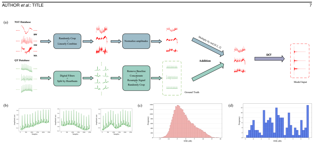
The load-bearing premise is that the filtered, baseline-corrected reference ECG recordings used as ground truth are faithful clean signals, and that amplitude-normalized mixtures of three archived noise excerpts at intensity λ∈[0.2,2] span real ambulatory noise.
What would settle it
Take raw multi-beat ECG with a simultaneously recorded reference clean lead (e.g., shoulder electrodes), apply TFCDiff without any preprocessing, and measure residual error with and without applying the paper's own bandpass/median/piecewise-baseline pipeline to the reference. If the model's advantage over the strongest non-diffusion baseline shrinks or reverses when the reference is not pipeline-processed, the claimed gain is at least partly an artifact of learning the preprocessing. Additionally, injecting a noise type absent from training (e.g., 50-Hz powerline interference) and observing wh
If this is right
- A single model can denoise raw 10-second multi-beat ECG without beat segmentation or padding, removing a barrier to wearable deployment.
- Training under flexible random mixed noise (random weights for baseline wander, muscle artifact, and electrode motion, intensity λ∈[0.2,2]) yields robustness across noise levels up to the hardest 1.5–2.0 intensity bin.
- With one generation, TFCDiff already outperforms the strongest non-diffusion baseline on an unseen real EMG-contaminated database, suggesting cross-dataset generalization rather than overfitting to synthesized noise.
- Averaging more generations (3, 5, 10) monotonically improves all metrics, so users can trade compute (0.17–1.63 s per 10-s clip) for fidelity.
- Denoised waveforms visually preserve T-wave morphology that time-domain diffusion smooths away, which matters for clinical interpretation of repolarization abnormalities.
Where Pith is reading between the lines
- If the DCT-domain advantage generalizes, the same truncated-DCT conditioning plus time-frequency fusion could apply to other quasi-periodic biosignals (PPG, EEG, EMG) where morphology preservation is diagnostically valuable.
- The claimed generalization to real EMG noise is tested only against EMG-type interference; testing against real electrode-motion or baseline-wander recordings would clarify whether the model's robustness extends beyond the noise type present in the unseen test set.
- The ground-truth preprocessing dependency suggests a discriminating experiment: compare TFCDiff on raw multi-beat signals against a simultaneously recorded clean lead without the paper's filtering/baseline pipeline; if the margin narrows, the model may be learning the preprocessing rather than true ECG structure.
- The authors note unsatisfactory results for intense EMG noise and suggest flow matching; a direct measure of residual noise power inside the QRS band as a function of input SNR would quantify this ceiling and guide whether a different generative formulation is needed.
Editorial analysis
A structured set of objections, weighed in public.
Referee Report
Summary. The paper proposes TFCDiff, a conditional diffusion model for denoising 10-second ambulatory ECG signals. The method operates in the DCT domain: clean and noisy signals are transformed by DCT, truncated to the first 1000 coefficients (≈50 Hz at fs=360 Hz), and used as diffusion training targets and conditioning inputs. The noise predictor is a 1D U-Net augmented with a Temporal Feature Enhancement Mechanism (TFEM) that interleaves time-domain and frequency-domain feature streams. Training data are QT Database segments preprocessed with bandpass/median filtering and piecewise-linear baseline removal, corrupted by flexible random mixtures of BW, MA, and EM noise from the NST Database. The model is evaluated on a synthesized QT/NST test set and on the external SimEMG database, with comparisons against eight baselines. Table III reports that TFCDiff-10 achieves the best mean values on all five metrics for the synthesized set, while Table IX shows TFCDiff-3 and TFCDiff-1 outperforming all benchmarks on the SimEMG set. The paper also includes ablations for the DCT-domain design and TFEM, plus generation-time measurements.
Significance. If the results hold, TFCDiff addresses a real gap: multi-beat, raw-sequence ECG denoising without beat segmentation, which is more clinically relevant than single-beat evaluation. The external SimEMG validation is a genuine strength, as is the public code release and the systematic ablation of the time-frequency design. The DCT-domain diffusion idea with TFEM is technically interesting and the reported computational costs are plausible. However, the central SOTA and generalization claims currently rest on a comparison that may be confounded by the hard 50-Hz band-limit inherent to the DCT truncation, and the abstract overstates the synthesized-set result by omitting the ensemble-size dependence. The underlying method is defensible and the issues are addressable with additional control experiments, so the contribution is potentially significant for wearable ECG denoising.
major comments (4)
- [§III-A, §V-B, Table IX] The DCT truncation to 50 Hz gives TFCDiff a structural advantage that is not controlled for. All inputs and outputs are hard band-limited by zeroing DCT coefficients above [50/Δf] (≈1000 of 3600 coefficients at fs=360 Hz). In contrast, all eight baselines operate on full-band time-domain signals. On SimEMG, which contains broadband real EMG noise, simply truncating the DCT of the noisy input and applying IDCT may remove a large fraction of the noise without any learned denoiser. The paper does not include a truncation-only baseline, nor does it band-limit the baselines. Consequently, the claim in Table IX that TFCDiff-3 outperforms all benchmarks on all metrics is confounded: the improvement could be largely due to the fixed 50-Hz cutoff rather than the learned conditional denoiser or TFEM. Please add a control that applies the same 50-Hz truncation to the noisy input (and optionally to
- [Abstract, §V-A, Table III] The state-of-the-art claim in the abstract ('achieves state-of-the-art performance across five evaluation metrics') is unqualified. Table III shows that TFCDiff-1 is worse than TCDAE on SSD, PRD, and CosSim; TFCDiff-3 lags on SSD, PRD, and CosSim; and only TFCDiff-10 leads on all five metrics. The ensemble size k is a critical hyperparameter that determines the headline result. Please qualify the SOTA claim with the ensemble size, and either state TFCDiff-10 explicitly or report the trade-off between k and performance. The conclusion also repeats the unqualified claim.
- [§IV-B] The ground-truth x0 used for training and for evaluating the synthesized set is not the raw QT recording but a surrogate: bandpass/median-filtered, then piecewise-linear baseline-removed within QRS-annotated segments, with Hermite interpolation at junctions. The paper does not validate this surrogate against the original signal or an independent reference. If residual baseline wander, filter ringing, or interpolation artifacts remain, the model is trained to reproduce those artifacts and the reported SSD/MAD/PRD/CosSim/ImSNR measure reconstruction to the processed surrogate, not to the true clean ECG. Please provide a quantitative or visual validation of the preprocessing (e.g., comparison of the surrogate with the raw signal in low-noise segments, or an expert review), and discuss how any residual artifacts affect the interpretation of the intra-dataset results.
- [§VI-A, Table X] The ablation in Table X does not isolate the effect of the 50-Hz truncation from the effect of DCT-domain modeling. The standalone DCT-diffusion row (SSD 146.8, CosSim 0.727) is much worse than the time-domain baseline (SSD 59.5, CosSim 0.889), and adding TFEM reverses this. This suggests that TFEM is compensating for information lost by truncation or by the skewed DCT-coefficient scaling, but the reader cannot tell how much of the final advantage comes from the hard band-limit versus from the learned denoiser. Please include a control where a time-domain model is trained on the same truncated-DCT-reconstructed signals (i.e., band-limited inputs without the DCT diffusion objective), or equivalently, where the time-domain baselines receive a 50-Hz low-passed version of the input.
minor comments (6)
- [§III-A] Duplicate sentence: 'Both transforms must be orthogonal to ensure invertibility.' appears twice consecutively.
- [Throughout] The name 'DesCod' is inconsistently written as 'DesCod' and 'DeScoD' (e.g., Section IV-D vs. Section V-A and reference [18]). Please unify.
- [Table III] CBAM-DAE reports PRD = 1910.425±877.430%, which is physically implausible for ECG denoising; consider explaining this outlier or omitting it from the table with a note, as is already done for later tables.
- [§IV-D] The text says all methods use input length 3600, with exceptions for FCN-DAE and CBAM-DAE at 3584. Please clarify how the outputs are aligned to the 3600-sample evaluation window (padding? cropping?) so that the metric comparison is unambiguous.
- [Fig. 4] The caption of Fig. 4(b) does not identify which curve corresponds to which SNR scaling factor c. Please add a legend.
- [§IV-C] The PRD definition in Eq. (19) uses the denoised signal's mean in the denominator; this differs from some prior definitions that normalize by the clean signal's mean. Please ensure this is intentional and state it clearly, since PRD values are otherwise hard to compare across papers.
Circularity Check
No significant circularity: TFCDiff's claims are empirical benchmark results, externally anchored by held-out test sets and the SimEMG database.
full rationale
The paper's central claims are empirical benchmark measurements, not derivations that reduce to their own inputs. TFCDiff is trained on 33,294 synthesized QT/NST pairs and evaluated on a held-out 4,296-pair test set as well as the external SimEMG database never used in training (Section IV-B, Tables III and IX). No reported metric is a fitted parameter of the model, and the k-generation averaging is an evaluation protocol rather than a prediction. The DCT truncation to 50 Hz and the scaling bound eta from Eq. 9 are transparent preprocessing/hyperparameter choices, with truncation fidelity independently checked by SSD reconstruction error. The choice of c=150 is described as an empirical search ('We empirically find that the diffusion model yields the best performance when c is set to 150'), not as a derived result. Citations such as [30] for DCT-space diffusion and [33] for cross-domain feature fusion provide architectural inspiration but are not invoked as an unverified uniqueness theorem that forces the conclusion. The skeptic's band-limiting concern is a fairness/threat-to-validity issue for baseline comparison, not circularity, because the SimEMG generalization claim is externally grounded. The paper's own stated limitations (Section VI-D) acknowledge gaps such as poor performance under intensive EMG noise and lack of device validation, which further supports that no hidden circular step is being asserted as a derivation.
Axiom & Free-Parameter Ledger
free parameters (7)
- SNR scaling factor c =
150
- DC scaling percentile tau =
1.75 (yields eta ~= 3)
- DCT truncation length =
1000 coefficients (~50 Hz cutoff)
- noise intensity factor lambda =
uniform in [0.2, 2]
- noise mixture weights r,m,n =
r+m+n=1 (sampling distribution unspecified)
- generation ensemble count k =
10 for the headline SOTA (tables also report k=1,3,5)
- diffusion schedule endpoints =
T=50, beta_1=1e-4, beta_T=0.5
axioms (6)
- standard math DDPM forward process, reverse process, and ELBO-derived L1 training objective (Eqs. 3-8)
- domain assumption Truncated DCT coefficients of ECG are adequately modeled as diffusible data corrupted by unit Gaussian noise
- domain assumption ECG diagnostic information lies below 50 Hz; coefficients beyond index 1000 are negligible
- domain assumption QT recordings after bandpass/median filtering and piecewise-linear baseline removal within QRS-annotated segments are clean ground truth
- domain assumption Amplitude-normalized linear mixtures of NST BW/MA/EM excerpts (Eq. 16) represent real ambulatory noise
- domain assumption Scaling bound eta computed from the DC component alone suffices to normalize the AC coefficients
read the original abstract
Ambulatory electrocardiogram (ECG) readings are prone to mixed noise from physical activities, including baseline wander (BW), muscle artifact (MA), and electrode motion artifact (EM). Developing a method to remove such complex noise and reconstruct high-fidelity signals is clinically valuable for diagnostic accuracy. However, denoising of multi-beat ECG segments remains understudied and poses technical challenges. To address this, we propose Time-Frequency Complementary Diffusion (TFCDiff), a novel approach that operates in the Discrete Cosine Transform (DCT) domain and uses the DCT coefficients of noisy signals as conditioning input. To refine waveform details, we incorporate Temporal Feature Enhancement Mechanism (TFEM) to reinforce temporal representations and preserve key physiological information. Comparative experiments on a synthesized dataset demonstrate that TFCDiff achieves state-of-the-art performance across five evaluation metrics. Furthermore, TFCDiff shows superior generalization on the unseen SimEMG Database, outperforming all benchmark models. Notably, TFCDiff processes raw 10-second sequences and maintains robustness under flexible random mixed noise (fRMN), enabling plug-and-play deployment in wearable ECG monitors for high-motion scenarios. Source code is available at https://github.com/Miroircivil/TFCDiff.
Figures




Reference graph
Works this paper leans on
-
[1]
Global Burden of Cardiovascular Diseases and Risks, 1990-2022,
G. A. Mensah et al., “Global Burden of Cardiovascular Diseases and Risks, 1990-2022,”J. Am. Coll. Cardiol., vol. 82, no. 25, pp. 2350–2473, Dec. 2023
1990
-
[2]
A survey on ECG analysis,
S. Kaplan Berkaya, A. K. Uysal, E. Sora Gunal, S. Ergin, S. Gunal, and M. B. Gulmezoglu, “A survey on ECG analysis,”Biomed. Signal Process. Control, vol. 43, pp. 216–235, May 2018
2018
-
[3]
A Systematic Review on the Use of Consumer-Based ECG Wearables on Cardiac Health Monitoring,
R. Wang, S. C. M. Veera, O. Asan, and T. Liao, “A Systematic Review on the Use of Consumer-Based ECG Wearables on Cardiac Health Monitoring,”IEEE J. Biomed. Health Inform., vol. 28, no. 11, pp. 6525–6537, Nov. 2024
2024
-
[4]
Advancing Sports Cardiology: Integrating Artificial Intelligence with Wearable Devices for Cardiovascular Health Manage- ment,
X. Zheng et al., “Advancing Sports Cardiology: Integrating Artificial Intelligence with Wearable Devices for Cardiovascular Health Manage- ment,”ACS Appl. Mater . Interfaces, vol. 17, no. 12, pp. 17895–17920, Mar. 2025
2025
-
[5]
State-of-the-art wearable sensors for cardiovascular health: a review,
H. Xie, L. Yang, B. Jiang, Z. Huang, and Y . Lin, “State-of-the-art wearable sensors for cardiovascular health: a review,”Npj Cardiovasc. Health, vol. 2, no. 1, Oct. 2025, Art. no. 53
2025
-
[6]
Opportunities and challenges of noise interference suppression algorithms for dynamic ECG signals in wearable devices: A review,
J. Zhang et al., “Opportunities and challenges of noise interference suppression algorithms for dynamic ECG signals in wearable devices: A review,”Measurement, vol. 250, Jun. 2025, Art. no. 117067
2025
-
[7]
Removal of noise from electrocardiogram using digital FIR and IIR filters with various meth- ods,
K. S. Kumar, B. Yazdanpanah, and P. R. Kumar, “Removal of noise from electrocardiogram using digital FIR and IIR filters with various meth- ods,” inProc. IEEE Int. Conf. Commun. Signal Process., Melmaruvathur, India, Apr. 2015, pp. 0157–0162
2015
-
[8]
Wavelet-domain group- sparse denoising method for ECG signals,
C. Chen, M. Shu, S. Zhou, Z. Liu, and R. Liu, “Wavelet-domain group- sparse denoising method for ECG signals,”Biomed. Signal Process. Control, vol. 83, May 2023, Art. no. 104702
2023
-
[9]
Power line noise and baseline wander removal from ECG signals using empirical mode decomposition and lifting wavelet transform technique,
S. A. Malik, S. A. Parah, and B. A. Malik, “Power line noise and baseline wander removal from ECG signals using empirical mode decomposition and lifting wavelet transform technique,”Health Technol., vol. 12, no. 4, pp. 745–756, July 2022
2022
-
[10]
Deep recurrent neural networks for ECG signal denoising,
K. Antczak, “Deep recurrent neural networks for ECG signal denoising,” 2018, arXiv:1807.11551
Pith/arXiv arXiv 2018
-
[11]
DeepFilter:An ECG baseline wander removal filter using deep learning techniques,
F. P. Romero, D. C. Pi ˜nol, and C. R. V´azquez-Seisdedos, “DeepFilter:An ECG baseline wander removal filter using deep learning techniques,” Biomed. Signal Process. Control, vol. 70, 2021, Art. no. 102992
2021
-
[12]
A lightweight U-Netmodel for denoising and noise localization of ECG signals,
L. Hu, W. Cai, Z. Chen, and M. Wang, “A lightweight U-Netmodel for denoising and noise localization of ECG signals,”Biomed.Signal Process. Control, vol. 88, Feb. 2024, Art. no. 105504
2024
-
[13]
Noise Reduction in ECG Signals Using Fully Convolutional Denoising Autoencoders,
H.-T. Chiang, Y .-Y . Hsieh, S.-W. Fu, K.-H. Hung, Y . Tsao, and S.- Y . Chien, “Noise Reduction in ECG Signals Using Fully Convolutional Denoising Autoencoders,”IEEE Access, vol. 7, pp. 60806–60813, 2019
2019
-
[14]
Convolutional block attention autoencoder for denoising electrocardiograms,
W. Chorney, H. Wang, L. He, S. Lee, and L.-W. Fan, “Convolutional block attention autoencoder for denoising electrocardiograms,”Biomed. Signal Process. Control, vol. 86, Sep. 2023, Art. no.105242
2023
-
[15]
Attention-Based Convolutional Denoising Au- toencoder for Two-Lead ECG Denoising and Arrhythmia Classification,
P. Singh and A. Sharma, “Attention-Based Convolutional Denoising Au- toencoder for Two-Lead ECG Denoising and Arrhythmia Classification,” IEEE Trans. Instrum. Meas., vol. 71, pp. 1–10, 2022
2022
-
[16]
Elimination of Random Mixed Noise in ECG Us- ing Convolutional Denoising Autoencoder With Transformer Encoder,
M. Chen et al., “Elimination of Random Mixed Noise in ECG Us- ing Convolutional Denoising Autoencoder With Transformer Encoder,” IEEE J. Biomed. Health Inform., vol. 28, no. 4, pp. 1993–2004, Apr. 2024
1993
-
[17]
An ECG Signal Denoising Method Using Conditional Generative Adversarial Net,
X. Wang et al., “An ECG Signal Denoising Method Using Conditional Generative Adversarial Net,”IEEE J. Biomed. Health Inform., vol. 26, no. 7, pp. 2929–2940, July 2022
2022
-
[18]
DeScoD-ECG: Deep Score- Based Diffusion Model for ECG Baseline Wander and Noise Removal,
H. Li, G. Ditzler, J. Roveda, and A. Li, “DeScoD-ECG: Deep Score- Based Diffusion Model for ECG Baseline Wander and Noise Removal,” IEEE J. Biomed. Health Inform., vol. 28, no. 9, pp. 5081–5091, Sept. 2024
2024
-
[19]
EDDM: A Novel ECG Denoising Method Using Dual-Path Diffusion Model,
Z. Li et al., “EDDM: A Novel ECG Denoising Method Using Dual-Path Diffusion Model,”IEEE Trans. Instrum. Meas., vol. 74, 2025, Art no. 2509815
2025
-
[20]
Leveraging conditional diffusion and pruning for optimal ECG signal denoising,
B. A. Hassoon, S. Xiong, M. A. Hasson, and A. O. Abdulsalami, “Leveraging conditional diffusion and pruning for optimal ECG signal denoising,”Comput. Biol. Med., vol. 193, July 2025, Art. no. 110341
2025
-
[21]
A Database of Simultaneously Recorded ECG Signals With and Without EMG Noise,
V . Atanasoski et al., “A Database of Simultaneously Recorded ECG Signals With and Without EMG Noise,”IEEE Open J. Eng. Med. Biol., vol. 4, pp. 222–225, 2023
2023
-
[22]
Deep Unsupervised Learning using Nonequilibrium Thermodynamics,
J. Sohl-Dickstein, E. A. Weiss, N. Maheswaranathan, and S. Ganguli, “Deep Unsupervised Learning using Nonequilibrium Thermodynamics,” inProc. Int. Conf. Mach. Learn., Lille, France, Jul. 2015, pp. 2256–2265
2015
-
[23]
Denoising diffusion probabilistic mod- els,
J. Ho, A. Jain, and P. Abbeel, “Denoising diffusion probabilistic mod- els,” inProc. Adv. Neural Inf. Process. Syst., Virtual, Jan. 2020, pp. 6840–6851
2020
-
[24]
Generative Modeling by Estimating Gradients of the Data Distribution,
Y . Song and S. Ermon, “Generative Modeling by Estimating Gradients of the Data Distribution,” inProc. Adv. Neural Inform. Process. Syst., Vancouver, Canada, Dec. 2019, pp. 11918-11930
2019
-
[25]
Classifier-free diffusion guidance,
J. Ho, and T. Salimans, “Classifier-free diffusion guidance,” 2022, arXiv:2207.12598. 14 IEEE TRANSACTIONS AND JOURNALS TEMPLATE
Pith/arXiv arXiv 2022
-
[26]
DPM-solver: A fast ode solver for diffusion probabilistic modelsampling in around 10 steps
C. Lu, Y . Zhou, F. Bao, J. Chen, C. Li, and J. Zhu, “DPM-solver: A fast ode solver for diffusion probabilistic modelsampling in around 10 steps.” inProc. Adv. Neural Inform. Process. Syst., New Orleans, LA, USA, 2022, pp. 5775–5787
2022
-
[27]
Flow matching for generative modeling,
Y . Lipman, R. T. Q. Chen, H. Ben-Hamu, M. Nickel, and M. Le, “Flow matching for generative modeling,” inProc. Int. Conf. Learn. Represent., Kigali, Rwanda, May 2023, pp. 22698-22725
2023
-
[28]
All are worth words: A ViT backbone for diffusion models,
F. Bao et al., “All are worth words: A ViT backbone for diffusion models,” inProc. IEEE Conf. Comput. Vis. Pattern Recognit., Vancouver, Canada, Jun. 2023, pp. 22669–22679
2023
-
[29]
Scalable diffusion models with transformers,
W. Peebles and S. Xie, “Scalable diffusion models with transformers,” inProc. IEEE Int. Conf. Comput. Vis., Paris, France, Oct. 2023, pp. 4195–4205
2023
-
[30]
DCTdiff: Intriguing Properties of Image Generative Modeling in the DCT Space,
M. Ning et al., “DCTdiff: Intriguing Properties of Image Generative Modeling in the DCT Space,” inProc. Int. Conf. Mach. Learn., Van- couver, Canada, Jul. 2025, pp. 46498–46524
2025
-
[31]
Fft-based dynamic token mixer for vision,
Y . Tatsunami and M. Taki, “Fft-based dynamic token mixer for vision,” inProc. Conf. AAAI Artif. Intell., Vancouver, Canada, Feb. 2024, pp. 15328–15336
2024
-
[32]
Efficient Frequency Domain- based Transformers for High-Quality Image Deblurring,
L. Kong, J. Dong, J. Ge, M. Li, and J. Pan, “Efficient Frequency Domain- based Transformers for High-Quality Image Deblurring,” inProc. IEEE Conf. Comput. Vis. Pattern Recognit., Vancouver, Canada, Jun. 2023, pp. 5886–5895
2023
-
[33]
Complementary advantages: Exploiting cross-field frequency correlation for NIR-assisted image denoising,
Y . Wang et al., “Complementary advantages: Exploiting cross-field frequency correlation for NIR-assisted image denoising,” inProc. IEEE Conf. Comput. Vis. Pattern Recognit., Nashville, TN, USA, Jun. 2025, pp. 12679-12689
2025
-
[34]
JPEG-LM: LLMs as Image Generators with Canonical Codec Representations,
X. Han, M. Ghazvininejad, P. W. Koh, and Y . Tsvetkov, “JPEG-LM: LLMs as Image Generators with Canonical Codec Representations,” 2024, arXiv:2408.08459
Pith/arXiv arXiv 2024
-
[35]
Fourier Image Transformer,
T.-O. Buchholz and F. Jug, “Fourier Image Transformer,” inProc. IEEE Conf. Comput. Vis. Pattern Recognit., New Orleans, LA, USA, Jun. 2022, pp. 1846–1854
2022
-
[36]
Discrete Cosine Transform,
N. Ahmed, T. Natarajan, and K. R. Rao, “Discrete Cosine Transform,” IEEE Trans. Comput., vol. C–23, no. 1, pp. 90–93, Jan. 1974
1974
-
[37]
The JPEG still picture compression standard,
G. K. Wallace, “The JPEG still picture compression standard,”IEEE Trans. Consum. Electron., vol. 38, no. 1, pp. 18-34, Feb. 1992
1992
-
[38]
A 12-lead electrocardiogram database for arrhythmia research covering more than 10,000 patients,
J. Zheng, J. Zhang, S. Danioko, H. Yao, H. Guo, and C. Rakovski, “A 12-lead electrocardiogram database for arrhythmia research covering more than 10,000 patients,”Sci. Data, vol. 7, Jan. 2020, Art. no. 48
2020
-
[39]
Understanding Diffusion Models: A Unified Perspective,
C. Luo, “Understanding Diffusion Models: A Unified Perspective,” 2022, arXiv:2208.11970
Pith/arXiv arXiv 2022
-
[40]
Wavegrad: Estimating gradients for waveform generation,
N. Chen, Y . Zhang, H. Zen, R. J. Weiss, M. Norouzi, and W. Chan, “Wavegrad: Estimating gradients for waveform generation,” inProc. Int. Conf. Learn. Represent., Vienna, Austria, May 2021, pp. 13188-13202
2021
-
[41]
U-Net: Convolutional net- works for biomedical image segmentation,
O. Ronneberger, P. Fischer, and T. Brox, “U-Net: Convolutional net- works for biomedical image segmentation,” inProc. Int. Conf. Med. Image Comput. Comput.-Assist. Interv., Munich, Germany, Oct. 2015, pp. 234–241
2015
-
[42]
Saharia, J
C. Saharia, J. Ho, W. Chan, T. Salimans, D. J. Fleet, and M. Norouzi, ”Image Super-Resolution via Iterative Refinement,”IEEE Trans. Pattern Anal. Mach. Intell., vol. 45, no. 4, pp. 4713-4726, Apr. 2023
2023
-
[43]
FiLM: visual reasoning with a general conditioning layer,
E. Perez, F. Strub, H. de Vries, V . Dumoulin, and A. Courville, “FiLM: visual reasoning with a general conditioning layer,” inProc. Conf. AAAI Artif. Intell., New Orleans, LA, USA, Feb. 2018, pp. 3942-3951
2018
-
[44]
Attention is all you need,
A. Vaswani et al., “Attention is all you need,” inAdv. Neural inform. Process. Syst., Long Beach, CA, USA, Dec. 2017, pp. 6000-6010
2017
-
[45]
A database for evaluation of algorithms for measurement of QT and other wave-form intervals in the ECG,
P. Laguna, R. G. Mark, A. Goldberg, and G. B. Moody, “A database for evaluation of algorithms for measurement of QT and other wave-form intervals in the ECG,” inProc. IEEE Comput. Cardiol., Lund, Sweden, 1997, pp. 673–676
1997
-
[46]
A noise stress test for arrhythmia detectors,
G. Moody, W. Muldrow, and R. Mark, “A noise stress test for arrhythmia detectors,” inProc. Comput. Cardiol., 1984, pp. 381–384
1984
-
[47]
PhysioBank, PhysioToolkit, and PhysioNet: Components of a New Research Resource for Complex Physiologic Signals,
A. L. Goldberger et al., “PhysioBank, PhysioToolkit, and PhysioNet: Components of a New Research Resource for Complex Physiologic Signals,”Circulation, vol. 101, no. 23, pp. e215–e220, 2000
2000
-
[48]
Y . Tao, B. Xu, and Y . Zhang, ”Refined Self-Attention Transformer Model for ECG-Based Arrhythmia Detection,”IEEE Trans. Instrum. Meas., vol. 73, pp. 1-14, 2024, Art no. 4007314
2024
-
[49]
Attention-refined U-Net with Skip Connections for Effective Brain Tumor Segmentation from MRI Images,
A. F. M. M. Rahman and Md. A. Hossain, “Attention-refined U-Net with Skip Connections for Effective Brain Tumor Segmentation from MRI Images,” inInt. Conf. Comput. Inform. Technol., Cox’s Bazar, Bangladesh, Dec. 2023, pp. 1-6
2023
-
[50]
Squeeze-and-excitation networks,
J. Hu, L. Shen, S. Albanie, G. Sun, and E. Wu, “Squeeze-and-excitation networks,” inProc. IEEE Conf. Comput. Vis. Pattern Recognit., Salt Lake City, UT, USA, Jun. 2018, pp. 7132–7141
2018
-
[51]
Dynamic convolution:Attention over convolution kernels,
Y . Chen, X. Dai, M. Liu, D. Chen, L. Yuan, and Z. Liu, “Dynamic convolution:Attention over convolution kernels,” inProc. IEEE Conf. Comput. Vis. Pattern Recognit., Seattle, W A, USA, Jun. 2020, pp. 11030–11039
2020
-
[52]
Axiomatic attribution for deep networks,
M. Sundararajan, A. Taly, and Q. Yan, “Axiomatic attribution for deep networks,” inProc. Int. Conf. Mach. Learn., Sydney, Australia, Aug. 2017, pp. 3319–3328
2017
discussion (0)
Sign in with ORCID, Apple, or X to comment. Anyone can read and Pith papers without signing in.