REVIEW 3 major objections 6 minor 41 references
The role of absorption in three-dimensional electron diffraction dynamical structure refinement
T0 review · 3 major / 6 minor · reviewed 2026-08-03 · deepseek-v4-flash
Pith's one-line read Absorption can be neglected in routine 3D electron diffraction refinement except for high-Z crystals approaching the extinction distance; zone-axis residuals once blamed on crystal quality are largely absorption.
desk verdict Practical conclusion likely right; the residual analysis has a definition inconsistency and a reused-fit prediction, but the paper is a solid, useful contribution that deserves refereeing. read the letter →
The pith
A machine-rendered reading of the paper's core claim, the machinery that carries it, and where it could break.
The reading
What carries the argument
The load-bearing mechanism is a complex optical potential added to the Bloch-wave dynamical equations: each Fourier coefficient of the potential becomes U_g + i U'_g, where U'_g is an absorptive form factor scaled by the Debye-Waller factor. The two-beam integrated-intensity ratio I_abs/I_no_abs ≈ e^{-2κ0t} [1 + (π² t ξ_g)/(2 ξ'^2_g)(1 - π² t²/(12ξ_g²))^{-1}] shows that absorption splits into a uniform thickness attenuation set by the mean absorptive potential and a reflection-specific anomalous part. The companion residual identity R1(t) ≈ √(2/π) t σ_λ/(2 λ̄²) links the refinement error directly to the mean absorption length λ̄ and its spread σ_λ, giving a testable predicted slope that matc
What would settle it
Refine a high-Z crystal, for example a lead-containing halide perovskite, at a thickness approaching its extinction distance, with and without absorption: the paper predicts a clear drop in Robs of several percent and a measurable change in refined thermal parameters when absorption is included. Observing no improvement would falsify the central claim; alternatively, measuring energy-filtered integrated intensities versus thickness at a zone axis and checking whether the elastic-versus-absorptive residual grows linearly with the predicted slope would also settle it.
Extended reading notes
Core claim
The central claim is that integrating intensities over a rotation series averages out most orientation-specific absorption contrast, so absorption exerts only a weak influence on refinement residuals in 3D electron diffraction. For t/ξ_g << 1, the two-beam integrated intensity factorizes into a uniform exponential decay set by the mean absorptive potential U'_0, plus a small anomalous term proportional to ξ_g/ξ'^2_g; many-beam simulations show the deviation between elastic-only and absorptive integrated intensities grows roughly linearly with thickness, with slope √(2/π) σ_λ / (2 λ̄²). Applying this to real refinement data, the absorptive model lowers the residual Robs for CsPbBr3 from 6.4%
Load-bearing premise
The analysis treats all inelastic scattering as a complex optical potential that removes intensity, and assumes background subtraction fully removes that absorbed signal; if inelastic intensity is redistributed back into the reflections or survives in the background, the residuals and the 'negligible' conclusion could change.
Editorial extensions
If this is right
- For most 3D electron diffraction datasets—crystals thinner than roughly 100 nm and mean atomic number below about 50—elastic-only dynamical refinement is not systematically biased by absorption.
- Zone-axis orientations with elevated residuals can be retained in refinement instead of excluded, because absorption accounts for part of the previously unexplained discrepancy.
- The size of the absorption bias grows linearly with thickness, set by the spread of absorption lengths relative to the mean, so thin specimens are safe while thick high-Z specimens are not.
- Integration over tilt angles suppresses absorption contrast; individual rocking curves show much stronger absorption effects than the integrated intensities used in 3D ED refinement.
- In high-Z materials approaching the extinction distance, absorption corrections become necessary and can alter refined displacement parameters as well as residuals.
Reading between the lines
- If the paper's conclusion holds, previously discarded zone-axis frames in published 3D ED refinements may have been dropped unnecessarily; re-analyzing those datasets with an absorptive model could recover usable data without harming accuracy.
- The same formalism implies that charge-density and bonding studies, which depend on low-order reflections with large U_g and U'_g, will be the first area where absorption matters even at moderate thickness.
- A direct testable extension is temperature: materials measured above their Debye temperature should show larger absorption effects, since the absorptive form factors are scaled by mean-squared displacements.
- The assumption that background subtraction removes all inelastic redistribution could be checked with energy-filtered 3D ED experiments; unfiltered measurements should show a larger apparent absorption than the model predicts.
Editorial analysis
A structured set of objections, weighed in public.
Referee Report
Summary. The paper investigates whether absorption (inelastic scattering treated through a complex optical potential) matters for dynamical refinement of 3D electron diffraction (3D ED) integrated intensities. It derives a two-beam analytic expression for the absorbed integrated intensity (Eq. 8), defines a residual R1(t) between absorptive and elastic-only simulated intensities (Eq. 9), and derives a closed-form linear approximation R1(t) ∝ t (Eqs. 10–11). Many-beam Bloch-wave simulations for CsPbBr3, α-quartz, and borane show that absorption effects on integrated intensities grow approximately linearly with thickness and are stronger near zone axes. Dynamical refinements with and without absorption on experimental data show a modest improvement for CsPbBr3 (Robs 6.4→5.3%) and negligible changes for quartz and borane. The paper concludes that absorption can be safely neglected for routine refinements except in high-Z materials at thicknesses approaching ξg.
Significance. If the conclusions hold, this is practically important: 3D ED practitioners would know when to include absorption and when to omit it, and high zone-axis residuals previously attributed to crystal quality could be reinterpreted. The paper’s strengths include a self-contained analytic derivation (Supplementary S1–S2), systematic many-beam simulations across materials and orientations, and refinements on real experimental datasets with code availability. However, the central quantitative claim—the linear R1(t) slope and the “safe to neglect” threshold—rests on a model that the paper itself identifies as approximate, and on a definition of R1 that is not the quantity actually plotted. The paper is a useful contribution, but the quantitative predictions need to be stated more carefully and validated against a more complete inelastic-scattering model before the practical guidance can be accepted at face value.
major comments (3)
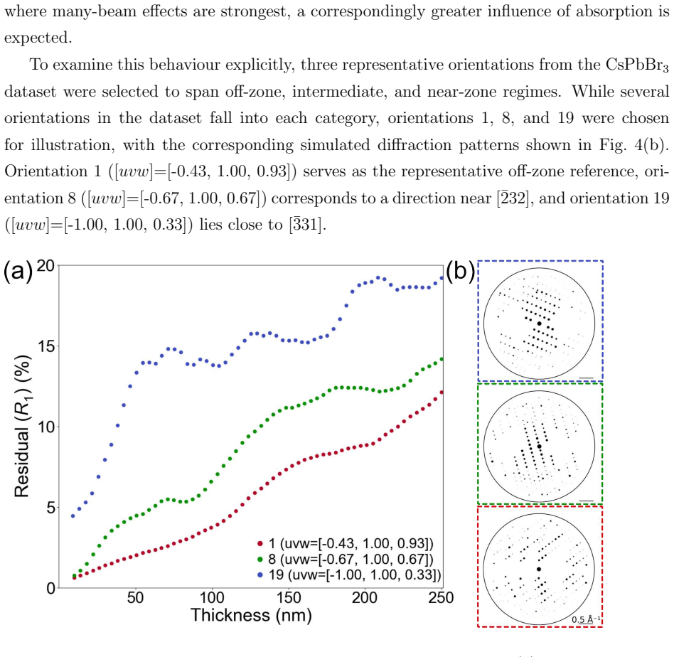
- [§3.2, Eq. (9) and Fig. 3(a)] The residual defined in Eq. (9) compares raw absorptive intensities with elastic-only intensities. If evaluated as written, the uniform attenuation e^{-t/λ̄} dominates R1(t), giving a slope of roughly 1/(2λ̄) ≈ 0.5 %/nm for λ̄ ≈ 90 nm—far larger than the reported ~0.05 %/nm. The actual analysis must have removed the uniform absorption component (as done in Supplementary Eq. S16 and Fig. S2). The main text should define the de-attenuated residual explicitly, otherwise the reader cannot reproduce Fig. 3(a) from Eq. (9). This is load-bearing because the closed-form slope in Eq. (11) is derived only for the de-attenuated quantity.
- [§3.2, Eq. (11) and Fig. 2(b)] The slope predicted by Eq. (11) uses λ̄ and σλ obtained by fitting exponential decays to the very same simulated I_abs/I_no_abs ratios (Fig. 2b) that generate the residual plotted in Fig. 3(a). The agreement between Eq. (11) and the best-fit slope is therefore a consistency check, not an independent prediction. The text should be reworded to avoid implying that the slope is predicted from first principles; at most it shows the fitted Gaussian-λ model reproduces the simulated residual.
- [§2.1 and Conclusion] The quantitative thresholds (e.g., ~2% at 50 nm for CsPbBr3, and the high-Z/ξg exception) are obtained within a model that assumes absorbed intensity is fully removed, uses isotropic Debye–Waller-scaled absorptive form factors, and neglects phonon correlations (Einstein model). The paper itself lists these as limitations, and the experimental improvement for CsPbBr3 (6.4→5.3%) is smaller than the simulated prediction (6.4→4.5%), implying the model may overestimate absorption in that case. That does not bound the error in the opposite direction. A frozen-phonon calculation, or at least an energy-filtered measurement for one representative material/orientation, would substantially increase confidence that the residual curves and the practical conclusion are not artifacts of the optical-potential approximation.
minor comments (6)
- [Supplementary Table S1, borane row] The third borane orientation lists dR1/dt = 0.007 %/nm and predicted contribution 0.17% at t_obs = 170 nm. This is arithmetically inconsistent (0.007×170 ≈ 1.19%); likely a typo for 0.0007 or 0.001. Please correct.
- [§3.2, Eq. (9)] The notation R1(t) conflicts with the experimental R1 defined just above. Consider using R_abs(t) or R_sim(t) to avoid confusion.
- [§3.1, Fig. 2 caption] The caption states “A decaying exponential was fitted to each hkl curve” and gives λ̄ and σλ. The text later calls the resulting slope a “prediction.” Please consistently use “fit” or “parameterization” rather than “prediction” for quantities derived from the same simulation ratios.
- [§2.2, Eq. (8)] Eq. (8) is presented as the weak-absorption limit of the integrated two-beam intensity. The derivation in Supplementary S1 is clear, but the main text would benefit from stating that the Lorentz correction is not included in this analytic expression; the numerical two-beam integration in §3.1 includes it. Otherwise readers may wonder why Eq. (8) does not have a Lorentz factor.
- [§4, Conclusion] The sentence “This work presents what is, to our knowledge, the first implementation of absorption in 3D ED dynamical refinement” is plausible but should be softened or substantiated by a more explicit comparison with prior use of constant absorptive potentials (e.g., Ref. [9]).
- [Fig. S2 caption] The term “de-attenuation factor exp(t/λ)” is used without deriving why λ=88.4 nm is chosen. A one-sentence explanation that this removes the uniform U′0 component would help.
Circularity Check
Supporting R1-slope 'prediction' reuses parameters fitted to the same simulation; central absorption-negligible conclusion has independent experimental support.
-
fitted input called prediction
[Section 3.2, Eq. (11), Fig. 3(a); fitted parameters from Section 3.1, Fig. 2(b)]
"Using the parameters ¯λ = 93.2 nm and σλ = 10.1 nm, the predicted slope from Eq. (11) is dR1/dt = 0.046 % nm−1, in close agreement with the best-fit slope of 0.048 % nm−1 shown in Fig. 3(a)."
The λ̄ and σλ used in Eq. (11) are obtained by fitting decaying exponentials to the same simulated I_abs/I_no_abs intensity-ratio curves from which R1(t) is then computed via Eq. (9). The paper states for Fig. 2(b): 'A decaying exponential was fitted to each hkl curve, yielding ... ¯λ = 93.2 nm, σλ = 10.1 nm (many-beam).' Thus the 'predicted' R1 slope is a function of parameters fitted to the very dataset being summarized; the agreement with the best-fit slope of the R1 curve is a consistency check of the Gaussian/exponential approximation, not an independent prediction of absorption effects. No new data or independent constraint enters.
full rationale
The central practical conclusion—absorption can safely be neglected for routine 3D-ED refinement except in high-Z materials approaching ξg—is not circular. It is supported by independent experimental refinements (CsPbBr3 Robs improving from 6.4% to 5.3%, with negligible changes for quartz and borane) and by a standard complex optical-potential model using literature absorptive form factors. The one genuinely circular-looking step is the quantitative 'prediction' of the R1(t) slope: λ̄ and σλ in Eq. (11) are fitted to the same simulated I_abs/I_no_abs ratios used to compute R1(t), so the close agreement between the Eq. (11) slope and the best-fit R1 slope is a self-consistency check rather than an independent validation. This does not undermine the main conclusion, because that conclusion rests primarily on the experimental refinement comparisons and on the relative insensitivity of integrated intensities, not on the fitted slope. No load-bearing self-citation, imported uniqueness theorem, or ansatz-smuggling was found; the citation of Malik et al. [7] is normal method reuse.
Assumptions & free parameters
free parameters (1)
- Fitted absorption lengths λ_i, λ̄, σλ =
λ̄=89.9 nm (two-beam), 93.2 nm (many-beam), σλ=2.0/10.1 nm; orientation-dependent values in Supp. Fig. S3
assumptions (6)
- domain assumption Complex optical potential with absorptive form factors f'_κ(s,B) from Thomas et al. [10] accurately represents inelastic scattering in 3D ED.
- domain assumption Absorbed electrons are removed from the measured integrated intensity; inelastic redistribution into diffuse background is negligible after background subtraction.
- domain assumption Isotropic Debye-Waller factors and the Einstein model adequately capture thermal vibrations for absorptive corrections.
- domain assumption The Bloch-wave code of Malik et al. [7] and the chosen 60-tilt/±1.5° sampling faithfully reproduce experimental 3D ED integrated intensities.
- ad hoc to paper Per-reflection absorption ratios follow I_abs/I_no_abs = e^{-t/λ_i} with Gaussian-distributed 1/λ_i, used to derive R1(t).
- domain assumption Plasmon/core-loss scattering, beam damage, and crystal imperfections do not dominate the observed residuals and do not prevent isolating absorption effects.
Cite this review
Pith. "Pith review of The role of absorption in three-dimensional electron diffraction dynamical structure refinement." pith.science (2026). https://pith.science/paper/2SYQKJT2
@misc{pith2026260208935,
  author       = {Pith},
  title        = {Pith review of: The role of absorption in three-dimensional electron diffraction dynamical structure refinement},
  year         = {2026},
  howpublished = {\url{https://pith.science/paper/2SYQKJT2}},
  note         = {Machine review of arXiv:2602.08935}
}
abstract
The role of absorption in 3D electron diffraction is established through analytical theory, simulation, and dynamical refinement. A two-beam expression for the absorbed integrated intensity in centrosymmetric crystals is derived, showing that for $t/\xi_g \ll 1$ reflections follow a uniform exponential decay set by the mean absorptive potential $U_0'$. Many-beam simulations of both centrosymmetric and non-centrosymmetric crystals reveal additional reflection-specific anomalous absorption beyond the uniform attenuation set by $U_0'$. Neglecting these effects in dynamical refinement of integrated intensities incurs an error that increases approximately linearly with thickness, with this error becoming more severe near zone axes. Dynamical refinements were performed on CsPbBr$_3$, quartz, and borane, with the inclusion of absorption yielding an improvement in $R_{\mathrm{obs}}$ from $6.4$ to $5.3$ \% for CsPbBr$_3$ and negligible improvements for quartz and borane. Anomalous absorption may therefore be ignored for routine refinement of integrated intensities except in high-$Z$ materials at thicknesses approaching $\xi_g $.
Figures


Figures from the paper (2 more)

Reference graph
Works this paper leans on
-
[1]
Double conical beam-rocking system for measurement of integrated electron diffraction intensities
R. Vincent and P. A. Midgley. “Double conical beam-rocking system for measurement of integrated electron diffraction intensities”. In: Ultramicroscopy 53.3 (1994), pp. 271– 282
1994
-
[2]
3D Electron Diffraction: The Nanocryst allography Revolution
Mauro Gemmi et al. “3D Electron Diffraction: The Nanocryst allography Revolution”. In: ACS Central Science 5.8 (2019). Open Access, pp. 1315–1329. doi: 10 . 1021 / acscentsci.9b00394
2019
-
[3]
In:Zeitschrift f¨ ur Kristallographie - Crystalline Materials238.7-8 (2023), pp
V´ aclav Petˇ r ´ ıˇ cek et al. In:Zeitschrift f¨ ur Kristallographie - Crystalline Materials238.7-8 (2023), pp. 271–282. doi: doi:10.1515/zkri-2023-0005
-
[4]
Hydrogen positions in single na nocrystals revealed by electron diffraction
Luk´ aˇ s Palatinus et al. “Hydrogen positions in single na nocrystals revealed by electron diffraction”. In: Science 355 (2017), pp. 166–169. doi: 10.1126/science.aal3003
-
[5]
Dynamical refinement with multipol ar electron scattering fac- tors
Barbara Olech et al. “Dynamical refinement with multipol ar electron scattering fac- tors”. In: IUCrJ 11 (2024), pp. 309–324. doi: 10.1107/S2052252524001763
-
[6]
Ionisation of atoms determined by ka ppa refinement against 3D electron diffraction data
Ashwin Suresh et al. “Ionisation of atoms determined by ka ppa refinement against 3D electron diffraction data”. In: Nature Communications 15 (2024), p. 9066. doi: 10.1038/s41467-024-53448-2
-
[7]
Hybrid Physics-Machine Learning Models for Quantitative Electron Diffraction Refinements
Shreshth A. Malik et al. “Hybrid Physics-Machine Learning Models for Quantitative Electron Diffraction Refinements”. In: arXiv preprint arXiv:2508.05908 (2025). doi: 10.48550/arXiv.2508.05908. arXiv: 2508.05908 [physics.comp-ph]
work page Pith review arXiv doi:10.48550/arxiv.2508.05908 2025
-
[8]
Electron Diffraction of 3D Molecula r Crystals
Ambarneil Saha et al. “Electron Diffraction of 3D Molecula r Crystals”. In: Chemi- cal Reviews 122.17 (2022). PMID: 35970513, pp. 13883–13914. doi: 10.1021/acs. chemrev.1c00879. eprint: https://doi.org/10.1021/acs.chemrev.1c00879
doi:10.1021/acs 2022
Show all 41 references
-
[9]
Structure refinement from precession electron diffraction data
Luk´ aˇ s Palatinus et al. “Structure refinement from precession electron diffraction data”. In: Acta Crystallographica Section A: Foundations of Crystallography 69 (2013), pp. 171–
2013
-
[10]
Parameterized absorptive electron sc attering factors
M. Thomas et al. “Parameterized absorptive electron sc attering factors”. In: Acta Crystallographica Section A: Foundations and Advances 80.2 (2024), pp. 146–150. doi: 10.1107/S2053273323010963
2024 doi
-
[11]
Aberration-corrected and en ergy-filtered precession elec- tron diffraction
Alexander S. Eggeman et al. “Aberration-corrected and en ergy-filtered precession elec- tron diffraction”. In: Zeitschrift f¨ ur Kristallographie228 (2013), pp. 43–50. doi: 10. 1524/zkri.2013.1565. 20
2013
-
[12]
Absorption parameters in e lectron diffraction theory
C. J. Humphreys and P. B. Hirsch. “Absorption parameters in e lectron diffraction theory”. In: The Philosophical Magazine: A Journal of Theoretical, Experimental and Applied Physics 18.151 (1968), pp. 115–122. doi: 10.1080/14786436808227313
1968 doi
-
[13]
The Absorption Factor in Crystal Spect roscopy
Gustav Albrecht. “The Absorption Factor in Crystal Spect roscopy”. In: Review of Scientific Instruments 10.8 (1939), pp. 221–222. doi: 10.1063/1.1751537
1939 doi
-
[14]
van Genderen et al
E. van Genderen et al. “Ab initio structure determinatio n of nanocrystals of or- ganic pharmaceutical compounds by electron diffraction at r oom temperature using a Timepix quantum area direct electron detector”. In: Acta Crystallographica Section A: Foundations and Advances 7...
2016 doi
-
[15]
Electron crystallography and de dicated electron-diffraction in- strumentation
Petra Simoncic et al. “Electron crystallography and de dicated electron-diffraction in- strumentation”. In: Acta Crystallographica Section E: Crystallographic Communica- tions 79 (2023), pp. 410–422. doi: 10.1107/S2056989023003109
2023 doi
-
[16]
From formulation to structure: 3D el ectron diffraction for the structure solution of a new indomethacin polymorph from an amorphous solid dispersion
Helen W. Leung et al. “From formulation to structure: 3D el ectron diffraction for the structure solution of a new indomethacin polymorph from an amorphous solid dispersion”. In: IUCrJ 11 (2024), pp. 744–748. doi: 10.1107/S2052252524005487
2024 doi
-
[18]
Automated structure factor refi nement from convergent- beam patterns
J. M. Zuo and J. C. H. Spence. “Automated structure factor refi nement from convergent- beam patterns”. In: Ultramicroscopy 35.3–4 (June 1991), pp. 185–196. doi: 10.1016/ 0304-3991(91)90071-D
1991
-
[19]
Energy-filtered convergent-beam di ffraction: examples and future prospects
P. A. Midgley et al. “Energy-filtered convergent-beam di ffraction: examples and future prospects”. In: Ultramicroscopy 59.1–4 (July 1995), pp. 1–13. doi: 10.1016/0304- 3991(95)00014-R
1995 doi
-
[20]
Direct observation of d-orbital holes an d Cu–Cu bonding in Cu 2O
J. M. Zuo et al. “Direct observation of d-orbital holes an d Cu–Cu bonding in Cu 2O”. In: Nature 401 (Sept. 1999), pp. 49–52. doi: 10.1038/43403
1999 doi
-
[21]
A refinable three-parameter e quation for phenomeno- logical absorption in quantitative electron microscopy – d etermining the equation
Philip N. H. Nakashima et al. “A refinable three-parameter e quation for phenomeno- logical absorption in quantitative electron microscopy – d etermining the equation”. In: Journal of Applied Crystallography 58.5 (Oct. 2025), pp. 1665–1676. doi: 10.1107/ S160057672500545X
2025
-
[22]
On the intensities of electron diffrac tion rings
Moses Blackman. “On the intensities of electron diffrac tion rings”. In: Proceedings of the Royal Society A 173.952 (Nov. 1939), pp. 68–82. doi: 10.1098/rspa.1939.0129. 21
1939
-
[23]
Anomalous electron absorption effects in metal foils: theory and comparison with experiment
H. Hashimoto et al. “Anomalous electron absorption effects in metal foils: theory and comparison with experiment”. In: Proceedings of the Royal Society of London. Series A. Mathematical and Physical Sciences 269.1336 (Aug. 1962), pp. 67–87. doi: 10 . 1098/rspa.1962.0164
1962
-
[24]
Dynamical Simulations of Energy-Filtered I nelastic Electron Diffraction Patterns
Z. L. Wang. “Dynamical Simulations of Energy-Filtered I nelastic Electron Diffraction Patterns”. In: Acta Crystallographica Section A 48 (1992), pp. 674–688. doi: 10.1107/ S0108767392005791
1992
-
[25]
Measurement of molecular mo tion in organic semicon- ductors by thermal diffuse electron scattering
Alexander S. Eggeman et al. “Measurement of molecular mo tion in organic semicon- ductors by thermal diffuse electron scattering”. In: Nature Materials 12 (July 2013), pp. 1045–1049. doi: 10.1038/nmat3690
2013 doi
-
[26]
Zur Theorie des Durchgangs schneller Korpusk ularstrahlen durch Materie
H. Bethe. “Zur Theorie des Durchgangs schneller Korpusk ularstrahlen durch Materie”. In: Annalen der Physik 392 (1928), pp. 55–129. doi: 10.1002/andp.19283920202
1928 doi
-
[27]
Effect of Inelastic Waves on Electron Diff raction
Hide Yoshioka. “Effect of Inelastic Waves on Electron Diff raction”. In: Journal of the Physical Society of Japan 12.6 (June 1957), pp. 618–628. doi: 10.1143/JPSJ.12.618
1957 doi
-
[28]
Absorptive Form Factors for High- Energy Electron Diffraction
D. M. Bird and Q. A. King. “Absorptive Form Factors for High- Energy Electron Diffraction”. In: Acta Crystallographica Section A: Foundations of Crystallography 46 (1990), pp. 202–208. doi: 10.1107/S0108767390000502
1990 doi
-
[29]
Anisotropic Thermal Vibrations and Dyna mical Electron Diffraction by Crystals
Lian-Mao Peng. “Anisotropic Thermal Vibrations and Dyna mical Electron Diffraction by Crystals”. In: Acta Crystallographica Section A: Foundations of Crystallography 53 (1997), pp. 663–672. doi: 10.1107/S0108767397004907
1997 doi
-
[30]
Elastic and Inelastic Scattering in Electron Diffraction and Imaging
Zhong Lin Wang. Elastic and Inelastic Scattering in Electron Diffraction and Imaging . English. Includes bibliographical references and index. Ne w York; London: Plenum Press, 1995
1995
-
[31]
Modelling the inelastic scattering of f ast electrons
L. J. Allen et al. “Modelling the inelastic scattering of f ast electrons”. In: Ultrami- croscopy 151 (Apr. 2015), pp. 11–22. doi: 10.1016/j.ultramic.2014.10.011
2015 doi
-
[32]
Bloch wave simulations in the f rozen lattice approximation
Takashi Yamazaki et al. “Bloch wave simulations in the f rozen lattice approximation”. In: Ultramicroscopy 135 (2013), pp. 16–23. doi: https : / / doi . org / 10 . 1016 / j . ultramic.2013.05.018
2013
-
[33]
A physical optics formulation of Bl och waves and its application to 4D STEM, 3D ED and inelastic scattering simulations
Budhika G. Mendis. “A physical optics formulation of Bl och waves and its application to 4D STEM, 3D ED and inelastic scattering simulations”. In: Acta Crystallographica Section A 81.2 (Mar. 2025), pp. 113–123. doi: 10.1107/S2053273325000142. 22
2025 doi
-
[34]
Dirac–Fock calculations of X-ray scatteri ng factors and contributions to the mean inner potential for electron scattering
D. Rez et al. “Dirac–Fock calculations of X-ray scatteri ng factors and contributions to the mean inner potential for electron scattering”. In: Acta Crystallographica Section A 50.4 (1994), pp. 481–497. doi: https://doi.org/10.1107/S0108767393013200 . eprint: https://onlinelib...
1994 doi
-
[35]
P. B. Hirsch et al. Electron Microscopy of Thin Crystals . London: Butterworths, 1965, p. 549
1965
-
[36]
A general Lorentz co rrection for single-crystal diffractometers
G. J. McIntyre and R. F. D. Stansfield. “A general Lorentz co rrection for single-crystal diffractometers”. In: Acta Crystallographica Section A 44.3 (May 1988), pp. 257–262. doi: 10.1107/S0108767387011656
1988 doi
-
[37]
In: Zeitschrift f¨ ur Kristallographie 225.2-3 (2010), pp
Daliang Zhang et al. In: Zeitschrift f¨ ur Kristallographie 225.2-3 (2010), pp. 94–102. doi: doi:10.1524/zkri.2010.1202
2010
-
[38]
Simple Bayesian method for improve d analysis of quasi-two- dimensional scattering data
Alexander T. Holmes. “Simple Bayesian method for improve d analysis of quasi-two- dimensional scattering data”. In: Phys. Rev. B 90 (2 July 2014), p. 024514. doi: 10. 1103/PhysRevB.90.024514
2014
-
[39]
Accurate structure models and absolu te configuration determi- nation using dynamical effects in continuous-rotation 3D el ectron diffraction data
Paul B. Klar et al. “Accurate structure models and absolu te configuration determi- nation using dynamical effects in continuous-rotation 3D el ectron diffraction data”. en. In: Nature Chemistry 15.6 (June 2023). Number: 6 Publisher: Nature Publishing Group, pp. 848–855. doi: 10....
2023 doi
-
[40]
Specifics of the data processing of precession electron diffraction tomography data and their implementation in the program PET S2.0
Luk´ aˇ s Palatinus et al. “Specifics of the data processing of precession electron diffraction tomography data and their implementation in the program PET S2.0”. In: Acta Crys- tallographica Section A: Structural Science, Crystal Engineering and Materials 75.4 (Aug. 2019), pp. ...
2019 doi
-
[41]
Electron diffraction determines mo lecular absolute configuration in a pharmaceutical nanocrystal
Petr Br´ azda et al. “Electron diffraction determines mo lecular absolute configuration in a pharmaceutical nanocrystal”. In: Science 364 (2019), pp. 667–669. doi: 10.1126/ science.aaw2560. 23 Supplementary Information: The role of absorption in dynamical three-dimensional elect...
2019
-
[188]
doi: 10.1107/S0108767312048171
Reviewed August 3, 2026 · model on record in the stance chip above.
Discussion (0). Continue with ORCID to comment.