Ptychographic Algorithms for Phase Recovery in 4D Scanning Transmission Electron Microscopy
Pith reviewed 2026-06-27 21:36 UTC · model grok-4.3
The pith
Ptychography recovers the electron probe wavefunction and specimen transmission function from 4D STEM diffraction patterns.
A machine-rendered reading of the paper's core claim, the machinery that carries it, and where it could break.
Core claim
Ptychography is a reconstruction algorithm that allows the extraction of the probe wavefunction and the multiplicative object transmission function of the specimen. It is implemented through direct and iterative schemes such as ePIE, WDD, and SSB. An SSB reconstruction was performed with an original script on simulated MoS2 monolayer data, and four-dimensional datasets were acquired on a STEM instrument.
What carries the argument
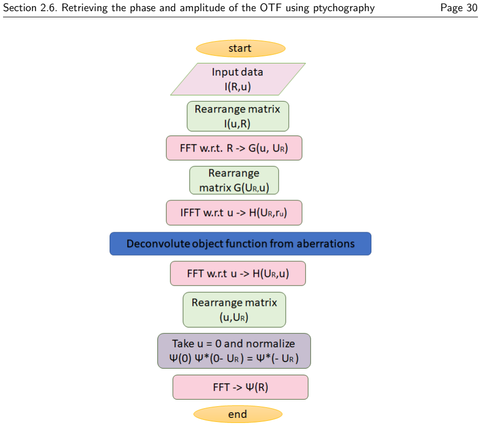
The Single Side-Band (SSB) reconstruction, a simplified form of Wigner distribution deconvolution that isolates phase information from the interference cross-terms in the 4D diffraction data.
If this is right
- The SSB method demonstrated on simulated data can be applied directly to the recorded experimental 4D STEM datasets for phase imaging.
- Implementation of the full WDD algorithm would recover additional information beyond the SSB approximation.
- The mathematical framework allows systematic comparison of iterative (ePIE) and direct (WDD/SSB) schemes on the same dataset.
- Atomic-resolution transmission functions become available for materials such as monolayer MoS2 without requiring conventional phase-contrast optics.
Where Pith is reading between the lines
- The recorded experimental 4D data sets could serve as a benchmark for testing noise robustness of other ptychographic variants not covered in the thesis.
- Extending the SSB script to thicker or defective specimens would test how well the multiplicative transmission model holds beyond the monolayer case.
- Combining the probe recovery from ptychography with conventional STEM imaging modes could reduce dose while maintaining resolution.
Load-bearing premise
The simulated MoS2 monolayer diffraction patterns used for the SSB test accurately reproduce the noise levels and experimental conditions of real 4D STEM measurements.
What would settle it
Applying the same SSB script to actual experimental 4D STEM data of MoS2 and obtaining a reconstructed object function that fails to show the expected atomic lattice positions would show the simulation does not capture real conditions.
Figures


read the original abstract
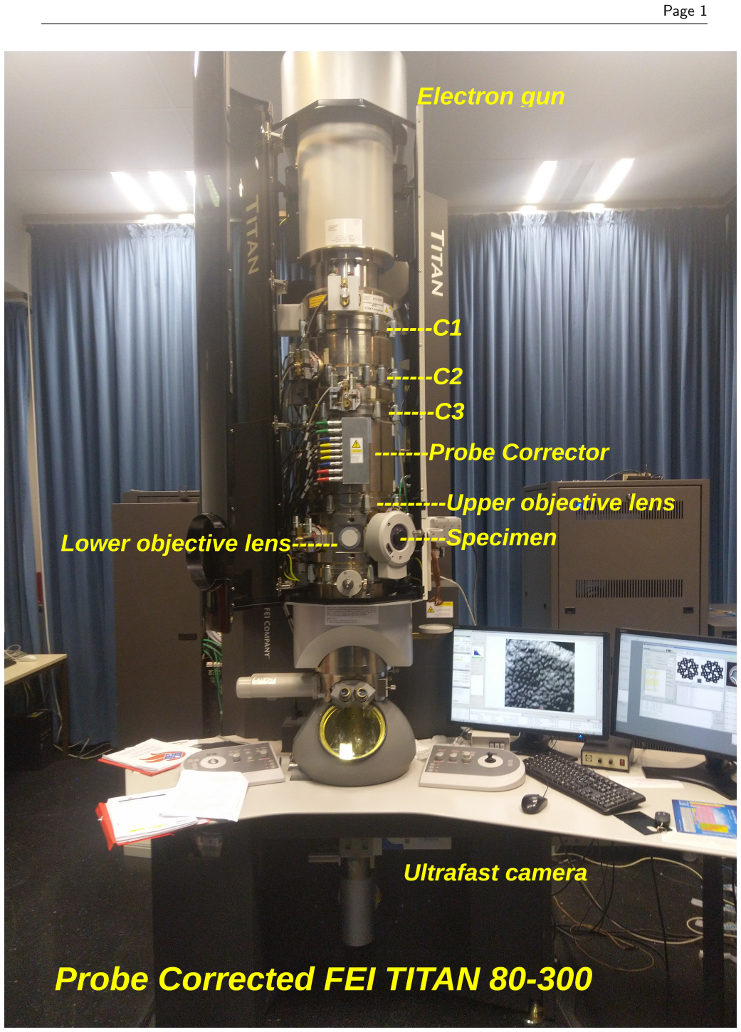
In Momentum-resolved Scanning Transmission Electron Microscopy (4D STEM), a convergent electron beam is raster-scanned across a think specimen in 2D in real space. The corresponding 2D diffraction pattern, in momentum space, to each point is recorded, forming a 4D data set. Information decoding process can follow thereafter to produce an image of the specimen in real space. Ptychography is reconstruction algorithm that allow the extraction of the probe wavefunction and the multiplicative object transmission function of the specimen. Ptychography is implemented through direct and iterative schemes. Some of which are the extended Ptychographic Iterative Engine (ePIE), the Wigner Distribution Deconvolution (WDD) and the simpler version of WDD, the Single Side-Band (SSB). This thesis gives an overview of STEM ptychography giving examples of its experimental and simulated implementations. The different ptychographic reconstruction methods are explored in a mathematical framework when applicable. Finally, an SSB reconstruction was made using an original script for simulated data of MoS2 monolayer. Moreover, four-dimensional data was recorded using a STEM instrument. A natural step following this research would be the implementation of the WDD algorithm.
Editorial analysis
A structured set of objections, weighed in public.
Referee Report
Summary. The manuscript provides an overview of ptychography in 4D-STEM, describing direct and iterative methods (ePIE, WDD, SSB) with mathematical frameworks where applicable. It reports an original-script SSB reconstruction performed on simulated MoS2 monolayer data and notes the acquisition of experimental 4D-STEM data, with WDD implementation listed as future work.
Significance. If the mathematical sections are rigorous and the SSB implementation is shown to be correct via validation, the work could function as an accessible introduction to 4D-STEM ptychography. The exclusive reliance on simulation without experimental processing or cross-checks against reference codes, however, restricts its contribution to practical phase recovery.
major comments (2)
- [Abstract] Abstract: the claim that an SSB reconstruction was performed supplies no equations, validation metrics, error analysis, or comparison to ground truth, so the correctness of the original script cannot be assessed.
- [Experimental data section] Section describing experimental data: experimental 4D-STEM data were recorded but left unprocessed, so the central claim that the method extracts probe and object functions in real 4D-STEM settings rests on the untested premise that the MoS2 simulation reproduces experimental noise, aberrations, and detector response.
minor comments (2)
- [Abstract] Abstract: 'think specimen' is a typo for 'thin specimen'.
- [Abstract] Abstract: 'Ptychography is reconstruction algorithm that allow' should read 'Ptychography is a reconstruction algorithm that allows'.
Simulated Author's Rebuttal
We thank the referee for the review and constructive feedback. We address the major comments point by point below, indicating planned revisions where appropriate.
read point-by-point responses
-
Referee: [Abstract] Abstract: the claim that an SSB reconstruction was performed supplies no equations, validation metrics, error analysis, or comparison to ground truth, so the correctness of the original script cannot be assessed.
Authors: The abstract is a concise summary; the mathematical framework for SSB (as a simplified WDD) and the implementation details of the original script are presented in the main text. We agree that quantitative validation would strengthen the work and allow direct assessment of correctness. In revision we will add explicit equations for the SSB reconstruction, validation metrics, error analysis, and comparison to the known ground-truth MoS2 structure in a dedicated results subsection. revision: yes
-
Referee: [Experimental data section] Section describing experimental data: experimental 4D-STEM data were recorded but left unprocessed, so the central claim that the method extracts probe and object functions in real 4D-STEM settings rests on the untested premise that the MoS2 simulation reproduces experimental noise, aberrations, and detector response.
Authors: The manuscript does not assert that probe and object functions have been extracted from real experimental data. It reports acquisition of the 4D-STEM dataset and explicitly identifies WDD implementation on that data as future work. The SSB demonstration is performed exclusively on simulated MoS2 data. We will revise the text to state these scope limitations more clearly and to note that the simulation does not claim to reproduce all experimental effects. revision: partial
Circularity Check
No significant circularity; paper is overview plus implementation of prior methods on simulated data
full rationale
The manuscript is an overview of established ptychographic algorithms (ePIE, WDD, SSB) with a single original-script SSB reconstruction performed exclusively on simulated MoS2 monolayer data. No derivations, parameter fits presented as predictions, self-citation load-bearing steps, or ansatzes smuggled via citation are present. The central contribution reduces to applying known SSB mathematics to synthetic input; the unprocessed experimental dataset is explicitly noted as future work and does not enter any claimed result. This satisfies the default expectation of a self-contained, non-circular implementation report.
Axiom & Free-Parameter Ledger
axioms (1)
- standard math Standard Fourier optics and multiplicative transmission function model for thin specimens in STEM
Reference graph
Works this paper leans on
-
[1]
URL https://doi.org/10.1107/S0365110X59001104
doi: 10.1107/S0365110X59001104. URL https://doi.org/10.1107/S0365110X59001104. Nellist P. D. Scanning Transmission Electron Microscopy , pages 49 –
-
[3]
URL http://www.opticsexpress.org/abstract.cfm?URI=oe-23-26-33812
doi: 10.1364/OE.23.033812. URL http://www.opticsexpress.org/abstract.cfm?URI=oe-23-26-33812. A. J. D’Alfonso, A. J. Morgan, A. W. C. Yan, P. Wang, H. Sawada, A. I. Kirkland, and L. J. Allen. Deterministic electron ptychography at atomic resolution. Phys. Rev. B , 89:064101, Feb
-
[4]
URL https://link.aps.org/doi/10.1103/PhysRevB.89.064101
doi: 10.1103/PhysRevB.89.064101. URL https://link.aps.org/doi/10.1103/PhysRevB.89.064101. H. Faulkner and J. Rodenburg. Movable aperturelensless transmission microscopy: A novel phaseretrieval algorithm. Physics Review Letters , 93(2)(023903),
-
[5]
doi: 10.1021/nn405938z. PMID: 24660756. Si Gao, Peng Wang, Fucai Zhang, Gerardo T. Martinez, Peter D. Nellist, Xiaoqing Pan, and Angus I. Kirkland. Electron ptychographic microscopy for three-dimensional imaging. Nat Commun., 8(163),
-
[6]
Martin Huth, Robert Ritz, Colum M
doi: 10.1038/ncomms1733. Martin Huth, Robert Ritz, Colum M. O’Leary, Ian Griffiths, Peter Nellist, and Heike Soltau. Ultrafast ptychography with 7500 frames per second. Microscopy and Microanalysis , 25(S2):40–41,
-
[7]
36 REFERENCES Page 37 Eugene S
doi: 10.1017/S143192761900093X. 36 REFERENCES Page 37 Eugene S. Kadantsev and Pawel Hawrylak. Electronic structure of a single mos2 monolayer. Solid State Communications, 152(10):909 – 913,
-
[8]
doi: https://doi.org/10.1016/j.ssc.2012.02.005
ISSN 0038-1098. doi: https://doi.org/10.1016/j.ssc.2012.02.005. URL http://www.sciencedirect.com/science/article/pii/S0038109812000889. Richard Kasprowicz, Rakesh Suman, and Peter O’Toole. Characterising live cell behaviour: Traditional label-free and quantitative phase imaging approaches. The International Journal of Biochemistry and Cell Biology , 84:89 – 95,
-
[9]
doi: https://doi.org/10.1016/j.biocel.2017.01.004
ISSN 1357-2725. doi: https://doi.org/10.1016/j.biocel.2017.01.004. URL http://www.sciencedirect.com/science/article/pii/S1357272517300055. Earl J. Kirkland. Advanced Computing in Electron Microscopy . Springer, second edition,
-
[10]
doi: 10.1007/978-1-4419-6533-2 . Peng Li, Tega B. Edo, and John M. Rodenburg. Ptychographic inversion via wigner distribution deconvolution: Noise suppression and probe design. Ultramicroscopy, 147:106 – 113, 2014a. Peng Li, Tega B. Edo, and John M. Rodenburg. Ptychographic inversion via wigner distribution deconvolution: Noise suppression and probe desig...
-
[11]
doi: \url{https://doi.org/10.1016/j.ultramic.2009.05.012}
ISSN 0304-3991. doi: \url{https://doi.org/10.1016/j.ultramic.2009.05.012}. Joanne Marrison, Lotta R¨ aty, Poppy Marriott, and Peter O’Toole. Ptychography – a label free, high-contrast imaging technique for live cells using quantitative phase information. Sci. Rep., 3 (2369),
-
[12]
doi: \url{https://doi.org/10.1038/srep02369}. M¨ uller-Caspary. Structure, composition, functionality: Advanced electron microscopy techniques in solid state physics,
-
[13]
doi: 10.1063/1.5143213. Stephen J. Pennycook and Peter D. Nellist, editors. Scanning Transmission Electron Microscopy Imaging and Analysis . Springer,
-
[14]
doi: 10.1007/978-1-4419-7200-2 . Timothy J. Pennycook, Gerardo T. Martinez, Peter D. Nellist, and Jannik C. Meyer. High dose efficiency atomic resolution imaging via electron ptychography. Ultramicroscopy, 196:131 – 135,
-
[15]
doi: https://doi.org/10.1016/j.ultramic.2018.10.005
ISSN 0304-3991. doi: https://doi.org/10.1016/j.ultramic.2018.10.005. URL http://www.sciencedirect.com/science/article/pii/S0304399118302316. Timothy J Pennycooka, Andrew R Lupinic, Hao Yangb, Matthew F Murfittd, Lewys Jonesb, and Peter D Nellist. Efficient phase contrast imaging in stem using a pixelated detector. part 1: Experimental demonstration at ato...
-
[16]
doi: https://doi.org/10.1016/0304-3991(93)90105-7
ISSN 0304-3991. doi: https://doi.org/10.1016/0304-3991(93)90105-7. URL http://www.sciencedirect.com/science/article/pii/0304399193901057. John Rodenburg and Andrew Maiden. Ptychography, pages 819 –
-
[18]
URL https://doi.org/10.1007/978-3-030-00069-1
-
[19]
A. Stevens, L. Luzi, H. Yang, L. Kovarik, B. L. Mehdi, A. Liyu, M. E. Gehm, and N. D. Browning. A sub-sampled approach to extremely low-dose stem. Applied Physics Letters , 112(4), 2018a. doi: 10.1063/1.5016192. Andrew Stevens. Compressive Sensing in Transmission Electron Microscopy . PhD thesis, Duke University, January
-
[20]
ISSN 2050-5698. doi: 10.1093/jmicro/dft042. Andrew Stevens, Hao Yang, Weituo Hao, Lewys Jones, Colin Ophus, Peter D. Nellist, and Nigel D. Browning. Subsampled stem-ptychography. Applied Physics Letters , 113(3):033104, 2018b. doi: 10.1063/1.5040496. URL https://doi.org/10.1063/1.5040496. A. Strauch. Internal communication
-
[21]
H Yang, L Jones, H Ryll, M Simson, H Soltau, Y Kondo, R Sagawa, H Banba, I Maclaren, and P D Nellist
doi: 10.1007/978-1-4757-2519-3 . H Yang, L Jones, H Ryll, M Simson, H Soltau, Y Kondo, R Sagawa, H Banba, I Maclaren, and P D Nellist. 4d stem: High efficiency phase contrast imaging using a fast pixelated detector. J. Phys.: Conf. Ser. 644 012032 ,
-
[22]
URL http://dx.doi.org/10.1038/ncomms12532
doi: 10.1038/ncomms12532. URL http://dx.doi.org/10.1038/ncomms12532. REFERENCES Page 39 Hao Yang, Ian MacLaren, Lewys Jones, Gerardo T. Martinez, Martin Simson, Martin Huth, Henning Ryll, Heike Soltau, Ryusuke Sagawa, Yukihito Kondo, Colin Ophus, Peter Ercius, Lei Jin, Andr´ as Kov´ acs, and Peter D. Nellist. Electron ptychographic phase imaging of light ...
-
[23]
doi: https://doi.org/10.1016/j.ultramic.2017.02.006
ISSN 0304-3991. doi: https://doi.org/10.1016/j.ultramic.2017.02.006. URL http://www.sciencedirect.com/science/article/pii/S0304399117300773. Ondrej Krivanek: A research life in EELS and aberration corrected STEM. Liqi Zhou, Jingdong Song, Judy S. Kim, Xudong Pei, Chen Huang, Mark Boyce, Luiza Mendon¸ ca, Daniel Clare, Alistair Siebert, Christopher S. Alle...
-
[24]
doi: \url{https://doi.org/10.1038/s41467-020-16391-6 }
discussion (0)
Sign in with ORCID, Apple, or X to comment. Anyone can read and Pith papers without signing in.