REVIEW 5 major objections 6 minor 2 cited by
HYATT-Net is Grand: A Hybrid Attention Network for Performant Anatomical Landmark Detection
T0 review · 5 major / 6 minor · reviewed 2026-08-11 · deepseek-v4-flash
Pith's one-line read A hybrid attention network beats prior bests on five X-ray landmark datasets.
desk verdict A workmanlike hybrid architecture with a SOTA claim that isn't yet backed by the experiments; the core idea is reasonable but the comparison table has errors and protocol-mixing problems. read the letter →
The pith
A machine-rendered reading of the paper's core claim, the machinery that carries it, and where it could break.
The reading
What carries the argument
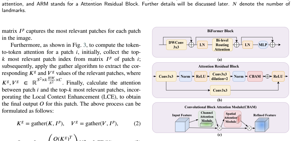
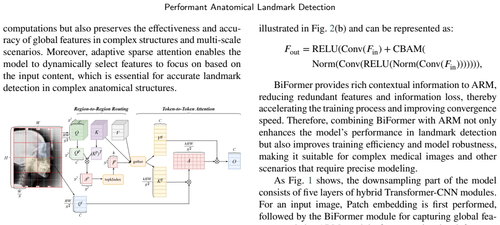
The carrying object is the BiFormer module built on Bi-Level Routing Attention (BRA), a two-stage sparse attention. BRA first computes patch-level queries and keys, forms a patch adjacency matrix, and applies a row-wise top-$k$ operator to select the $k$ most relevant patches per region; it then gathers only those key-value pairs and computes token-to-token attention with a depth-wise convolution (kernel size 5) as Local Context Enhancement. This mechanism is what makes global context modeling tractable at high resolution, and it is paired with two supporting modules: the Attention Residual Module, which wraps a dilated-convolution residual block with CBAM-style channel and spatial attention, and the Feature Fusion Correction Module, which concatenates global-pooled image features with the upsampled feature map to recover resolution. The deep-supervision scheme (MSE losses on three heatmap scales) ties the multi-scale predictions together.
What would settle it
Retrain HTC, CeLDA, FARNet, and HYATT-Net from scratch with identical preprocessing, augmentation, and splits on ISBI2015 and CephAdoAdu using the released code and data, and compare MRE; if any baseline matches or beats the reported HYATT-Net numbers, the claimed state-of-the-art margins are not established.
Extended reading notes
Core claim
On the paper's own terms, the discovery is that query-aware dynamic sparsity in attention is sufficient to push anatomical landmark detection past prior state-of-the-art methods. HYATT-Net embeds the BiFormer module, which uses Bi-Level Routing Attention: the feature map is partitioned into patches, a coarse patch-level adjacency matrix is built from average queries and keys, and a row-wise top-$k$ operator keeps only the $k$ most relevant patches per query; token-to-token attention then runs on the gathered key-value pairs, supplemented by a depth-wise convolution for local context. This global feature is fed into an Attention Residual Module whose channel and spatial attention (CBAM) refine local features, and a Feature Fusion Correction Module pools global image context to restore resolution lost in downsampling. Training uses deep supervision with a mean-square error loss on heatmaps at three resolutions ($\sigma=2,2,4$; weights $1,3,3$). The reported outcome is a mean radial error of 1.13 mm on ISBI2015 (HTC: 1.19 mm), 1.05 mm on ISBI2023, 0.98 mm on CephAdoAdu (CeLDA: 1.05 mm), 0.53 mm on the hand dataset, and 6.64 px on the pelvic dataset, with success detection rates higher than or close to the best baselines at most thresholds.
Load-bearing premise
The claimed margins of improvement assume that the previously published baseline numbers cited with asterisks in Tables 1-3 were obtained under the same image resizing, augmentation, landmark sets, and train/test splits as the authors' own runs.
Editorial extensions
If this is right
- On the five evaluated X-ray benchmarks, HYATT-Net sets a new best mean radial error, so the method transfers across cephalometric, hand, and pelvic imaging with different resolutions and annotation counts.
- Because the routing attention concentrates computation on the most relevant regions, high-resolution inputs (1024 by 1216) become affordable, which implies the approach can scale to other high-detail medical images without aggressive downsampling.
- Ablations replacing BiFormer with a Swin or basic Transformer backbone lose over 1% SDR and more than 4% relative MRE, so the bi-level routing attention itself, not merely the transformer structure, drives the gain.
- Adding CBAM and the feature fusion correction module each produces a small but consistent MRE reduction on ISBI2015, supporting the claim that global-guided local refinement and multi-scale fusion are complementary.
- The released code and data allow the reported numbers to be reproduced and reused as a new baseline for later landmark detection work.
Reading between the lines
- If the gains replicate under a common training protocol, the same recipe could transfer to other sparse-target dense prediction tasks such as fetal ultrasound landmark detection or surgical tool localization, where global geometry matters and images are high resolution; this goes beyond the paper's experiments.
- A natural extension would be to condition the top-$k$ routing on an anatomical prior or a coarse landmark proposal map, which could further cut computation while holding accuracy; the paper does not test this.
- Because the method helps most on the smallest training set (ISBI2015), a plausible but untested corollary is that dynamic attention's benefit grows as data become scarcer.
Editorial analysis
A structured set of objections, weighed in public.
Referee Report
Summary. The manuscript proposes HYATT-Net, a U-shaped hybrid CNN-Transformer architecture for anatomical landmark detection from X-ray images. The main components are a BiFormer module using bi-level routing attention for efficient global context, an Attention Residual Module with CBAM for local feature refinement, a Feature Fusion Correction Module that combines multi-scale features with global pooling, and deep supervision with MSE loss on multi-resolution heatmaps. The authors evaluate on five datasets (ISBI2015, ISBI2023, CephAdoAdu, Hand X-rays, Pelvic X-rays) and report MRE/SDR improvements over several baselines, e.g., MRE 1.13 mm on ISBI2015 versus 1.19 mm for HTC and 0.98 mm on CephAdoAdu versus 1.05 mm for CeLDA. Code and data are released on GitHub.
Significance. If the reported results are taken at face value and the baseline comparisons are protocol-matched, the work offers a modest but consistent improvement over prior state of the art across several anatomies, with released code that would benefit the community. The architecture itself is a combination of existing components (BiFormer, CBAM, deep supervision, feature fusion), so the novelty lies in the specific integration for ALD rather than in a new attention mechanism. The main strengths are the breadth of datasets, the public release of code/data, and the consistent direction of the reported improvements. However, the claimed margins over baselines (0.03–0.07 mm) are comparable to or smaller than the ablation differences within the paper and well below the reported standard deviations; the reliability of the quantitative claims is also undercut by text/table inconsistencies and missing protocol documentation. The paper is a plausible contribution if these issues are resolved.
major comments (5)
- [Section 4.4.1, Table 1] The text states that on ISBI2023 'the SDR at 2.0mm and 2.5mm reaches 84.78% and 89.92%', but Table 1 reports 87.43% and 91.43% for ISBI2023; the 84.78/89.92 values are the ISBI2015 numbers. This mismatch makes it impossible for the reader to determine which dataset the quoted margins refer to, and it affects the claimed superiority over HTC and FARNet on ISBI2023. Please correct the text or the table and re-verify all quoted differences after the correction.
- [Table 1, CELDA (Average) row] The reported CeLDA SDR values are 89.13% at 2 mm, 93.60% at 2.5 mm, 99.67% at 3 mm, and 98.67% at 4 mm. Since SDR is a cumulative success rate within a radius, it must be non-decreasing in the threshold; 99.67% at 3 mm and 98.67% at 4 mm is impossible and indicates an entry error or an inconsistent definition. Please correct this row and run the same monotonicity check on all SDR columns, as a non-monotonic SDR also appears in the CELDA (Teenager) row where 96.90% at 3 mm is followed by 98.33% at 4 mm (this is allowed) but the average row is not.
- [Section 4.2 and Tables 1–3] The SOTA comparisons mix published numbers (marked with *) with apparently in-house numbers without stating which baselines were rerun and under what protocol. Differences in image resizing (1024×1216 vs 1024×1024 vs 512×512), augmentation, train/test splits, landmark sets, metric conventions, and the 50 mm hand-scaling assumption can plausibly shift MRE by more than the claimed 0.03–0.07 mm margins. Because the central claim is comparative, please document the exact protocol for every baseline or rerun all baselines under the same pipeline; otherwise the SOTA claim is not established.
- [Section 4.5, Table 4] The ablation attributes 0.007 mm improvement to CBAM and 0.005 mm to FFCM on ISBI2015. Given that the reported standard deviations are about 1.1–1.2 mm and no significance test or repeated-run statistics are provided, these differences are indistinguishable from noise. The claim that these modules 'further enhance SDR and reduce MRE' is therefore not supported by the evidence as presented. Please add variance across seeds, confidence intervals, or a paired significance test, or rephrase the attribution to avoid overclaiming.
- [Abstract and Section 4.4.3] The paper claims superiority in 'accuracy, robustness, and efficiency,' but Section 4 contains no efficiency measurements (parameters, FLOPs, inference time) and no explicit robustness experiments (e.g., noise, corruption, or domain shift beyond the five datasets). The multi-dataset results support generalizability, not the stated robustness and efficiency claims. Please either add the corresponding experiments and metrics or limit the claims to accuracy.
minor comments (6)
- [Equation (1)] The normalization factor in the 2D Gaussian heatmap is written as 1/(√(2π)σ), which is the 1D normalization; for a 2D isotropic Gaussian it should be 1/(2πσ²). As written the heatmap is not volume-normalized, although for training this may not matter in practice.
- [Section 3.2] The dimensions of Q, K, V and Q_p, K_p are inconsistent: after partitioning an H×W image into S×S patches, the number of patches is HW/S², so Q should be in R^{HW/S² × C} rather than R^{S² × HW/S² × C}. Please clarify the notation.
- [Section 3.4 vs Section 4.3] Equation (4) defines the total loss with weights α₁ and α₂, and the text says α₁ = 3 and α₂ = 3; Section 4.3 instead reports w₁ = 1, w₂ = 3, w₃ = 3. Please reconcile these notations and values.
- [Section 4.4.1] In the CephAdoAdu paragraph, the adolescent MRE is reported as 0.85 px, but the dataset and all other MRE values in that section are in mm; this is presumably a typo and should read 0.85 mm.
- [Table 1 and Section 4.1.1] The dataset is referred to inconsistently as 'CephAdoAdu' in the text and 'CELDA' in Table 1; please use a consistent naming convention and clarify whether 'CELDA (Average)' refers to the full CephAdoAdu test set.
- [References] References [8] and [28] are the same paper (Lowe, 1999), and references [9] and [31] are the same paper (Liu et al., 2010); please merge the duplicates.
Circularity Check
No significant circularity: the paper's claims rest on external test-set comparisons and independently cited components, not on self-referential derivations.
full rationale
HYATT-Net's central claim is empirical: it reports MRE and SDR numbers on five public or externally provided datasets, computed from ground-truth landmark annotations that are independent of the model architecture. No equation in the paper defines a proposed component in terms of the target metric, and no fitted parameter is renamed as a prediction; the loss weights and heatmap sigmas are fixed hyperparameters, and the ablations (CBAM, FFCM, backbone choice) are reported as measured differences on held-out test data. The BiFormer module is adopted from an external CVPR 2023 paper (Zhu et al. [41]), and CBAM is an external module [43], so the core architectural choices are not justified by a self-citation chain. The authors' own prior works (GU2Net [36], DATR [23], PELE [48]) appear only as comparison baselines or as providers of dataset/evaluation conventions, not as premises that force the conclusion. Concerns about protocol matching of starred baseline numbers, the non-monotonic CeLDA SDR row in Table 1, and the text/table mismatch for ISBI2023 SDR values are correctness and verification risks, not circularity. The derivation chain is therefore self-contained with respect to circularity.
Assumptions & free parameters
free parameters (4)
- Deep supervision loss weights (alpha1, alpha2) =
alpha1 = 3, alpha2 = 3
- Heatmap standard deviations (sigma1, sigma2, sigma3) =
2, 2, 4
- BiFormer patch size and top-k routing parameter =
not specified in the paper
- Wrist distance assumption for Hand dataset =
50 mm
assumptions (4)
- domain assumption Heatmap regression with MSE loss and Gaussian targets is a valid surrogate objective for minimizing landmark localization error.
- domain assumption Published baseline results marked with * are directly comparable to the authors' runs despite possible differences in training setup, preprocessing, and splits.
- domain assumption The private CephAdoAdu dataset annotations are reliable and the train/test split matches the source paper.
- domain assumption Bi-Level Routing Attention retains enough global context for landmark detection while reducing computation.
Cite this review
Pith. "Pith review of HYATT-Net is Grand: A Hybrid Attention Network for Performant Anatomical Landmark Detection." pith.science (2026). https://pith.science/paper/PCVQTYK3
@misc{pith2026241206499,
  author       = {Pith},
  title        = {Pith review of: HYATT-Net is Grand: A Hybrid Attention Network for Performant Anatomical Landmark Detection},
  year         = {2026},
  howpublished = {\url{https://pith.science/paper/PCVQTYK3}},
  note         = {Machine review of arXiv:2412.06499}
}
read the original abstract
Anatomical landmark detection (ALD) from a medical image is crucial for a wide array of clinical applications. While existing methods achieve quite some success in ALD, they often struggle to balance global context with computational efficiency, particularly with high-resolution images, thereby leading to the rise of a natural question: where is the performance limit of ALD? In this paper, we aim to forge performant ALD by proposing a {\bf HY}brid {\bf ATT}ention {\bf Net}work (HYATT-Net) with the following designs: (i) A novel hybrid architecture that integrates CNNs and Transformers. Its core is the BiFormer module, utilizing Bi-Level Routing Attention for efficient attention to relevant image regions. This, combined with Attention Residual Module(ARM), enables precise local feature refinement guided by the global context. (ii) A Feature Fusion Correction Module that aggregates multi-scale features and thus mitigates a resolution loss. Deep supervision with a mean-square error loss on multi-resolution heatmaps optimizes the model. Experiments on five diverse datasets demonstrate state-of-the-art performance, surpassing existing methods in accuracy, robustness, and efficiency. The HYATT-Net provides a promising solution for accurate and efficient ALD in complex medical images. Our codes and data are already released at: \url{https://github.com/ECNUACRush/HYATT-Net}.
Figures

Figures from the paper (5 more)


Forward citations
Cited by 2 Pith papers
-
SimCroP: Radiograph Representation Learning with Similarity-driven Cross-granularity Pre-training
SimCroP learns chest-CT representations by aligning each report sentence to its most similar visual patches and fusing whole-scan and word-patch features, reporting higher classification and segmentation scores than s...
-
U-RWKV: Lightweight medical image segmentation with direction-adaptive RWKV
U-RWKV is a lightweight U-shaped medical image segmenter that combines multi-directional RWKV scanning with stage-adaptive channel recalibration, reporting competitive Dice scores with about three million parameters.
Reference graph
Works this paper leans on
-
[1]
Handbook of medical image computing and computer assisted intervention
SKevinZhou,DanielRueckert,andGaborFichtinger. Handbook of medical image computing and computer assisted intervention. Academic Press, 2019
work page 2019
-
[2]
Percutaneous vertebral surgery
J Chiras, C Depriester, A Weill, MT Sola-Martinez, and H Deramond. Percutaneous vertebral surgery. technics and indications.Journal of neuroradiology= Journal de neuroradiologie, 24(1):45–59, 1997
work page 1997
-
[3]
Ching-WeiWang,Cheng-TaHuang,Meng-CheHsieh, Chung-Hsing Li, Sheng-Wei Chang, Wei-Cheng Li, Rémy Vandaele, Raphaël Marée, Sébastien Jodogne, Pierre Geurts, et al. Evaluation and comparison of anatomical landmark detection methods for cephalo- metric x-ray images: a grand challenge.IEEE trans- actions on medical imaging, 34(9):1890–1900, 2015
work page 1900
-
[4]
Robust anatomical land- mark detection for mr brain image registration
Dong Han, Yaozong Gao, Guorong Wu, Pew-Thian Yap, and Dinggang Shen. Robust anatomical land- mark detection for mr brain image registration. In Medical Image Computing and Computer-Assisted X. Zhou et al.:Preprint submitted to IEEE Page 12 of 15 Performant Anatomical Landmark Detection Intervention–MICCAI 2014: 17th International Con- ference, Boston, MA,...
work page 2014
-
[5]
IEEE transactions on medical imaging, 36(1):332– 342, 2016
Ozan Oktay, Wenjia Bai, Ricardo Guerrero, Martin Rajchl, Antonio De Marvao, Declan P O’Regan, Stu- art A Cook, Mattias P Heinrich, Ben Glocker, and DanielRueckert.Stratifieddecisionforestsforaccurate anatomical landmark localization in cardiac images. IEEE transactions on medical imaging, 36(1):332– 342, 2016
work page 2016
-
[6]
Diffuseex- pand: Expanding dataset for 2d medical image seg- mentation using diffusion models
Shitong Shao, Xiaohan Yuan, Zhen Huang, Ziming Qiu, Shuai Wang, and Kevin Zhou. Diffuseex- pand: Expanding dataset for 2d medical image seg- mentation using diffusion models. arXiv preprint arXiv:2304.13416, 2023
arXiv 2023
-
[7]
Parametric modelling and segmentation of vertebralbodiesin3dctandmrspineimages
DarkoŠtern,BoštjanLikar,FranjoPernuš,andTomaž Vrtovec. Parametric modelling and segmentation of vertebralbodiesin3dctandmrspineimages. Physics in Medicine & Biology, 56(23):7505, 2011
work page 2011
-
[10]
Robust and accurate shape model matchingusingrandomforestregression-voting
Claudia Lindner, Paul A Bromiley, Mircea C Ionita, and Tim F Cootes. Robust and accurate shape model matchingusingrandomforestregression-voting. IEEE transactions on pattern analysis and machine intelli- gence, 37(9):1862–1874, 2014
work page 2014
Show all 48 references
-
[11]
U-net: Convolutional networks for biomedical image segmentation
Olaf Ronneberger, Philipp Fischer, and Thomas Brox. U-net: Convolutional networks for biomedical image segmentation. In Medical image computing and computer-assisted intervention–MICCAI 2015: 18th international conference, Munich, Germany, October 5-9, 2015, proceedings, part ...
2015
-
[12]
Regressing heatmaps for multiple land- mark localization using cnns
Christian Payer, Darko Štern, Horst Bischof, and Mar- tin Urschler. Regressing heatmaps for multiple land- mark localization using cnns. In International con- ference on medical image computing and computer- assisted intervention, pages 230–238. Springer, 2016
2016
-
[13]
Cephalometric landmark detection in dental x-ray im- ages using convolutional neural networks
Hansang Lee, Minseok Park, and Junmo Kim. Cephalometric landmark detection in dental x-ray im- ages using convolutional neural networks. InMedi- cal imaging 2017: Computer-aided diagnosis, volume 10134, pages 494–499. SPIE, 2017
2017
-
[14]
Attaininghuman-levelper- formance with atlas location autocontext for anatomi- callandmarkdetectionin3dctdata
Alison Q O’Neil, Antanas Kascenas, Joseph Henry, Daniel Wyeth, Matthew Shepherd, Erin Beveridge, Lauren Clunie, Carrie Sansom, Evelina Seduikyte KeithMuir,andIanPoole. Attaininghuman-levelper- formance with atlas location autocontext for anatomi- callandmarkdetectionin3dctdata...
2018
-
[15]
Integrating spatial configuration into heatmap regression based cnns for landmark localiza- tion
Christian Payer, Darko Štern, Horst Bischof, and Mar- tin Urschler. Integrating spatial configuration into heatmap regression based cnns for landmark localiza- tion. Medical image analysis, 54:207–219, 2019
2019
-
[16]
Feature aggregation and refinement network for 2d anatomical landmark detection
Yueyuan Ao and Hong Wu. Feature aggregation and refinement network for 2d anatomical landmark detection. JournalofDigitalImaging ,36(2):547–561, 2023
2023
-
[17]
Attention is all you need.Advances in Neural Information Processing Systems, 2017
A Vaswani. Attention is all you need.Advances in Neural Information Processing Systems, 2017
2017
-
[18]
An image is worth 16x16 words: Transformers for image recognition at scale
Alexey Dosovitskiy. An image is worth 16x16 words: Transformers for image recognition at scale. arXiv preprint arXiv:2010.11929, 2020
2010 arXiv
-
[19]
Utnet: a hybrid transformer architecture for medical image segmentation
YunheGao,MuZhou,andDimitrisNMetaxas. Utnet: a hybrid transformer architecture for medical image segmentation. InMedicalImageComputingandCom- puterAssistedIntervention–MICCAI2021:24thInter- national Conference, Strasbourg, France, September 27–October 1, 2021, Proceedings, Part...
2021
-
[20]
Transunet:Transformersmakestrongen- codersformedicalimagesegmentation
Jieneng Chen, Yongyi Lu, Qihang Yu, Xiangde Luo, Ehsan Adeli, Yan Wang, Le Lu, Alan L Yuille, and YuyinZhou. Transunet:Transformersmakestrongen- codersformedicalimagesegmentation. arXivpreprint arXiv:2102.04306, 2021
2021 arXiv
-
[21]
nnformer: Interleaved transformer for volumetric segmentation
Hong-Yu Zhou, Jiansen Guo, Yinghao Zhang, Lequan Yu, Liansheng Wang, and Yizhou Yu. nnformer: Interleaved transformer for volumetric segmentation. arXiv preprint arXiv:2109.03201, 2021
2021 arXiv
-
[22]
Spinehrformer: A transformer-based deep learning model for automatic spine deformity assessment with prospective validation
Moxin Zhao, Nan Meng, Jason Pui Yin Che- ung, Chenxi Yu, Pengyu Lu, and Teng Zhang. Spinehrformer: A transformer-based deep learning model for automatic spine deformity assessment with prospective validation. Bioengineering, 10(11):1333, 2023
2023
-
[23]
Datr: Domain-adaptive transformer for multi-domain land- mark detection
Heqin Zhu, Qingsong Yao, and S Kevin Zhou. Datr: Domain-adaptive transformer for multi-domain land- mark detection. arXiv preprint arXiv:2203.06433, 2022
2022 arXiv
-
[24]
In International Conference on Medical X
Thanaporn Viriyasaranon, Serie Ma, and Jang-Hwan Choi.Anatomicallandmarkdetectionusingamultires- olution learning approach with a hybrid transformer- cnn model. In International Conference on Medical X. Zhou et al.:Preprint submitted to IEEE Page 13 of 15 Performant Anatomical...
2023
-
[25]
Cephalformer: incorporat- ing global structure constraint into visual features for general cephalometric landmark detection
Yankai Jiang, Yiming Li, Xinyue Wang, Yubo Tao, Jun Lin, and Hai Lin. Cephalformer: incorporat- ing global structure constraint into visual features for general cephalometric landmark detection. In Inter- national Conference on Medical Image Computing and Computer-Assisted Int...
2022
-
[26]
Medical transformer: Gatedaxial-attentionformedicalimagesegmentation
Jeya Maria Jose Valanarasu, Poojan Oza, Ilker Haci- haliloglu, and Vishal M Patel. Medical transformer: Gatedaxial-attentionformedicalimagesegmentation. In Medical image computing and computer assisted intervention–MICCAI2021:24thinternationalconfer- ence, Strasbourg, France, ...
2021
-
[27]
A multi-stage en- semble network system to diagnose adolescent idio- pathic scoliosis
ChaoWu,GedongMeng,JieLian,JingxuXu,Mingjie Gao, Chencui Huang, Shu Zhang, Yunfeng Zhang, Yizhou Yu, Haiyan Wang, et al. A multi-stage en- semble network system to diagnose adolescent idio- pathic scoliosis. European Radiology, 32(9):5880– 5889, 2022
2022
-
[28]
Object recognition from local scale- invariant features
David G Lowe. Object recognition from local scale- invariant features. In Proceedings of the seventh IEEE international conference on computer vision, volume 2, pages 1150–1157. Ieee, 1999
1999
-
[29]
Automatic computerized radiographic identification of cephalo- metric landmarks
DJRudolph,PMSinclair,andJMCoggins. Automatic computerized radiographic identification of cephalo- metric landmarks. American Journal of Orthodontics and Dentofacial Orthopedics, 113(2):173–179, 1998
1998
-
[30]
Automatic localization of cephalometric landmarks
Vicente Grau, M Alcaniz, MC Juan, Carlos Mon- serrat, and Christian Knoll. Automatic localization of cephalometric landmarks. Journal of Biomedical Informatics, 34(3):146–156, 2001
2001
-
[31]
Search strategies for multiple land- mark detection by submodular maximization
David Liu, Kevin S Zhou, Dominik Bernhardt, and Dorin Comaniciu. Search strategies for multiple land- mark detection by submodular maximization. In2010 IEEE computer society conference on computer vi- sionandpatternrecognition ,pages2831–2838.IEEE, 2010
2010
-
[32]
An image processingsystemforlocatingcraniofaciallandmarks
John Cardillo and Maher A Sid-Ahmed. An image processingsystemforlocatingcraniofaciallandmarks. IEEE transactions on medical imaging, 13(2):275– 289, 1994
1994
-
[33]
Automatic localization of craniofacial land- marks for assisted cephalometry.Pattern Recognition, 37(3):609–621, 2004
Idris El-Feghi, Maher A Sid-Ahmed, and Majid Ah- madi. Automatic localization of craniofacial land- marks for assisted cephalometry.Pattern Recognition, 37(3):609–621, 2004
2004
-
[34]
Automated cephalo- metric landmark identification using shape and local appearancemodels.In 201020thInternationalConfer- enceonPatternRecognition ,pages2464–2467.IEEE, 2010
JohannesKeustermans,WouterMollemans,DirkVan- dermeulen, and Paul Suetens. Automated cephalo- metric landmark identification using shape and local appearancemodels.In 201020thInternationalConfer- enceonPatternRecognition ,pages2464–2467.IEEE, 2010
2010
-
[35]
AA Saad, A El-Bialy, AH Kandil, and AA Sayed. Automaticcephalometricanalysisusingactiveappear- ance model and simulated annealing.ICGST Int J on Graphics, Vision and Image Processing, Special Issue onImageRetrievalandRepresentation ,6:51–67,2006
2006
-
[36]
You only learn once: Universal anatomical landmark detection
HeqinZhu,QingsongYao,LiXiao,andSKevinZhou. You only learn once: Universal anatomical landmark detection. In Medical Image Computing and Com- puterAssistedIntervention–MICCAI2021:24thInter- national Conference, Strasbourg, France, September 27–October 1, 2021, Proceedings, Part...
2021
-
[37]
Anatomical landmark detection in chest x-ray images using transformer-based networks
Akhil Kasturi, Ali Vosoughi, Nathan Hadjiyski, Larry Stockmaster, William J Sehnert, and Axel Wismüller. Anatomical landmark detection in chest x-ray images using transformer-based networks. InMedical Imag- ing 2024: Computer-Aided Diagnosis, volume 12927, pages 647–660. SPIE, 2024
2024
-
[38]
Swin- unet: Unet-like pure transformer for medical image segmentation
Hu Cao, Yueyue Wang, Joy Chen, Dongsheng Jiang, Xiaopeng Zhang, Qi Tian, and Manning Wang. Swin- unet: Unet-like pure transformer for medical image segmentation. In European conference on computer vision, pages 205–218. Springer, 2022
2022
-
[39]
Swin transformer com- bined with convolutional encoder for cephalometric landmarksdetection
Ao Yueyuan and Wu Hong. Swin transformer com- bined with convolutional encoder for cephalometric landmarksdetection. In 202118thInternationalCom- puter Conference on Wavelet Active Media Technol- ogyandInformationProcessing(ICCWAMTIP) ,pages 184–187. IEEE, 2021
2021
-
[40]
Swin transformer: Hierarchical vision transformer us- ingshiftedwindows
Ze Liu, Yutong Lin, Yue Cao, Han Hu, Yixuan Wei, Zheng Zhang, Stephen Lin, and Baining Guo. Swin transformer: Hierarchical vision transformer us- ingshiftedwindows. In ProceedingsoftheIEEE/CVF international conference on computer vision, pages 10012–10022, 2021
2021
-
[41]
Biformer: Vision transformer with bi-level routing attention
LeiZhu,XinjiangWang,ZhanghanKe,WayneZhang, and Rynson WH Lau. Biformer: Vision transformer with bi-level routing attention. InProceedings of the IEEE/CVF conference on computer vision andpattern recognition, pages 10323–10333, 2023
2023
-
[42]
Visiontransformerwithdeformableatten- tion
Zhuofan Xia, Xuran Pan, Shiji Song, Li Erran Li, and GaoHuang. Visiontransformerwithdeformableatten- tion. In Proceedings of the IEEE/CVF conference on computer vision and pattern recognition, pages 4794– 4803, 2022. X. Zhou et al.:Preprint submitted to IEEE Page 14 of 15 Perf...
2022
-
[43]
Cbam: Convolutional block attention module
Sanghyun Woo, Jongchan Park, Joon-Young Lee, and In So Kweon. Cbam: Convolutional block attention module. In Proceedings of the European conference on computer vision (ECCV), pages 3–19, 2018
2018
-
[44]
A benchmark for comparison of dentalradiographyanalysisalgorithms
Ching-Wei Wang, Cheng-Ta Huang, Jia-Hong Lee, Chung-Hsing Li, Sheng-Wei Chang, Ming-Jhih Siao, Tat-Ming Lai, Bulat Ibragimov, Tomaž Vrtovec, Olaf Ronneberger, et al. A benchmark for comparison of dentalradiographyanalysisalgorithms. Medicalimage analysis, 31:63–76, 2016
2016
-
[45]
Cepha29: Automatic cephalometric land- mark detection challenge 2023
Muhammad Anwaar Khalid, Kanwal Zulfiqar, Ul- fat Bashir, Areeba Shaheen, Rida Iqbal, Zarnab Rizwan, Ghina Rizwan, and Muhammad Moazam Fraz. Cepha29: Automatic cephalometric land- mark detection challenge 2023. arXiv preprint arXiv:2212.04808, 2022
2023 arXiv
-
[46]
In International Conference on Medical Image ComputingandComputer-AssistedIntervention ,pages 155–165
HanWu,ChongWang,LanzhujuMei,TongYang,Min Zhu,DinggangShen,andZhimingCui.Cephalometric landmark detection across ages with prototypical net- work. In International Conference on Medical Image ComputingandComputer-AssistedIntervention ,pages 155–165. Springer, 2024
2024
-
[47]
A scalable physician-level deep learning algorithm de- tects universal trauma on pelvic radiographs.Nature communications, 12(1):1066, 2021
Chi-Tung Cheng, Yirui Wang, Huan-Wu Chen, Po- Meng Hsiao, Chun-Nan Yeh, Chi-Hsun Hsieh, Shun Miao, Jing Xiao, Chien-Hung Liao, and Le Lu. A scalable physician-level deep learning algorithm de- tects universal trauma on pelvic radiographs.Nature communications, 12(1):1066, 2021
2021
-
[48]
Pele scores: pelvic x-ray landmark detection with pelvis extraction and enhancement
ZhenHuang,HanLi,ShitongShao,HeqinZhu,Huijie Hu, Zhiwei Cheng, Jianji Wang, and S Kevin Zhou. Pele scores: pelvic x-ray landmark detection with pelvis extraction and enhancement. International Journal of Computer Assisted Radiology and Surgery, 19(5):939–950, 2024
2024
-
[49]
Cascade r-cnn: Delvingintohighqualityobjectdetection
Zhaowei Cai and Nuno Vasconcelos. Cascade r-cnn: Delvingintohighqualityobjectdetection. In Proceed- ings of the IEEE conference on computer vision and pattern recognition, pages 6154–6162, 2018
2018
-
[50]
Revisiting cephalometric landmark detection from the viewofhumanposeestimationwithlightweightsuper- resolution head
Qian Wu, Si Yong Yeo, Yufei Chen, and Jun Liu. Revisiting cephalometric landmark detection from the viewofhumanposeestimationwithlightweightsuper- resolution head. arXiv preprint arXiv:2309.17143, 2023. X. Zhou et al.:Preprint submitted to IEEE Page 15 of 15
2023 arXiv
Reviewed August 11, 2026 · model on record in the stance chip above.
Discussion (0). Continue with ORCID to comment.