REVIEW 5 major objections 4 minor 49 references
Poisson Flow Joint Model for Multiphase contrast-enhanced CT
T0 review · 5 major / 4 minor · reviewed 2026-08-09 · deepseek-v4-flash
Pith's one-line read PFJM, a Poisson-flow joint model, learns the three CT phases as a single joint distribution and reports average MAE of 8.99 HU, beating PFGM, DDIM, EDM, and PFGM++.
desk verdict A plausible PFGM++ extension for multiphase CT denoising that shows consistent gains, but the key conditional-sampling weight is never disclosed, making the central improvement unreproducible. read the letter →
The pith
A machine-rendered reading of the paper's core claim, the machinery that carries it, and where it could break.
The reading
What carries the argument
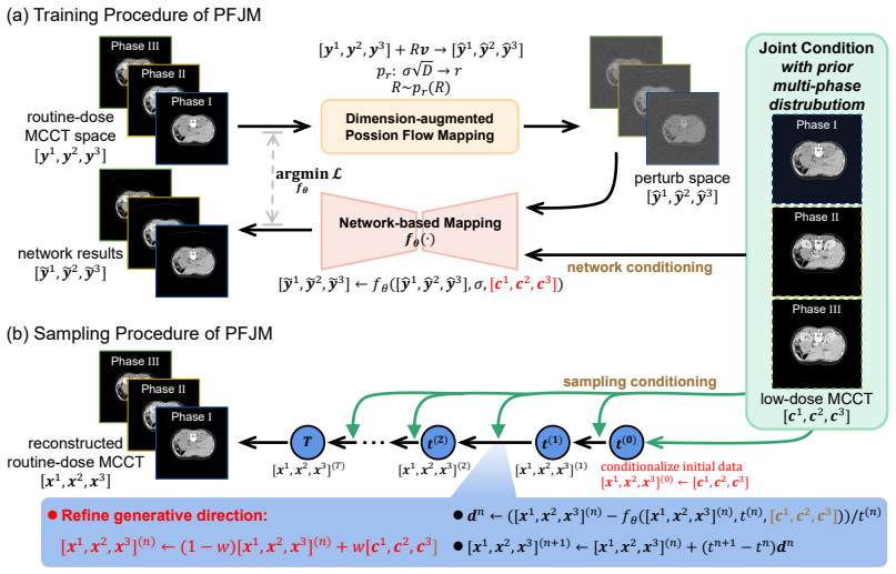
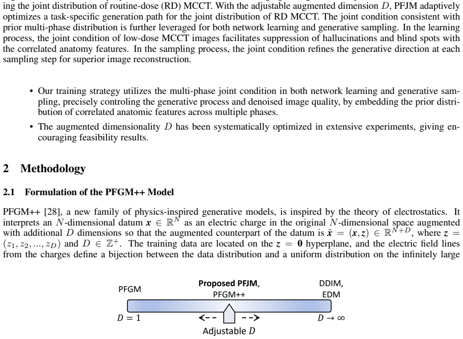
The central machinery is PFGM++, a physics-inspired generative framework that treats each $N$-dimensional data point as an electric charge in an $(N+D)$-dimensional augmented space and generates samples by following the electric field lines back to the data hyperplane; the augmented dimension $D$ interpolates between Poisson flow ($D=1$) and diffusion ($D\to\infty$). PFJM wraps this machinery around the three-phase MCCT problem: it learns the joint distribution of routine-dose images $[\mathbf{y}_1,\mathbf{y}_2,\mathbf{y}_3]$, perturbs them through the field, and trains a network $f_\theta$ to map back, with the joint low-dose images $[\mathbf{c}_1,\mathbf{c}_2,\mathbf{c}_3]$ injected as a condition. At sampling, the same joint condition initializes the trajectory and is blended into each intermediate step through $(1-w)[\mathbf{x}_1,\mathbf{x}_2,\mathbf{x}_3]^{(n)} + w[\mathbf{c}_1,\mathbf{c}_2,\mathbf{c}_3]$ before the field-guided update. The adjustable $D$ is the task-specific dial that lets the generative path be optimized; $D=128$ is reported as the optimal setting.
What would settle it
Acquire real low-dose MCCT scans at 10% dose with paired routine-dose ground truth from the same patients, run the trained PFJM on them, and compare average MAE and FID against PFGM++ under identical conditions; if the reported margins (MAE 8.99 HU, FID 4.30) do not reproduce, the central claim that PFJM robustly drives the trajectory to the routine-dose domain is refuted.
Extended reading notes
Core claim
On the paper's own terms, the discovery is that a PFGM++-based generative model can take the joint low-dose MCCT images of the three phases as a condition and robustly drive the generative trajectory toward the routine-dose MCCT domain. The model learns the joint distribution of routine-dose multiphase images, with the augmented dimensionality $D$ of the Poisson flow optimized as a task-specific control; $D=128$ is the reported sweet spot. During sampling, the low-dose joint condition is re-injected at every step to refine the generation direction, and the ablations credit this conditional sampling with the largest gains: removing it degrades MAE by 7.58 HU, SSIM by 3.97%, and PSNR by 2.40 dB on average across $D$ settings. The paper's experiments give average MAE of 8.99 HU, SSIM of 98.75%, PSNR of 48.24 dB, and FID of 4.30, with PFJM surpassing PFGM, DDIM, EDM, and PFGM++ on every phase.
Load-bearing premise
The results rest on the simulated 10%-dose low-dose images faithfully reproducing the noise and artifact behavior of real reduced-dose CT scans; if that simulation is not representative, the reported quality gains may not transfer to patients.
Editorial extensions
If this is right
- A 10%-dose MCCT acquisition could be reconstructed to near routine-dose quality across all three phases, substantially cutting the radiation dose of multiphase exams.
- Because $D$ is adjustable, the same joint Poisson-flow framework could be re-tuned for other imaging tasks; for this CT problem the optimized setting is $D=128$.
- The ablation shows joint conditional sampling is the main driver: removing it degrades MAE by 7.58 HU, SSIM by 3.97%, and PSNR by 2.40 dB on average, and it rescues even poor settings like $D=2$ and $D=8$.
- With only 10 sampling steps, PFJM beats PFGM at 30 steps, DDIM and EDM at 20 steps, making the method faster than the baselines it outperforms.
Reading between the lines
- Editorial inference: the $D=128$ sweet spot hints that the optimal augmented dimension tracks the effective dimensionality of the data distribution, so other denoising tasks might pick $D$ by a short validation sweep rather than a full grid.
- Editorial inference: the large ablation gap suggests that a conventional conditional diffusion model with the same joint conditioning might close part of the gap, isolating whether the Poisson-flow path itself or the joint condition carries the improvement.
- Editorial inference: the simulated low-dose evaluation means the strongest next test is a real low-dose clinical dataset; the paper lists generalizability and fairness as planned follow-up work.
- Editorial inference: the joint-condition design could be extended to use one phase (for example, the non-contrast phase) as a structural prior for the later contrast phases, a temporal ordering not tested here.
Editorial analysis
A structured set of objections, weighed in public.
Referee Report
Summary. The manuscript proposes PFJM, a conditional PFGM++ variant for denoising low-dose multiphase contrast-enhanced CT (MCCT). The model learns a joint distribution of routine-dose images across the non-contrast, arterial, and venous phases, conditioned on the corresponding low-dose joint images. During sampling, PFJM initializes from the low-dose condition and, at each step, blends the current iterate with the condition via a weight w (Algorithm 2, line 4). Experiments on the public VinDr-Multiphase dataset with simulated 10%-dose data report improved MAE, SSIM, PSNR, and FID over PFGM, DDIM, EDM, and PFGM++ across a range of augmented dimensions D, with D=128 as the best setting. The paper claims averaged MAE of 8.99 HU, SSIM of 98.75%, and PSNR of 48.24 dB over all phases.
Significance. If the reported results are reproducible, the paper offers a useful application of PFGM++ to multiphase low-dose CT, with a systematic D-sweep and a clearly described conditional-sampling mechanism. Strengths include the use of a public dataset, comparisons to several generative baselines, qualitative phase-by-phase evaluation, and an ablation of D across eight settings. The D-sweep is hyperparameter selection rather than circular reasoning, and the one self-citation (JCCS-PFGM) is not load-bearing. However, the central sampling mechanism depends on an unreported weight w, the quantitative claims lack variance estimates, and the D selection appears to use the test set; these are fixable issues but currently block full confidence in the headline gains.
major comments (5)
- [Algorithm 2, line 4] The blending weight w in the conditional-sampling update [x1,x2,x3]^(n) <- (1-w)[x1,x2,x3]^(n) + w[c1,c2,c3] is never specified anywhere in the paper, and no sensitivity analysis or ablation over w is reported. This update is the only algorithmic difference between PFJM and PFGM++, and the ablation study in Section 3.2 attributes the performance gains entirely to this conditional sampling. Without the value or schedule of w, the central claim is not reproducible and the gains cannot be attributed to the proposed mechanism. Please report the exact value or schedule, provide a sensitivity sweep, and justify why this convex combination preserves the PFGM++ ODE trajectory rather than acting as a simple interpolation toward the input condition.
- [Section 3.2, Table 1] The augmented dimension D is selected using what appears to be the same test set that then produces the headline numbers. The text does not state that D=128 was chosen on the validation split; if Table 1 reports test-set metrics for all D values, then the final comparison is a test-set-tuned result. This introduces optimistic bias in the claimed advantage over PFGM++ at each D. Please select D on the validation set and report test-set results only for the chosen configuration, or otherwise quantify the selection bias.
- [Tables 1 and 2] No error bars, confidence intervals, or repeated-seed results are reported for any quantitative claim. Differences as small as 0.5 HU (e.g., Phase I MAE 8.85 for PFJM versus 9.44 for PFGM++ at D=128) are presented as evidence of superiority, but without variance estimates these differences could lie within run-to-run or patient-subset noise. Please add standard deviations over test patients or multiple training runs, and state the number of independent runs used.
- [Section 3.1] The low-dose MCCT images are simulated at 10% dose using the method of Yu et al. [48], but the paper provides no validation or quantitative characterization of the simulated noise relative to real low-dose acquisitions. The translational claim in Section 4, that PFJM reduces radiation dose for patients, depends on this simulation being representative. Please add a comparison of simulated versus real low-dose noise statistics (or a clearly scoped claim that the evaluation is on simulated low-dose data only).
- [Section 3.2] There is an internal inconsistency in the reported PSNR values. The text states 'PSNR up to 46.3 dB' for PFJM, while Table 1 and the abstract report values around 48.2-48.3 dB for D=128, and the per-phase rows give 48.29, 48.28, and 48.15 dB. Please correct the typo and reconcile the averaged headline metrics ('MAE down to 8.99 HU, SSIM up to 98.75%, PSNR up to 48.24 dB') with the per-phase table entries.
minor comments (4)
- [Throughout] The manuscript contains numerous typographical and grammatical errors, including 'Poission flow', 'pathologial', 'contemporay', 'distoring', 'illustred', 'MPCT', and 'PDFM++'. A careful proofread is needed.
- [Figures 4-6 captions] In each of the qualitative figure captions, the entries for PFGM++ and PFJM are both labeled '(f1) and (f2)', which makes it ambiguous which panel corresponds to which method. Please correct the labels.
- [Section 3.2] The phrase 'MAE decreased by 49.17 HU' for D=2 does not exactly match the Table 1 difference between PFJM (11.65 HU) and PFGM++ (60.79 HU) in Phase I, which is 49.14 HU; if another averaging convention is used, please state it.
- [Algorithm 1] The perturbation step uses the notation 'R_i v' in line 7, but only R is sampled in line 5; the subscript is unexplained. Please unify the notation.
Circularity Check
No significant circularity: PFJM is an empirical PFGM++ variant benchmarked against external baselines, and the conditional-sampling step is a method component rather than a fitted input renamed as a prediction.
full rationale
This paper does not claim a first-principles derivation of its denoising result. PFJM is presented as an application of the external PFGM++ framework [28] with two modifications: multi-phase joint conditioning in training (Algorithm 1) and a conditional pull in sampling (Algorithm 2, line 4). The quantitative claims are benchmarked against PFGM, DDIM, EDM, and PFGM++ on the public VinDr-Multiphase dataset, so the comparisons are self-contained and externally checkable. The author self-citations ([10], [17], [41]) occur in literature surveys and are not used to justify the correctness of the proposed method. The D sweep is hyperparameter selection, not fitting a parameter to the target quantity; selecting D=128 by best test metrics is an evaluation-protocol concern rather than circularity. The ablation removing conditional sampling in Section 3.2 confirms that the conditional pull is the operative difference from PFGM++, but this is exactly the method being proposed, not an input renamed as a prediction. The undisclosed value of w in Algorithm 2 is a reproducibility limitation, not a circular derivation. Therefore, no circular step meets the quoted-reduction bar.
Assumptions & free parameters
free parameters (2)
- Augmented dimension D =
128
- Conditioning weight w
assumptions (4)
- standard math PFGM++ theory: the augmented electric field defines a bijection between the data distribution on the z=0 hyperplane and a uniform distribution on a large hemisphere, with the ODE in Eq. (2) generating samples.
- domain assumption The low-dose CT simulation of Yu et al. (2012) produces realistic low-dose MCCT images from routine-dose images, including bowtie filter, automatic exposure control, and electronic noise effects.
- domain assumption The VinDr-Multiphase dataset provides paired multiphase images with consistent phase alignment, and the random split into 65/21/45 patients is representative.
- domain assumption Joint conditioning on low-dose images across phases is a valid way to guide the reverse generative trajectory to the routine-dose distribution.
Cite this review
Pith. "Pith review of Poisson Flow Joint Model for Multiphase contrast-enhanced CT." pith.science (2026). https://pith.science/paper/TKPIBF7X
@misc{pith2026250203079,
  author       = {Pith},
  title        = {Pith review of: Poisson Flow Joint Model for Multiphase contrast-enhanced CT},
  year         = {2026},
  howpublished = {\url{https://pith.science/paper/TKPIBF7X}},
  note         = {Machine review of arXiv:2502.03079}
}
read the original abstract
In clinical practice, multiphase contrast-enhanced CT (MCCT) is important for physiological and pathological imaging with contrast injection, which undergoes non-contrast, venous, and delayed phases. Inevitably, the accumulated radiation dose to a patient is higher for multiphase scans than for a plain CT scan. Low-dose CECT is thus highly desirable, but it often leads to suboptimal image quality due to reduced radiation dose. Recently, a generalized Poisson flow generative model (PFGM++) was proposed to unify the diffusion model and the Poisson flow generative models (PFGM), and outperform either of them with an optimized dimensionality of the augmentation data space, holding a significant promise for generic or conditional image generation. In this paper, we propose a Poisson flow joint model (PFJM) for low-dose MCCT to suppress image noise and preserve clinical features. Our model is built on the PFGM++ architecture to transform the multiphase imaging problem into learning the joint distribution of routine-dose MCCT images by optimizing a task-specific generation path with respect to the dimensionality D of the augmented data space. Then, our PFJM model takes the joint low-dose MCCT images as the condition and robustly drives the generative trajectory towards the solution in the routine-dose MCCT domain. Extensive experiments demonstrate that our model is favorably compared with competing models, with MAE of 8.99 HU, SSIM of 98.75% and PSNR of 48.24db, as averaged over all the phases.
Figures

Figures from the paper (6 more)


Reference graph
Works this paper leans on
-
[48]
L. Yu, M. Shiung, D. Jondal, C. H. McCollough, Development and validation of a practical lower-dose- simulation tool for optimizing computed tomography scan protocols , Journal of computer assisted tomography, 2012, 36(4): 477-487
work page 2012
-
[1]
X.P.Meng, Y .C.Wang, S.Ju, et al.,Radiomics analysis on multiphase contrast-enhanced CT: a survival prediction tool in patients with hepatocellular carcinoma undergoing transarterial chemoembolization , Frontiers in Oncol- ogy, 2020, 10: 1196
work page 2020
-
[2]
Smithuis, CT contrast injection and protocols
R. Smithuis, CT contrast injection and protocols. Radiology Assistant , https://radiologyassistant. nl/more/ct- protocols/ct-contrast-injection-and-protocols, 2014
work page 2014
-
[3]
D.J. Brenner, E.J. Hall, Computed tomography an increasing source of radiation exposure, New England journal of medicine, 2007, 357(22): 2277-2284
work page 2007
-
[4]
S. Rastogi, R. Singh, R. Borse, et al., Use of multiphase CT protocols in 18 countries: appropriateness and radiation doses, Canadian Association of Radiologists Journal, 2021, 72(3): 381-387
work page 2021
-
[5]
K.N. Prasad, W.C.Cole, G.M. Haase, Radiation protection in humans: extending the concept of as low as rea- sonably achievable (ALARA) from dose to biological damage , The British journal of radiology, 2004, 77(914): 97-99
work page 2004
-
[6]
H. Chen, Y . Zhang, M. K. Kalra, F. Lin, Y . Chen, P. Liao, J. Zhou, G. Wang, Low-dose CT with a residual encoder-decoder convolutional neural network, IEEE transactions on medical imaging, 2017, 36(12): 2524-2535
work page 2017
-
[7]
X. Yin, Q. Zhao, J. Liu, et al., Domain progressive 3D residual convolution network to improve low-dose CT imaging, IEEE transactions on medical imaging, 2019, 38(12): 2903-2913. 12
work page 2019
Show all 49 references
-
[8]
F. Fan, H. Shan, M.K. Kalra, et al., Quadratic autoencoder (Q-AE) for low-dose CT denoising, IEEE transactions on medical imaging, 2019, 39(6): 2035-2050
2019
-
[9]
Z. Lu, W. Xia, Y . Huang, et al., M3NAS: Multi-scale and multi-level memory-efficient neural architecture search for low-dose ct denoising, IEEE Transactions on Medical Imaging, 2022, 42(3): 850-863
2022
-
[10]
L. Yang, Z. Li, R. Ge, et al.,Low-dose CT denoising via sinogram inner-structure transformer, IEEE transactions on medical imaging, 2022, 42(4): 910-921
2022
-
[11]
Q. Yang, P. Yan, Y . Zhang, H. Yu, Y . Shi, X. Mou, M.K. Kalra, Y . Zhang, L. Sun, G. Wang, Low-dose CT image denoising using a generative adversarial network with Wasserstein distance and perceptual loss , IEEE transactions on medical imaging, 37(6):1348-1357, 2018
2018
-
[12]
Bera, P.K
S. Bera, P.K. Biswas, Biswas Noise conscious training of non local neural network powered by self attentive spectral normalized Markovian patch GAN for low dose CT denoising , IEEE Transactions on Medical Imaging, 2021, 40(12): 3663-3673
2021
-
[13]
Kwon, J.C
T. Kwon, J.C. Ye, Cycle-free CycleGAN using invertible generator for unsupervised low-dose CT denoising , IEEE Transactions on Computational Imaging, 2021, 7: 1354-1368
2021
-
[14]
Moghari, L
M.D. Moghari, L. Zhou, B. Yu, et al. Efficient radiation dose reduction in whole-brain CT perfusion imaging using a 3D GAN: performance and clinical feasibility, Physics in Medicine & Biology, 2021, 66(7): 075008
2021
-
[15]
H. Shan, Y . Zhang, Q. Yang, et al., 3-D convolutional encoder-decoder network for low-dose CT via transfer learning from a 2-D trained network, IEEE transactions on medical imaging, 2018, 37(6): 1522-1534
2018
-
[16]
Huang, J
Z. Huang, J. Zhang, Y . Zhang, H. Shan, DU-GAN: Generative adversarial networks with dual-domain U-Net- based discriminators for low-dose CT denoising, IEEE Transactions on Instrumentation and Measurement, 71:1- 12, 2021
2021
-
[17]
R. Ge, G. Yang, C. Xu, Y . Chen, L. Luo, S. Li, Stereo-correlation and noise-distribution aware ResVoxGAN for dense slices reconstruction and noise reduction in thick low-dose CT, In International Conference of Medical Image Computing and Computer Assisted Intervention–MICCAI,...
2019
-
[18]
Y . Fu, S. Dong, M. Niu, et al., AIGAN: Attention–encoding Integrated Generative Adversarial Network for the reconstruction of low-dose CT and low-dose PET images, Medical Image Analysis, 2023, 86: 102787
2023
-
[19]
Goodfellow, J
I. Goodfellow, J. Pouget-Abadie, M. Mirza, B. Xu, D. Warde-Farley, S. Ozair, A. Courville, Y . Bengio,Genera- tive adversarial nets, Advances in neural information processing systems, pp. 2672-2680, 2014
2014
-
[20]
Salimans, I
T. Salimans, I. Goodfellow, W. Zaremba, V . Cheung, A. Radford, X. Chen, Improved techniques for training gans, Advances in Neural Information Processing Systems, pp. 2226-2234, 2016
2016
-
[21]
L. Metz, B. Poole, D. Pfau, J. Sohl-Dickstein, Unrolled Generative Adversarial Networks , 5th International Conference on Learning Representations, 2017
2017
-
[22]
S. Zhao, H. Ren, A. Yuan, J. Song, N. Goodman, S. Ermon, Bias and generalization in deep generative models: An empirical study, Advances in Neural Information Processing Systems, 2018
2018
-
[23]
Q. Gao, Z. Li, J. Zhang, Y . Zhang, H. Shan, CoreDiff: Contextual error-modulated generalized diffusion model for low-dose CT denoising and generalization, IEEE Transactions on Medical Imaging, 43(2):745-759, 2024
2024
-
[24]
Y . Song, S. Ermon, Improved techniques for training score-based generative models, Advances in neural infor- mation processing systems, 33: 12438-12448, 2020
2020
-
[25]
Karras, M
T. Karras, M. Aittala, T. Aila, S. Laine, Elucidating the design space of diffusion-based generative models , Advances in neural information processing systems, 35: 26565-26577, 2022
2022
-
[26]
J. Song, C. Meng, S. Ermon, Denoising Diffusion Implicit Models. In International Conference on Learning Representations
-
[27]
Y . Xu, Z. Liu, M. Tegmark, T. Jaakkola, Poisson flow generative models, Advances in Neural Information Pro- cessing Systems, 35, pp.16782-16795, 2022
2022
-
[28]
Y . Xu, Z. Liu, Y . Tian, S. Tong, M. Tegmark, T. Jaakkola,Pfgm++: Unlocking the potential of physics-inspired generative models, In International Conference on Machine Learning, pp. 38566-38591, 2023
2023
-
[29]
J. Ho, A. Jain, P. Abbeel, Denoising diffusion probabilistic models, Advances in neural information processing systems, 33: 6840-6851, 2020
2020
-
[30]
Nichol, P
A.Q. Nichol, P. Dhariwal, Improved denoising diffusion probabilistic models , International conference on ma- chine learning, 2021: 8162-8171. 13
2021
-
[31]
J. Song, C. Meng, S. Ermon, Denoising diffusion implicit models , in International Conference on Learning Representations, 2021, pp. 1–22
2021
-
[32]
W. Zhao, Y . Rao, W. Shi, et al., Diffswap: High-fidelity and controllable face swapping via 3d-aware masked diffusion, Proceedings of the IEEE/CVF Conference on Computer Vision and Pattern Recognition, pp. 8568-8577, 2023
2023
-
[33]
Y . Song, J. Sohl-Dickstein,D.P. Kingma, et al.,Score-Based Generative Modeling through Stochastic Differential Equations, International Conference on Learning Representations, 2021
2021
-
[34]
Saharia, W
C. Saharia, W. Chan, H. Chang, et al., Palette: Image-to-image diffusion models, in Proceedings of ACM SIG- GRAPH Conference, pp. 1-10, 2022
2022
-
[35]
Saharia, J
C. Saharia, J. Ho, W. Chan, et al., Image super-resolution via iterative refinement, IEEE transactions on pattern analysis and machine intelligence, 2022, 45(4): 4713-4726
2022
-
[36]
L.Zhang, A. Rao, M. Agrawala, Adding conditional control to text-to-image diffusion models, in Proceedings of the IEEE/CVF International Conference on Computer Vision, pp.3836-3847, 2023
2023
-
[37]
J. Ho, C. Saharia, W. Chan, D.J. Fleet, M. Norouzi, T. Salimans, Cascaded diffusion models for high fidelity image generation, Journal of Machine Learning Research, 23(47): 1-33, 2022
2022
-
[38]
Kazerouni, E.K
A. Kazerouni, E.K. Aghdam, M. Heidari, R. Azad, M. Fayyaz, I. Hacihaliloglu, D. Merhof, Diffusion models in medical imaging: A comprehensive survey, Medical Image Analysis, 88: 102846, 2023
2023
-
[39]
D. Hein, A. Bozorgpour, D. Merhof, G. Wang, Physics-Inspired Generative Models in Medical Imaging: A Review, arXiv preprint arXiv:2407.10856, 2024
2024 arXiv
-
[40]
W. Du, H.H. Cui, L.C. He, et al. Structure-aware diffusion for low-dose CT imaging , Physics in Medicine & Biology, 2024, 69(15): 155008
2024
-
[41]
R. Ge, Y . He, C. Xia, Y . Chen, D. Zhang, G. Wang,JCCS-PFGM: A novel circle-supervision based poisson flow generative model for multiphase CECT progressive low-dose reconstruction with joint condition, in International Conference on Medical Image Computing and Computer-Assist...
2023
-
[42]
D. Hein, S. Holmin, T. Szczykutowicz, J.S. Maltz, M. Danielsson, G. Wang, M. Persson,PPFM: Image denoising in photon-counting CT using single-step posterior sampling Poisson flow generative models , IEEE Transactions on Radiation and Plasma Medical Sciences, 2024
2024
-
[43]
C. M. Restrepo-Galeano, G. R. Arce, Super-Resolution in Low Dose X-Ray CT Via Focal Spot Mitigation with Generative Diffusion Networks, IEEE Transactions on Computational Imaging, 2024
2024
-
[44]
Karageorgos, J
G.M. Karageorgos, J. Zhang, N. Peters, W. Xia, C. Niu, H. Paganetti, G. Wang, B.De Man,A denoising diffusion probabilistic model for metal artifact reduction in CT, IEEE Transactions on Medical Imaging, 2024
2024
-
[45]
Guan, , C
B. Guan, , C. Yang, L. Zhang, S. Niu, M. Zhang, Y . Wang, W. Wu, Q. Liu, Generative modeling in sinogram domain for sparse-view CT reconstruction, IEEE Transactions on Radiation and Plasma Medical Sciences, 2023
2023
-
[46]
J. Liu, R. Anirudh, J.J. Thiagarajan, S. He, K.A. Mohan, U.S. Kamilov, H. Kim, DOLCE: A model-based prob- abilistic diffusion framework for limited-angle ct reconstruction , In Proceedings of the IEEE/CVF International Conference on Computer Vision, pp. 10498-10508, 2023
2023
-
[47]
Vincent, A connection between score matching and denoising autoencoders, Neural computation, 23(7): 1661- 1674, 2011
P. Vincent, A connection between score matching and denoising autoencoders, Neural computation, 23(7): 1661- 1674, 2011
2011
-
[49]
B. T. Dao, T. V . Nguyen, H. H. Pham, H. Q. Nguyen, Phase recognition in contrast-enhanced CT scans based on deep learning and random sampling, Medical Physics, 2022, 49(7), 4518-4528. 14
2022
Reviewed August 9, 2026 · model on record in the stance chip above.
Discussion (0). Continue with ORCID to comment.