REVIEW 5 major objections 5 minor 38 references
ZeroReg3D: A Zero-shot Registration Pipeline for 3D Consecutive Histopathology Image Reconstruction
T0 review · 5 major / 5 minor · reviewed 2026-08-06 · deepseek-v4-flash
Pith's one-line read A zero-shot pipeline aligns serial kidney slices for 3D reconstruction with no retraining or fine-tuning.
desk verdict Useful zero-shot pairwise registration pipeline, but the 3D reconstruction claim rests on 2D pairwise evidence alone. read the letter →
The pith
A machine-rendered reading of the paper's core claim, the machinery that carries it, and where it could break.
The reading
What carries the argument
The carrying mechanism is the pipeline itself: a rotation-preprocessing step that reorients the moving image to increase the number of valid keypoint correspondences, affine registration computed from keypoint matches that survive RANSAC outlier rejection, and B-spline non-rigid registration where displacements of control points are optimized by minimizing a loss that combines local normalized cross-correlation with a diffusion regularizer that penalizes non-smooth displacement fields. XFeat, a lightweight learned keypoint detector and descriptor, is the named zero-shot matcher that supplies initial correspondences; the affine and B-spline stages supply the global and local deformation models that turn those correspondences into a registered stack.
What would settle it
Register a long serial section series with ZeroReg3D, then compare the reconstructed stack against an independent 3D reference of the same tissue block, such as block-face images or micro-CT taken before sectioning, by measuring 3D distances between corresponding anatomical landmarks; if those 3D errors grow beyond the pairwise 2D rTRE values as the number of sections increases, the pairwise-proxy assumption fails.
Extended reading notes
Core claim
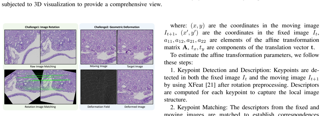
The paper's central claim is that a cascade of three stages—rotation-aware zero-shot keypoint extraction and matching, RANSAC-filtered affine registration, and B-spline free-form deformation optimized against a local normalized cross-correlation loss with a diffusion regularizer—can register serial kidney sections accurately across stains, illumination changes, and local tissue distortion. On the two mouse datasets the method achieves the lowest AMrTRE and AMean landmark distances among the methods compared, and on the human needle-biopsy dataset it shows the lowest median landmark distance with the least variability across 20 cases. The authors interpret these pairwise 2D registration results as the evidence that the stacked volume supports 3D reconstruction, and they summarize the gain as about 10% over the strongest baseline.
Load-bearing premise
The load-bearing premise is that accurate pairwise 2D landmark alignment is a sufficient proxy for a faithful 3D reconstruction, even though the authors acknowledge that chaining many pairwise registrations can accumulate and propagate errors along the sequence.
Editorial extensions
If this is right
- A lab with no annotated training data can apply the pipeline directly to a new serial-section dataset and obtain alignments comparable to or better than published registration methods, as demonstrated on mouse and human kidney whole-slide images.
- Because the method works without stain-specific training, it can be applied to consecutive sections with different stains in the same sequence, such as the H&E–PAS–Jones repeated staining cycles in the human biopsy protocol.
- The rotation-preprocessing step measurably increases the number of valid keypoint pairs (e.g., from 12,965 to 16,882 on the mouse set), so orientation normalization is a directly useful enhancement for keypoint-based histology registration.
- The reported landmark-error improvements are paired with a complete open-source implementation, making the pipeline reproducible and immediately usable for downstream 3D spatial analysis.
Reading between the lines
- Beyond the paper's claims, pairwise 2D landmark error alone does not certify 3D reconstruction fidelity; a direct geometric check against an independent volume reference of the same tissue block would clarify how much of the 3D claim follows from the pairwise results.
- A testable extension is to run the same cascade on non-kidney organs or on sections with large gaps, where keypoint density drops; observing how accuracy degrades would delimit the method's true zero-shot range.
- If cumulative stacking error is the dominant failure mode, as the authors note, then replacing the chained pairwise transforms with a joint multi-section optimization is a natural next experiment that could reduce drift without changing the matching stage.
Editorial analysis
A structured set of objections, weighed in public.
Referee Report
Summary. The manuscript proposes ZeroReg3D, a zero-shot registration pipeline for serial histological sections. The method combines zero-shot XFeat keypoint extraction and matching (with a rotation preprocessing step), affine registration with RANSAC outlier rejection, and B-spline based non-rigid registration optimized by a local NCC plus diffusion regularization loss. The authors evaluate the pipeline on normal and db/db diabetic mouse kidney sections and on human kidney needle biopsy cases, reporting landmark-based metrics such as AMrTRE, MMrTRE, AMxrTRE, average robustness, and AMean D. They conclude that the method achieves approximately 10% improvement over state-of-the-art baselines and enables accurate 3D reconstruction of serial histology, and they release the code publicly.
Significance. If fully supported, the paper would offer a practically useful contribution: a public, zero-shot pipeline that requires no retraining or fine-tuning, with broad comparison against established registration methods and evaluation using external landmarks. Strengths include the open-source release, the inclusion of both mouse and human datasets, and the use of Wilcoxon signed-rank testing for the human data. The main weakness is that the central '3D reconstruction' claim is not supported by the reported evidence, which consists entirely of 2D pairwise registration metrics. The numerical summary in the conclusion is also inconsistent with the tables. These issues are localizable and fixable, but they are load-bearing for the paper's headline claims.
major comments (5)
- [Section V and Section V-A] The abstract and conclusion claim accurate 3D reconstruction, but every quantitative result in Tables I-III and Figures 4-6 is a 2D pairwise registration metric computed on adjacent section pairs. No experiment reconstructs a 3D volume, measures 3D landmark positions, evaluates cumulative drift along the stack, or compares the reconstructed volume against any reference. The Discussion (Section V-A) explicitly concedes that 'potential inaccuracies introduced during cumulative stacking of pairwise registration... potentially propagating registration errors along the sequence.' This is not a cosmetic issue: favorable pairwise rTRE does not imply an accurate 3D stack. Either add direct 3D validation (e.g., 3D landmarks, surface continuity, loop-closure error, or comparison to a reference volume) or revise the title, abstract, and conclusion to describe a pairwise registration pipeline for serial sections and remove the 'accurate 3D reconstruction' claim.
- [Conclusion and Tables I-II] The statement in the Conclusion that the method achieves 'approximately 10% improvement in registration accuracy compared to state-of-the-art baseline methods' is not supported by the tables. Against the closest baseline DeeperHistReg on the normal mouse dataset (Table I), AMrTRE improves from 0.0026 to 0.0024, a 7.7% relative improvement, while AMean D improves from 3.1353 to 2.6952, a 14.0% improvement. On the db/db mouse dataset (Table II), AMrTRE improves from 0.0064 to 0.0030, a 53.1% improvement, while AMean D improves from 4.9953 to 4.2919, a 14.1% improvement. There is no single 10% figure that follows from these numbers. Please specify the exact metric and baseline used for the summary claim, or remove it.
- [Tables I-II and Section V] The mouse results are reported as single point estimates with no variance, confidence intervals, or statistical significance tests. It is also unclear whether Tables I and II each summarize one representative slice or an aggregate over the full set of 29 and 39 sections, and how many landmark pairs are used. Since the central claim is that ZeroReg3D outperforms the compared baselines, the authors should report per-pair error distributions, standard deviations or confidence intervals, and preferably a paired test across image pairs. Without this, the robustness of the reported advantage over DeeperHistReg in particular cannot be assessed.
- [Section III-A and Table III] The rotation preprocessing step is described as improving keypoint matching and registration accuracy, but Table III reports only the number of keypoint pairs and processing time before and after rotation, not the effect on final registration error. Given the substantial computational cost (76 seconds per mouse slice and 281 seconds per human slice), an ablation comparing AMrTRE, AMean D, and robustness with and without the rotation step is needed to justify this design choice and to support the attributions made in Section V-A.
- [Section V and Figure 6] The human dataset evaluation is reported only through a boxplot in Figure 6 with asterisks indicating Wilcoxon signed-rank test significance. No numeric AMean D values, no p-values, and no effect sizes are provided, and it is unclear how many landmark pairs per case are used and whether the three different stains within each case are treated as separate registration pairs. These omissions make it difficult to verify the claimed statistical superiority on human data. Please provide a table of per-case or aggregate values, p-values, and a description of the landmark set construction.
minor comments (5)
- [References] Reference [19] is listed as 'A. Author and B. Coauthor, "Fully automatic and robust 3D registration of serial-section microscopy images"' with no real author or venue; this placeholder reference must be replaced or removed.
- [Section IV-A and Table III] The text states that the mouse dataset contains 29 normal and 39 db/db sections, which sums to 68, but Table III reports 66 slices for the mice dataset. This discrepancy should be reconciled, as it affects the timing and keypoint-count statistics.
- [Section III-B] After step 4 of the affine registration procedure, the text says the transformation parameters are estimated 'by solving the following system of equations,' but no system of equations is actually shown. Please include the equation or rephrase the sentence.
- [Section III-A] The sentence 'We employed XFeat as our image matching method for initial registration technique for the initial registration' contains a typo and should be rewritten for clarity.
- [Section IV-C] The image spacing is given as 1 mm x 1 mm x 8 mm, but the relationship between this spacing, the pixel size of 0.25 µm, and the section thickness of 2 µm is not explained. Please clarify these units to avoid confusion in interpreting the 3D visualization.
Circularity Check
No significant circularity: the zero-shot pipeline is evaluated against independent landmarks and external baselines; the 3D-reconstruction gap is a validity concern, not a definitional one.
full rationale
The paper's derivation chain is self-contained rather than circular. ZeroReg3D applies a fixed, zero-shot pipeline (XFeat keypoint matching, affine registration, and B-spline non-rigid registration) with no parameters fitted to the evaluation data. The reported metrics (rTRE, AMrTRE, AMean D, Ravg) use manually annotated landmarks that are not used in the optimization objective, so the evaluation is not an input to the method. The self-citations in the manuscript, most notably Map3D as a baseline, are not load-bearing for the central claim; the comparison set also includes many external methods (SuperGlue, LoFTR, SIFT, ORB, ANTs, DeeperHistReg). The acknowledged limitation about cumulative stacking errors and the mismatch between pairwise 2D evaluation and the 3D reconstruction claim are substantive external-validity or correctness concerns, but they are not cases where a prediction reduces by construction to a fitted input or to a self-citation chain. The unsupported 'approximately 10%' summary statement is likewise an accuracy/consistency issue rather than circularity. Therefore, the appropriate circularity score is 0.
Assumptions & free parameters
free parameters (4)
- Regularization weight lambda
- B-spline control point grid
- Optimizer settings (learning rate alpha, max iterations Nmax)
- Rotation search range and step for preprocessing
assumptions (4)
- domain assumption XFeat features trained on natural images transfer to histopathology without fine-tuning
- domain assumption Annotated glomerulus centers are reliable ground truth and rTRE is a valid accuracy measure
- domain assumption Pairwise alignment accuracy is a proxy for 3D reconstruction quality
- standard math Standard B-spline and affine transformation models can represent sectioning deformations
Cite this review
Pith. "Pith review of ZeroReg3D: A Zero-shot Registration Pipeline for 3D Consecutive Histopathology Image Reconstruction." pith.science (2026). https://pith.science/paper/YUYZ342H
@misc{pith2026250621923,
  author       = {Pith},
  title        = {Pith review of: ZeroReg3D: A Zero-shot Registration Pipeline for 3D Consecutive Histopathology Image Reconstruction},
  year         = {2026},
  howpublished = {\url{https://pith.science/paper/YUYZ342H}},
  note         = {Machine review of arXiv:2506.21923}
}
read the original abstract
Histological analysis plays a crucial role in understanding tissue structure and pathology. While recent advancements in registration methods have improved 2D histological analysis, they often struggle to preserve critical 3D spatial relationships, limiting their utility in both clinical and research applications. Specifically, constructing accurate 3D models from 2D slices remains challenging due to tissue deformation, sectioning artifacts, variability in imaging techniques, and inconsistent illumination. Deep learning-based registration methods have demonstrated improved performance but suffer from limited generalizability and require large-scale training data. In contrast, non-deep-learning approaches offer better generalizability but often compromise on accuracy. In this study, we introduced ZeroReg3D, a novel zero-shot registration pipeline tailored for accurate 3D reconstruction from serial histological sections. By combining zero-shot deep learning-based keypoint matching with optimization-based affine and non-rigid registration techniques, ZeroReg3D effectively addresses critical challenges such as tissue deformation, sectioning artifacts, staining variability, and inconsistent illumination without requiring retraining or fine-tuning. The code has been made publicly available at https://github.com/hrlblab/ZeroReg3D
Figures


Figures from the paper (3 more)


Reference graph
Works this paper leans on
-
[1]
J. Xiong, Y . Liu, R. Deng, R. N. Tyree, H. Correa, G. Hiremath, Y . Wang, and Y . Huo, “Deep learning-based open source toolkit for eosinophil detection in pediatric eosinophilic esophagitis,” in Medical Imaging 2024: Digital and Computational Pathology , vol. 12933. SPIE, 2024, pp. 231–237
work page 2024
-
[2]
Asign: An anatomy-aware spatial imputation graphic network for 3d spatial transcriptomics,
J. Zhu, R. Deng, T. Yao, J. Xiong, C. Qu, J. Guo, S. Lu, M. Yin, Y . Wang, S. Zhao, H. Yang, and Y . Huo, “Asign: An anatomy-aware spatial imputation graphic network for 3d spatial transcriptomics,” in Proceedings of the IEEE/CVF Conference on Computer Vision and Pattern Recognition (CVPR), June 2025, pp. 30 829–30 838
work page 2025
-
[3]
Hats: Hierarchical adaptive taxonomy segmentation for panoramic pathology image anal- ysis,
R. Deng, Q. Liu, C. Cui, T. Yao, J. Xiong, S. Bao, H. Li, M. Yin, Y . Wang, S. Zhao, Y . Tang, H. Yang, and Y . Huo, “Hats: Hierarchical adaptive taxonomy segmentation for panoramic pathology image anal- ysis,” in International Conference on Medical Image Computing and Computer-Assisted Intervention. Springer, 2024
work page 2024
-
[4]
Prpseg: Universal proposition learning for panoramic renal pathology segmentation,
R. Deng, Q. Liu, C. Cui, T. Yao, J. Yue, J. Xiong, L. Yu, Y . Wu, M. Yin, Y . Wanget al. , “Prpseg: Universal proposition learning for panoramic renal pathology segmentation,” in Proceedings of the IEEE/CVF con- ference on computer vision and pattern recognition , 2024, pp. 11 736– 11 746
work page 2024
-
[5]
Deep learning as a tool for increased accuracy and efficiency of histopathological diagnosis,
G. Litjens, C. I. S ´anchez, N. Timofeeva, M. Hermsen, I. Nagtegaal, I. Kovacs, C. Hulsbergen-Van De Kaa, P. Bult, B. Van Ginneken, and J. Van Der Laak, “Deep learning as a tool for increased accuracy and efficiency of histopathological diagnosis,”Scientific reports, vol. 6, no. 1, p. 26286, 2016
work page 2016
-
[6]
J. N. Kather, A. T. Pearson, N. Halama, D. J ¨ager, J. Krause, S. H. Loosen, A. Marx, P. Boor, F. Tacke, U. P. Neumann et al. , “Deep learning can predict microsatellite instability directly from histology in gastrointestinal cancer,” Nature medicine, vol. 25, no. 7, pp. 1054–1056, 2019
work page 2019
-
[7]
T. J. Brinker, L. Kiehl, M. Schmitt, T. B. Jutzi, E. I. Krieghoff-Henning, D. Krahl, H. Kutzner, P. Gholam, S. Haferkamp, J. Klode et al., “Deep learning approach to predict sentinel lymph node status directly from routine histology of primary melanoma tumours,” European Journal of Cancer, vol. 154, pp. 227–234, 2021
work page 2021
-
[8]
Deep learning can predict lymph node status directly from histology in colorectal cancer,
L. Kiehl, S. Kuntz, J. H ¨ohn, T. Jutzi, E. Krieghoff-Henning, J. N. Kather, T. Holland-Letz, A. Kopp-Schneider, J. Chang-Claude, A. Brobeil et al., “Deep learning can predict lymph node status directly from histology in colorectal cancer,” European Journal of Cancer , vol. 157, pp. 464–473, 2021
work page 2021
Show all 38 references
-
[9]
Assessment of cell nuclei ai foundation models in kidney pathology,
J. Guo, S. Lu, C. Cui, R. Deng, T. Yao, Z. Tao, Y . Lin, M. Lionts, Q. Liu, J. Xiong et al., “Assessment of cell nuclei ai foundation models in kidney pathology,” arXiv preprint arXiv:2408.06381 , 2024
2024 arXiv
-
[10]
How good are we? evaluating cell ai foundation models in kidney pathology with human-in-the-loop enrichment,
——, “How good are we? evaluating cell ai foundation models in kidney pathology with human-in-the-loop enrichment,” arXiv preprint arXiv:2411.00078, 2024
2024
-
[11]
Magnet: Multi-level attention graph network for predicting high-resolution spatial transcriptomics,
J. Zhu, R. Deng, T. Yao, J. Xiong, C. Qu, J. Guo, S. Lu, Y . Tang, D. Xu, M. Yin et al. , “Magnet: Multi-level attention graph network for predicting high-resolution spatial transcriptomics,” arXiv preprint arXiv:2502.21011, 2025
2025 arXiv
-
[12]
Circle rep- resentation for medical instance object segmentation,
J. Xiong, E. H. Nguyen, Y . Liu, R. Deng, R. N. Tyree, H. Correa, G. Hiremath, Y . Wang, H. Yang, A. B. Fogo et al. , “Circle rep- resentation for medical instance object segmentation,” arXiv preprint arXiv:2403.11507, 2024
2024
-
[13]
Weighted circle fusion: ensembling circle representation from different object detection results,
J. Yue, T. Yao, R. Deng, Q. Liu, J. Xiong, J. Guo, H. Yang, and Y . Huo, “Weighted circle fusion: ensembling circle representation from different object detection results,” in Medical Imaging 2025: Image Perception, Observer Performance, and Technology Assessment, vol. 13409. ...
2025
-
[14]
Three-dimensional imaging and quantitative analysis in clarity processed breast cancer tissues,
Y . Chen, Q. Shen, S. L. White, Y . Gokmen-Polar, S. Badve, and L. J. Goodman, “Three-dimensional imaging and quantitative analysis in clarity processed breast cancer tissues,” Scientific reports, vol. 9, no. 1, p. 5624, 2019
2019
-
[15]
High- resolution three-dimensional imaging for precise staging in melanoma,
S. F. Merz, P. Jansen, R. Ulankiewicz, L. Bornemann, T. Schimming, K. Griewank, Z. Cibir, A. Kraus, I. Stoffels, T. Aspelmeier et al., “High- resolution three-dimensional imaging for precise staging in melanoma,” European Journal of Cancer , vol. 159, pp. 182–193, 2021
2021
-
[16]
3d microscopy and deep learning reveal the heterogeneity of crown-like structure microen- vironments in intact adipose tissue,
J. Geng, X. Zhang, S. Prabhu, S. H. Shahoei, E. R. Nelson, K. S. Swanson, M. A. Anastasio, and A. M. Smith, “3d microscopy and deep learning reveal the heterogeneity of crown-like structure microen- vironments in intact adipose tissue,” Science advances, vol. 7, no. 8, p. eabe...
2021
-
[17]
A survey of methods for 3d histology reconstruction,
J. Pichat, J. E. Iglesias, T. Yousry, S. Ourselin, and M. Modat, “A survey of methods for 3d histology reconstruction,” Medical image analysis , vol. 46, pp. 73–105, 2018
2018
-
[18]
Elastic volume reconstruction from series of ultra-thin microscopy sections,
S. Saalfeld, R. Fetter, A. Cardona, and P. Tomancak, “Elastic volume reconstruction from series of ultra-thin microscopy sections,” Nature methods, vol. 9, no. 7, pp. 717–720, 2012
2012
-
[19]
Fully automatic and robust 3d registration of serial-section microscopy images,
A. Author and B. Coauthor, “Fully automatic and robust 3d registration of serial-section microscopy images,”Scientific Reports, vol. 5, p. 15051, 2015
2015
-
[20]
Comparison of consecutive and re-stained sections for image registration in histopathol- ogy,
J. Lotz, N. Weiss, J. van der Laak, and S. Heldmann, “Comparison of consecutive and re-stained sections for image registration in histopathol- ogy,” arXiv preprint arXiv:2106.13150 , 2021
2021 arXiv
-
[21]
Xfeat: Accelerated features for lightweight image matching,
G. Potje, F. Cadar, A. Araujo, R. Martins, and E. R. Nascimento, “Xfeat: Accelerated features for lightweight image matching,” 2024. [Online]. Available: https://arxiv.org/abs/2404.19174
2024 arXiv
-
[22]
Superglue: Learning feature matching with graph neural networks,
P. Sarlin, D. DeTone, T. Malisiewicz, and A. Rabinovich, “Superglue: Learning feature matching with graph neural networks,” CoRR, vol. abs/1911.11763, 2019. [Online]. Available: http://arxiv.org/abs/1911. 11763
1911 arXiv
-
[23]
Omniglue: Generalizable feature matching with foundation model guidance,
H. Jiang, A. Karpur, B. Cao, Q. Huang, and A. Araujo, “Omniglue: Generalizable feature matching with foundation model guidance,” 2024. [Online]. Available: https://arxiv.org/abs/2405.12979
2024 arXiv
-
[25]
Deep image homogra- phy estimation,
D. DeTone, L. Malisiewicz, and A. Rabinovich, “Deep image homogra- phy estimation,” in Proceedings of the IEEE Conference on Computer Vision and Pattern Recognition (CVPR) , 2016
2016
-
[26]
Alignment by maximization of mutual informa- tion,
P. Viola and W. Wells, “Alignment by maximization of mutual informa- tion,” in Proceedings of IEEE International Conference on Computer Vision, 1995, pp. 16–23
1995
-
[27]
Image matching by normalized cross-correlation,
F. Zhao, Q. Huang, and W. Gao, “Image matching by normalized cross-correlation,” in 2006 IEEE International Conference on Acoustics Speech and Signal Processing Proceedings , vol. 2, 2006, pp. II–II
2006
-
[28]
Image quality assess- ment: from error visibility to structural similarity,
Z. Wang, A. Bovik, H. Sheikh, and E. Simoncelli, “Image quality assess- ment: from error visibility to structural similarity,” IEEE Transactions on Image Processing , vol. 13, no. 4, pp. 600–612, 2004
2004
-
[29]
Distinctive image features from scale-invariant keypoints,
D. G. Lowe, “Distinctive image features from scale-invariant keypoints,” International Journal of Computer Vision , vol. 60, pp. 91–110, 2004. [Online]. Available: https://api.semanticscholar.org/CorpusID:174065
2004
-
[30]
Orb: An efficient alternative to sift or surf,
E. Rublee, V . Rabaud, K. Konolige, and G. Bradski, “Orb: An efficient alternative to sift or surf,” in2011 International Conference on Computer Vision, 2011, pp. 2564–2571
2011
-
[31]
Nonrigid registration using free-form deformations: application to breast mr images,
D. Rueckert, L. I. Sonoda, C. Hayes, D. L. G. Hill, M. O. Leach, and D. J. Hawkes, “Nonrigid registration using free-form deformations: application to breast mr images,” IEEE Transactions on Medical Imaging , vol. 18, pp. 712–721, 1999. [Online]. Available: https://api.semanti...
1999
-
[32]
Image matching as a diffusion process: an analogy with maxwell’s demons,
J.-P. Thirion, “Image matching as a diffusion process: an analogy with maxwell’s demons,” Medical Image Analysis, vol. 2, no. 3, pp. 243–260, 1998
1998
-
[33]
Principal warps: thin-plate splines and the decomposition of deformations,
F. Bookstein, “Principal warps: thin-plate splines and the decomposition of deformations,” IEEE Transactions on Pattern Analysis and Machine Intelligence, vol. 11, no. 6, pp. 567–585, 1989
1989
-
[34]
Advanced normalization tools (ants),
B. B. Avants, N. Tustison, G. Song et al., “Advanced normalization tools (ants),” Insight j, vol. 2, no. 365, pp. 1–35, 2009
2009
-
[35]
Map3d: Registration based multi-object tracking on 3d serial whole slide images,
R. Deng, H. Yang, A. Jha, Y . Lu, P. Chu, A. B. Fogo, and Y . Huo, “Map3d: Registration based multi-object tracking on 3d serial whole slide images,” 2021. [Online]. Available: https://arxiv.org/abs/2006.06038
2021 arXiv
-
[36]
Deeperhistreg: Robust whole slide images registration framework,
M. Wodzinski, N. Marini, M. Atzori, and H. M ¨uller, “Deeperhistreg: Robust whole slide images registration framework,” 2024. [Online]. Available: https://arxiv.org/abs/2404.14434
2024 arXiv
-
[37]
V oxelmorph: A learning framework for deformable medical image registration,
G. Balakrishnan, A. Zhao, M. R. Sabuncu, J. V . Guttag, and A. V . Dalca, “V oxelmorph: A learning framework for deformable medical image registration,” CoRR, vol. abs/1809.05231, 2018. [Online]. Available: http://arxiv.org/abs/1809.05231
2018 arXiv
-
[38]
Anhir: Automatic non-rigid histological image registration challenge,
J. Borovec, J. Kybic, I. Arganda-Carreras, D. V . Sorokin, G. Bueno, A. V . Khvostikov, S. Bakas, E. I.-C. Chang, S. Heldmann, K. Kartasalo, L. Latonen, J. Lotz, M. Noga, S. Pati, K. Punithakumar, P. Ruusuvuori, A. Skalski, N. Tahmasebi, M. Valkonen, L. Venet, Y . Wang, N. Wei...
2020
-
[2021]
Available: https://arxiv.org/abs/2104.00680
[Online]. Available: https://arxiv.org/abs/2104.00680
Reviewed August 6, 2026 · model on record in the stance chip above.
Discussion (0). Sign in to comment.