REVIEW 3 major objections 6 minor 84 references
Automatic Fine-grained Segmentation-assisted Report Generation
T0 review · 3 major / 6 minor · reviewed 2026-08-06 · deepseek-v4-flash
Pith's one-line read Adding specialist segmentation maps and intermediate features to LLaVA's projector raises chest X-ray report CE F1 by 2.77 points over the two-stage baseline.
desk verdict Simple modular fusion method with a clean ablation, but the grounding claim is undercut by the paper's own shuffled-map control, and test-set selection makes the p-values optimistic. read the letter →
The pith
A machine-rendered reading of the paper's core claim, the machinery that carries it, and where it could break.
The reading
What carries the argument
ASaRG's load-bearing machinery is a concatenation-based modification of LLaVA's multi-modal projector. LLaVA normally computes $O=\mathrm{LLM}(\mathrm{CAT}(P(\mathbf{F}_I), \mathbf{F}_T))$; ASaRG instead computes $O=\mathrm{LLM}(\mathrm{CAT}(P^*(\mathbf{F}_I,\mathbf{F}_{\mathrm{new}}), \mathbf{F}_T))$, where $\mathbf{F}_{\mathrm{new}}$ is built from a learnable class embedding $\mathbf{C}$, LVM-Med intermediate features $\mathbf{R}_I$, and down-pooled segmentation maps $\mathbf{S}_I$. The auxiliary inputs are aligned with small linear layers (and adaptive pooling plus a 1D convolution for the maps), then concatenated along the channel and sequence axes with the original vision features $\mathbf{F}_I$ before the pretrained projector $P$ and a frozen LLM. Concatenation is the decisive choice: the original vision-to-language alignment is kept intact, and any number of additional specialist modalities can be appended with negligible parameter cost.
What would settle it
Take a fixed set of test images and replace the CXAS maps with maps from a segmentation model with substantially higher per-class pathology recall, or with radiologist-corrected maps; if CE F1 and the shuffled-map gap do not rise, the mechanism is not map accuracy. Alternatively, train ASaRG with the 54 new pathological and foreign-object classes removed: if CE F1 on the pathology-related classes is unchanged, the extension's central premise fails.
Extended reading notes
Core claim
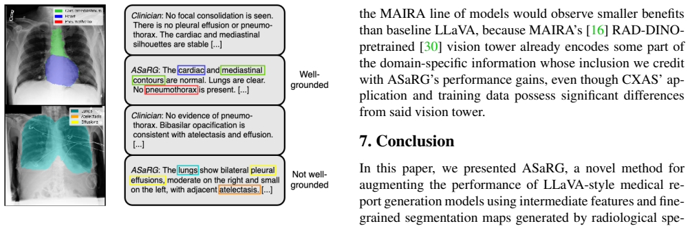
The paper's central claim is that a LLaVA-based report generator can be improved by simply concatenating two additional visual modalities into its projector: intermediate embeddings from LVM-Med, a medical segmentation foundation model, and 212 full-size segmentation maps from an extended CXAS segmenter covering anatomy, pathologies, and foreign objects. In the ASaRG configuration, the new sequence of vision features passes through LLaVA's pretrained projector, so the language model can attend to specialist local cues without changing the pretrained weights. On MIMIC-CXR the authors report CE F1 rising from 0.3872 for the two-stage LLaVA baseline to 0.4149 (+2.77 points, p<0.001), beating a fully finetuned LLaVA by 0.94 points (p=0.007) despite freezing the vision tower and the LLM. They further state that because the segmentation maps are explicit inputs, elements of the generated report can be checked against the corresponding maps, demonstrably allowing tracing of assessments to segmentation evidence and verification of their groundedness.
Load-bearing premise
The load-bearing premise is that the offline CXAS segmentation maps, including the 54 newly added pathological and foreign-object classes, are accurate enough on MIMIC-CXR to deliver usable local cues; if the maps are too noisy, the reported gain is mostly extra model capacity or feature interaction, and the grounding claim is not demonstrated.
Editorial extensions
If this is right
- Concatenation is the best of the four tested fusion strategies, beating learned mixing, weighted addition, and full replacement, while adding about 0.06% parameters in the one-epoch setting.
- Adding the 212 segmentation maps on top of the intermediate features gives a further +1.88 percentage points of CE F1, for +2.77 points over the two-stage LLaVA baseline with roughly 0.09% added projector parameters.
- ASaRG outperforms the two prior segmentation-guided report generators, COMG and ORID, by 6.98 and 6.28 CE F1 points, even though its lexical similarity scores are lower.
- Because the method freezes the vision tower and LLM and only touches the projector, it is compatible with other LLaVA-based advances, such as better encoders or language backbones, without retraining from scratch.
- The 20-superclass version matches the 212-class version within 0.01 CE F1, so the reported benefit appears insensitive to segmentation granularity.
Reading between the lines
- Editorial inference: the shuffled-map control implies the map-specific grounding contribution is only about +0.33 of the +2.77 points; the remainder likely comes from feature-map interaction or extra projection capacity, so future work should separate these before crediting the maps.
- Editorial inference: because 212 classes and 20 superclasses perform identically, investing in cheaper, cleaner segmentation (fewer but more accurate classes) may capture most of the benefit while reducing compute and memory.
- Editorial inference: the authors' own figure shows the segmenter missing atelectasis and pleural effusions; improving pathology recall in the segmenter, not just adding more classes, should convert part of the capacity-driven gain into genuinely grounded gain, which is testable by swapping in a higher-recall segmenter and checking whether the shuffled-map gap grows.
Editorial analysis
A structured set of objections, weighed in public.
Referee Report
Summary. The paper presents ASaRG, an extension of LLaVA for chest X-ray report generation. In addition to the standard LLaVA image features, ASaRG concatenates intermediate embeddings from the LVM-Med segmentation foundation model and 212-class segmentation maps from a CXAS-based segmenter into the LLaVA multimodal projector, using a learnable class embedding and lightweight linear layers. The method is evaluated on a VQA-formatted version of MIMIC-CXR, with four repeated runs per configuration and Welch's t-tests on the Clinical Efficacy F1 score. The authors report a +0.89 percentage-point gain when adding intermediate features alone and a +2.77 percentage-point gain when adding features plus fine-grained segmentation maps over a two-stage LLaVA baseline, with only about 0.09% added parameters. They also claim that the segmentation maps enable tracing elements of the generated report to the corresponding maps, and present a shuffled-map control in Section 6.1 to assess how much of the gain is due to the specific map content.
Significance. If the reported performance gain and the grounding capability were both substantiated, ASaRG would be a valuable lightweight contribution: it injects specialist knowledge into a LLaVA-style model without fine-tuning the backbone, maintains most of the original architecture, and opens a route toward visually grounded report generation. The paper has notable strengths in experimental practice: four repetitions per configuration, explicit p-values, a shuffled-map control that is uncommon and informative, and a careful accounting of parameter overhead. However, the shuffled-map control in Section 6.1 shows that only about +0.33 percentage points of the headline +2.77 gain survive randomizing the class-to-map correspondence, meaning the paper's central claim about the specific content of the fine-grained segmentation maps is not strongly supported. The grounding claim in the abstract and Section 6.2 is correspondingly weakened. The finding that a simple concatenation of extra features yields a stable CE F1 improvement is still of interest, but the stated contribution of the 212-class fine-grained maps needs better evidence.
major comments (3)
- [Section 6.1, Table 2] The shuffled-map control shows that randomizing the order of the segmentation maps on the test set removes only about +0.33 percentage points from the +2.77 percentage-point gain over the two-stage LLaVA baseline. This leaves about +2.44 percentage points of improvement under shuffled maps, which the authors attribute to projection capacity, features-map interaction, or generic map statistics. Given this result, the abstract's and Section 6.2's claim that the method 'demonstrably allows tracing elements of the report to the corresponding segmentation maps and verifying the groundedness of assessments' is not supported: if destroying the class-to-map correspondence leaves most of the gain intact, the generator is not shown to use the local, class-specific cues that grounding requires. Please either provide a direct grounding evaluation (for example, human raters judging whether report phrases correspond to the correct segmentation maps in sorted versus shuffled conditions, or an automatic per-class localization metric), or substantially temper the grounding claim to describe the architecture as enabling manual verification of correspondences rather than demonstrating that the model's outputs are actually grounded.
- [Section 4.1.1 and Tables 1-2] The concatenation fusion method was selected by comparing four fusion variants on the same holdout test set that is later used to compute the p-values against the LLaVA baselines. Because the test set was used for model selection, the reported p<0.001 and p=0.007 are selection-optimistic: the test statistic is effectively the maximum over the four configurations, so the probability of observing at least one significant improvement by chance is larger than the nominal level. The effect size is large and may well survive a proper treatment, but the p-values as reported are not statistically valid. Please either reserve a separate validation set for fusion selection and evaluate only the chosen configuration on the test set, or apply a multiple-comparison correction and state that the p-values are post-selection.
- [Section 6.2 and Figure 2] The two qualitative grounding examples are the only direct evidence for the grounding claim, and the second example explicitly shows the segmentation model missing atelectasis and pleural effusions that appear in the generated report. This is a false negative in the proposed verification procedure for two of the most clinically important findings. One or two curated examples, one of which fails on central pathologies, cannot support the general claim that ASaRG demonstrably allows verifying the groundedness of assessments. A systematic analysis is needed: for a random sample of test-set reports, how often do generated pathological findings have corresponding segmentation maps, and how does that correspondence rate compare on sorted versus shuffled maps? Without such evaluation, the grounding assertion should be removed or reduced to a suggestion for future work.
minor comments (6)
- [Section 3.3, Eq. (4)-(9)] The notation for the fusion functions is inconsistent: Eq. (4) defines Fnew = f(C,R,S), but the text then denotes the features after mixing as RI and the segmentation-derived features as SI, and f and g are used without precise signatures. Please state explicitly the dimensions of C, Rstack, and the inputs to Lin1, Lin2, and the segmentation branch, so the described variants can be reimplemented without ambiguity.
- [Abstract and Table 2] The abstract reports '+0.89% performance gain' for the features-only condition, while Table 2 shows +0.89 percentage points (0.3961 vs 0.3872) and Table 1 shows +0.87 percentage points for the one-epoch features-only comparison. Please use percentage points consistently and indicate which table corresponds to the abstract value.
- [Section 6.1 and Table 2] The shuffled-map result is reported as a difference of about +0.33% without a standard error or p-value. Since each run was repeated four times, please report the mean and standard deviation of the sorted-minus-shuffled difference and state whether +0.33% is statistically significant across the runs. This is important because the paper interprets this small difference as the entire interpretable contribution of the segmentation maps.
- [Section 4.1.2 and Supplementary Materials] The two-stage training recipe differs between the features-only and features-plus-segmentation conditions not only in the presence of segmentation maps but also in the number of trainable stages and the parameter count. The paper argues convincingly in Section 6.1 that parameter count alone cannot explain the full gain, but the comparisons in Table 2 would be cleaner if the two-stage features-only condition and the two-stage features-plus-maps condition had identical training schedules except for the added branch. Please state explicitly whether this is the case.
- [Reproducibility Statement] The code is promised 'at a later date' and the CXAS additional-class training data are not included. For the field to build on the 212-class segmentation setup, the extended CXAS class definitions and the finetuning data (or a description sufficient to reproduce them) should be released alongside the paper.
- [Section 6.3 and Supplementary Figures 4-5] The supplementary material reports that the per-class advantage of ASaRG is diffuse and varies across repeated runs, and that differences on some classes could not be determined due to class imbalance. Please mention this in the main text's limitations section, since it qualifies the clinical-efficacy interpretation of the aggregate CE F1 improvement.
Circularity Check
No significant circularity: the headline CE F1 gain rests on a held-out empirical comparison, not on a self-referential derivation; the shuffled-map control directly probes map contribution.
full rationale
The central claim—that concatenating LVM-Med intermediate features and CXAS segmentation maps into LLaVA's projector improves CE F1 on MIMIC-CXR—is an empirical finetuning result evaluated on the holdout test set. The report generator is trained on reference reports, not on the segmentation model's outputs, so the reported metric is not a by-construction match to the extra inputs. The only self-citation is CXAS [36], co-developed by two present authors, used to generate the input maps; the maps' contribution is not asserted by citation but measured via the shuffled-map control in Section 6.1, which shows only about +0.33% of the gain depends on map order/content. That control is the correct empirical probe and its outcome weakens the paper's grounding interpretation, but a weak or negative control result is not circular reasoning. The paper also candidly lists residual explanations (projection capacity, features–map interaction, generic map statistics). A separate statistical concern is that the concatenation fusion was chosen after evaluating on the same holdout test set, making the p-values selection-optimistic; this is a validity limitation, not a definitional reduction. No equation in the paper reduces the claimed gain to its inputs by construction, and no uniqueness or ansatz result is imported from the authors' prior work.
Assumptions & free parameters
free parameters (3)
- Number of finetuning epochs =
1 or 2
- Segmentation class granularity =
212 classes or 20 superclasses
- Learnable class embedding dimensions =
width b*256, depth 512
assumptions (4)
- domain assumption MIMIC-CXR, converted to a VQA format with 33 paraphrased prompts and GPT-3.5-generated multi-turn chats, preserves the clinical content of the original findings reports.
- domain assumption CheXbert-based Clinical Efficacy F1 is a valid proxy for report quality.
- domain assumption Concatenating foreign embeddings into LLaVA's projector preserves the pretrained alignment well enough for 1-2 epochs of fine-tuning to be meaningful.
- domain assumption The offline LVM-Med and CXAS models produce informative inputs for MIMIC-CXR images, including the 54 newly added CXAS classes.
Cite this review
Pith. "Pith review of Automatic Fine-grained Segmentation-assisted Report Generation." pith.science (2026). https://pith.science/paper/ZSLTMI3G
@misc{pith2026250716623,
  author       = {Pith},
  title        = {Pith review of: Automatic Fine-grained Segmentation-assisted Report Generation},
  year         = {2026},
  howpublished = {\url{https://pith.science/paper/ZSLTMI3G}},
  note         = {Machine review of arXiv:2507.16623}
}
abstract
Reliable end-to-end clinical report generation has been a longstanding goal of medical ML research. The end goal for this process is to alleviate radiologists' workloads and provide second opinions to clinicians or patients. Thus, a necessary prerequisite for report generation models is a strong general performance and some type of innate grounding capability, to convince clinicians or patients of the veracity of the generated reports. In this paper, we present ASaRG (\textbf{A}utomatic \textbf{S}egmentation-\textbf{a}ssisted \textbf{R}eport \textbf{G}eneration), an extension of the popular LLaVA architecture that aims to tackle both of these problems. ASaRG proposes to fuse intermediate features and fine-grained segmentation maps created by specialist radiological models into LLaVA's multi-modal projection layer via simple concatenation. With a small number of added parameters, our approach achieves a +0.89\% performance gain ($p=0.012$) in CE F1 score compared to the LLaVA baseline when using only intermediate features, and +2.77\% performance gain ($p<0.001$) when adding a combination of intermediate features and fine-grained segmentation maps. Compared with COMG and ORID, two other report generation methods that utilize segmentations, the performance gain amounts to 6.98\% and 6.28\% in F1 score, respectively. ASaRG is not mutually exclusive with other changes made to the LLaVA architecture, potentially allowing our method to be combined with other advances in the field. Finally, the use of an arbitrary number of segmentations as part of the input demonstrably allows tracing elements of the report to the corresponding segmentation maps and verifying the groundedness of assessments. Our code will be made publicly available at a later date.
Figures


Figures from the paper (2 more)


Reference graph
Works this paper leans on
-
[1]
Bottom-up and top-down attention for image captioning and visual question answering
Peter Anderson, Xiaodong He, Chris Buehler, Damien Teney, Mark Johnson, Stephen Gould, and Lei Zhang. Bottom-up and top-down attention for image captioning and visual question answering. In Proceedings of the IEEE con- ference on computer vision and pattern recognition , pages 6077–6086, 2018. 2
2018
-
[2]
Jinze Bai, Shuai Bai, Yunfei Chu, Zeyu Cui, Kai Dang, Xi- aodong Deng, Yang Fan, Wenbin Ge, Yu Han, Fei Huang, Binyuan Hui, Luo Ji, Mei Li, Junyang Lin, Runji Lin, Day- iheng Liu, Gao Liu, Chengqiang Lu, Keming Lu, Jianxin Ma, Rui Men, Xingzhang Ren, Xuancheng Ren, Chuanqi Tan, Sinan Tan, Jianhong Tu, Peng Wang, Shijie Wang, Wei Wang, Shengguang Wu, Benfe...
arXiv 2023
-
[3]
Shruthi Bannur, Kenza Bouzid, Daniel C. Castro, An- ton Schwaighofer, Anja Thieme, Sam Bond-Taylor, Max- imilian Ilse, Fernando P ´erez-Garc´ıa, Valentina Salvatelli, 8 Harshita Sharma, Felix Meissen, Mercy Ranjit, Shaury Srivastav, Julia Gong, Noel C. F. Codella, Fabian Falck, Ozan Oktay, Matthew P. Lungren, Maria Teodora Wetscherek, Javier Alvarez-Valle...
arXiv 2024
-
[4]
Baselines for Chest X-Ray Report Generation
William Boag, Tzu-Ming Harry Hsu, Matthew Mcdermott, Gabriela Berner, Emily Alesentzer, and Peter Szolovits. Baselines for Chest X-Ray Report Generation. In Proceed- ings of the Machine Learning for Health NeurIPS Workshop, pages 126–140. PMLR, 2020. 5
2020
-
[5]
Language models are few-shot learners
Tom Brown, Benjamin Mann, Nick Ryder, Melanie Sub- biah, Jared D Kaplan, Prafulla Dhariwal, Arvind Neelakan- tan, Pranav Shyam, Girish Sastry, Amanda Askell, Sand- hini Agarwal, Ariel Herbert-V oss, Gretchen Krueger, Tom Henighan, Rewon Child, Aditya Ramesh, Daniel Ziegler, Jeffrey Wu, Clemens Winter, Chris Hesse, Mark Chen, Eric Sigler, Mateusz Litwin, S...
1901
-
[6]
ImageNet: A large-scale hierarchical im- age database
Jia Deng, Wei Dong, Richard Socher, Li-Jia Li, Kai Li, and Li Fei-Fei. ImageNet: A large-scale hierarchical im- age database. In 2009 IEEE Conference on Computer Vision and Pattern Recognition, Miami, FL, 2009. IEEE. 12
2009
-
[7]
An Image is Worth 16x16 Words: Transformers for Image Recognition at Scale, 2021
Alexey Dosovitskiy, Lucas Beyer, Alexander Kolesnikov, Dirk Weissenborn, Xiaohua Zhai, Thomas Unterthiner, Mostafa Dehghani, Matthias Minderer, Georg Heigold, Syl- vain Gelly, Jakob Uszkoreit, and Neil Houlsby. An Image is Worth 16x16 Words: Transformers for Image Recognition at Scale, 2021. arXiv:2010.11929 [cs]. 3, 4
arXiv 2021
-
[8]
Abhimanyu Dubey, Abhinav Jauhri, Abhinav Pandey, Ab- hishek Kadian, Ahmad Al-Dahle, Aiesha Letman, Akhil Mathur, Alan Schelten, Amy Yang, Angela Fan, Anirudh Goyal, Anthony Hartshorn, Aobo Yang, Archi Mitra, Archie Sravankumar, Artem Korenev, Arthur Hinsvark, Arun Rao, Aston Zhang, Aur ´elien Rodriguez, Austen Gregerson, Ava Spataru, Baptiste Rozi `ere, B...
arXiv 2024
Show all 84 references
-
[9]
PhysioBank, PhysioToolkit, and PhysioNet: components of a new research resource for complex physio- logic signals
A L Goldberger, L A Amaral, L Glass, J M Hausdorff, P C Ivanov, R G Mark, J E Mietus, G B Moody, C K Peng, and H E Stanley. PhysioBank, PhysioToolkit, and PhysioNet: components of a new research resource for complex physio- logic signals. Circulation, 101(23):E215–20, 2000. 3
2000
-
[10]
Complex Organ Mask Guided Radiology Report Generation
Tiancheng Gu, Dongnan Liu, Zhiyuan Li, and Weidong Cai. Complex Organ Mask Guided Radiology Report Generation. In 2024 IEEE/CVF Winter Conference on Applications of Computer Vision (WACV), pages 7980–7989, Waikoloa, HI, USA, 2024. IEEE. 2, 3, 5, 6
2024
-
[11]
ORID: Organ-Regional In- formation Driven Framework for Radiology Report Genera- tion
Tiancheng Gu, Kaicheng Yang, Xiang An, Ziyong Feng, Dongnan Lin, and Weidong Cai. ORID: Organ-Regional In- formation Driven Framework for Radiology Report Genera- tion. In 2025 IEEE/CVF Winter Conference on Applications of Computer Vision (WACV) , pages 378–387, Tucson, AZ, US...
2025
-
[12]
Regiongpt: Towards region understanding vision lan- guage model
Qiushan Guo, Shalini De Mello, Hongxu Yin, Wonmin Byeon, Ka Chun Cheung, Yizhou Yu, Ping Luo, and Sifei Liu. Regiongpt: Towards region understanding vision lan- guage model. In Proceedings of the IEEE/CVF Conference on Computer Vision and Pattern Recognition, pages 13796– 1380...
2024
-
[13]
CT2Rep: Automated Radiology Report Generation for 3D Medical Imaging
Ibrahim Ethem Hamamci, Sezgin Er, and Bjoern Menze. CT2Rep: Automated Radiology Report Generation for 3D Medical Imaging . In proceedings of Medical Image Com- puting and Computer Assisted Intervention – MICCAI 2024. Springer Nature Switzerland, 2024. 1
2024
-
[14]
Swin unetr: Swin transformers for semantic segmentation of brain tumors in mri images
Ali Hatamizadeh, Vishwesh Nath, Yucheng Tang, Dong Yang, Holger R Roth, and Daguang Xu. Swin unetr: Swin transformers for semantic segmentation of brain tumors in mri images. In International MICCAI brainlesion workshop, pages 272–284. Springer, 2021. 4
2021
-
[15]
Arbitrary style transfer in real-time with adaptive instance normalization
Xun Huang and Serge Belongie. Arbitrary style transfer in real-time with adaptive instance normalization. In Proceed- ings of the IEEE international conference on computer vi- sion, pages 1501–1510, 2017. 4
2017
-
[16]
Hyland, Shruthi Bannur, Kenza Bouzid, Daniel C
Stephanie L. Hyland, Shruthi Bannur, Kenza Bouzid, Daniel C. Castro, Mercy Ranjit, Anton Schwaighofer, Fer- nando P ´erez-Garc´ıa, Valentina Salvatelli, Shaury Srivas- tav, Anja Thieme, Noel Codella, Matthew P. Lungren, Maria Teodora Wetscherek, Ozan Oktay, and Javier Alvarez-...
2024 arXiv
-
[17]
Alistair E. W. Johnson, Tom J. Pollard, Seth J. Berkowitz, Nathaniel R. Greenbaum, Matthew P. Lungren, Chih-ying Deng, Roger G. Mark, and Steven Horng. MIMIC-CXR, a de-identified publicly available database of chest radio- graphs with free-text reports. Sci Data, 6(1), 2019. P...
2019
-
[18]
Why does 9 my medical AI look at pictures of birds? Exploring the ef- ficacy of transfer learning across domain boundaries
Frederic Jonske, Moon Kim, Enrico Nasca, Janis Evers, Jo- hannes Haubold, Ren ´e Hosch, Felix Nensa, Michael Kamp, Constantin Seibold, Jan Egger, and Jens Kleesiek. Why does 9 my medical AI look at pictures of birds? Exploring the ef- ficacy of transfer learning across domain ...
-
[19]
Kingma and Jimmy Ba
Diederik P. Kingma and Jimmy Ba. Adam: A Method for Stochastic Optimization, 2017. arXiv:1412.6980 [cs]. 12
2017 arXiv
-
[20]
Meteor: an automatic met- ric for MT evaluation with high levels of correlation with human judgments
Alon Lavie and Abhaya Agarwal. Meteor: an automatic met- ric for MT evaluation with high levels of correlation with human judgments. In Proceedings of the Second Workshop on Statistical Machine Translation , pages 228–231, USA,
-
[21]
AutoRG-Brain: Grounded Report Generation for Brain MRI, 2024
Jiayu Lei, Xiaoman Zhang, Chaoyi Wu, Lisong Dai, Ya Zhang, Yanyong Zhang, Yanfeng Wang, Weidi Xie, and Yue- hua Li. AutoRG-Brain: Grounded Report Generation for Brain MRI, 2024. arXiv:2407.16684 [eess]. 1
2024 arXiv
-
[22]
LLaV A-med: training a large language- and-vision assistant for biomedicine in one day
Chunyuan Li, Cliff Wong, Sheng Zhang, Naoto Usuyama, Haotian Liu, Jianwei Yang, Tristan Naumann, Hoifung Poon, and Jianfeng Gao. LLaV A-med: training a large language- and-vision assistant for biomedicine in one day. In Proceed- ings of the 37th International Conference on Neu...
2023
-
[23]
A structure-aware relation network for thoracic diseases detection and segmentation
Jie Lian, Jingyu Liu, Shu Zhang, Kai Gao, Xiaoqing Liu, Dingwen Zhang, and Yizhou Yu. A structure-aware relation network for thoracic diseases detection and segmentation. IEEE Transactions on Medical Imaging , 40(8):2042–2052,
-
[24]
ROUGE: A Package for Automatic Evalu- ation of Summaries
Chin-Yew Lin. ROUGE: A Package for Automatic Evalu- ation of Summaries. In Text Summarization Branches Out, pages 74–81, Barcelona, Spain, 2004. Association for Com- putational Linguistics. 5
2004
-
[25]
Visual Instruction Tuning
Haotian Liu, Chunyuan Li, Qingyang Wu, and Yong Jae Lee. Visual Instruction Tuning. In Advances in Neural Informa- tion Processing Systems, pages 34892–34916. Curran Asso- ciates, Inc., 2023. 2, 3
2023
-
[26]
Improved Baselines with Visual Instruction Tuning
Haotian Liu, Chunyuan Li, Yuheng Li, and Yong Jae Lee. Improved Baselines with Visual Instruction Tuning. In 2024 IEEE/CVF Conference on Computer Vision and Pat- tern Recognition (CVPR), pages 26286–26296, Seattle, W A, USA, 2024. IEEE. 3
2024
-
[27]
LLaV A-NeXT: Im- proved reasoning, OCR, and world knowledge, 2024
Haotian Liu, Chunyuan Li, Yuheng Li, Bo Li, Yuanhan Zhang, Sheng Shen, and Yong Jae Lee. LLaV A-NeXT: Im- proved reasoning, OCR, and world knowledge, 2024. 3
2024
-
[28]
Duy M. H. Nguyen, Hoang Nguyen, Nghiem Diep, Tan Ngoc Pham, Tri Cao, Binh Nguyen, Paul Swoboda, Nhat Ho, Shadi Albarqouni, Pengtao Xie, Daniel Sonntag, and Mathias Niepert. LVM-Med: Learning Large-Scale Self- Supervised Vision Models for Medical Imaging via Second- order Graph...
2023
-
[29]
BLEU: a method for automatic evaluation of ma- chine translation
Kishore Papineni, Salim Roukos, Todd Ward, and Wei-Jing Zhu. BLEU: a method for automatic evaluation of ma- chine translation. In Proceedings of the 40th Annual Meet- ing on Association for Computational Linguistics - ACL ’02, page 311, Philadelphia, Pennsylvania, 2001. Associ...
2001
-
[30]
Castro, Anton Schwaighofer, Matthew P
Fernando P ´erez-Garc´ıa, Harshita Sharma, Sam Bond- Taylor, Kenza Bouzid, Valentina Salvatelli, Maximilian Ilse, Shruthi Bannur, Daniel C. Castro, Anton Schwaighofer, Matthew P. Lungren, Maria Teodora Wetscherek, Noel Codella, Stephanie L. Hyland, Javier Alvarez-Valle, and Oz...
2025
-
[31]
Learning transferable visual models from natural language supervision
Alec Radford, Jong Wook Kim, Chris Hallacy, Aditya Ramesh, Gabriel Goh, Sandhini Agarwal, Girish Sastry, Amanda Askell, Pamela Mishkin, Jack Clark, and others. Learning transferable visual models from natural language supervision. In International conference on machine learn- ...
2021
-
[32]
ReXErr-v1: Clini- cally Meaningful Chest X-Ray Report Errors Derived from MIMIC-CXR
Vishwanatha Rao, Serena Zhang, Julian Acosta, Subathra Adithan, and Pranav Rajpurkar. ReXErr-v1: Clini- cally Meaningful Chest X-Ray Report Errors Derived from MIMIC-CXR. 2
-
[33]
Glamm: Pixel grounding large multimodal model
Hanoona Rasheed, Muhammad Maaz, Sahal Shaji, Abdel- rahman Shaker, Salman Khan, Hisham Cholakkal, Rao M Anwer, Eric Xing, Ming-Hsuan Yang, and Fahad S Khan. Glamm: Pixel grounding large multimodal model. In Pro- ceedings of the IEEE/CVF Conference on Computer Vision and Patter...
2024
-
[34]
DeepSpeed: System Optimizations Enable Training Deep Learning Models with Over 100 Billion Pa- rameters
Jeff Rasley, Samyam Rajbhandari, Olatunji Ruwase, and Yuxiong He. DeepSpeed: System Optimizations Enable Training Deep Learning Models with Over 100 Billion Pa- rameters. In Proceedings of the 26th ACM SIGKDD Interna- tional Conference on Knowledge Discovery & Data Mining, pag...
2020
-
[35]
ZeRO-Offload: Democratizing Billion- Scale model training
Jie Ren, Samyam Rajbhandari, Reza Yazdani Aminabadi, Olatunji Ruwase, Shuangyan Yang, Minjia Zhang, Dong Li, and Yuxiong He. ZeRO-Offload: Democratizing Billion- Scale model training. In 2021 USENIX Annual Technical Conference (USENIX ATC 21) , pages 551–564. USENIX Associatio...
2021
-
[36]
Fink, Moon Kim, Simon Reiß, Ken Herrmann, Jens Kleesiek, and Rainer Stiefelhagen
Constantin Seibold, Alexander Jaus, Matthias A. Fink, Moon Kim, Simon Reiß, Ken Herrmann, Jens Kleesiek, and Rainer Stiefelhagen. Accurate Fine-Grained Segmentation of Human Anatomy in Radiographs via V olumetric Pseudo- Labeling, 2023. arXiv:2306.03934 [eess]. 2, 3
2023 arXiv
-
[37]
Combining Automatic Labelers and Expert Annotations for Accurate Radiology Report Labeling Using BERT
Akshay Smit, Saahil Jain, Pranav Rajpurkar, Anuj Pareek, Andrew Ng, and Matthew Lungren. Combining Automatic Labelers and Expert Annotations for Accurate Radiology Report Labeling Using BERT. In Proceedings of the 2020 Conference on Empirical Methods in Natural Language Pro- c...
2020
-
[38]
CLiP, catheter and line position dataset
Jennifer S N Tang, Jarrel C Y Seah, Adil Zia, Jay Gajera, Richard N Schlegel, Aaron J N Wong, Dayu Gai, Shu Su, Tony Bose, Marcus L Kok, Alex Jarema, George N Hari- sis, Chris-Tin Cheng, Helen Kavnoudias, Wayland Wang, Anouk Stein, George Shih, Frank Gaillard, Andrew Dixon, an...
2021
-
[39]
Interactive and Explainable Region-guided Radi- ology Report Generation
Tim Tanida, Philip M ¨uller, Georgios Kaissis, and Daniel Rueckert. Interactive and Explainable Region-guided Radi- ology Report Generation. In 2023 IEEE/CVF Conference on Computer Vision and Pattern Recognition (CVPR), pages 7433–7442, Vancouver, BC, Canada, 2023. IEEE. 2, 3
2023
-
[40]
Ryutaro Tanno, David G. T. Barrett, Andrew Sellergren, Sumedh Ghaisas, Sumanth Dathathri, Abigail See, Johannes Welbl, Charles Lau, Tao Tu, Shekoofeh Azizi, Karan Sing- hal, Mike Schaekermann, Rhys May, Roy Lee, SiWai Man, Sara Mahdavi, Zahra Ahmed, Yossi Matias, Joelle Barral...
2025
-
[41]
Sara Mah- davi, Bradley Green, Ewa Dominowska, Blaise Aguera Y Arcas, Joelle Barral, Dale Webster, Greg S
Tao Tu, Shekoofeh Azizi, Danny Driess, Mike Schaeker- mann, Mohamed Amin, Pi-Chuan Chang, Andrew Carroll, Charles Lau, Ryutaro Tanno, Ira Ktena, Anil Palepu, Basil Mustafa, Aakanksha Chowdhery, Yun Liu, Simon Korn- blith, David Fleet, Philip Mansfield, Sushant Prakash, Renee W...
2024
-
[42]
Lawrence Zitnick, and Devi Parikh
Ramakrishna Vedantam, C. Lawrence Zitnick, and Devi Parikh. CIDEr: Consensus-based image description evalua- tion. In 2015 IEEE Conference on Computer Vision and Pat- tern Recognition (CVPR) , pages 4566–4575, Boston, MA, USA, 2015. IEEE. 5
2015
-
[43]
Visionllm: Large language model is also an open- ended decoder for vision-centric tasks
Wenhai Wang, Zhe Chen, Xiaokang Chen, Jiannan Wu, Xizhou Zhu, Gang Zeng, Ping Luo, Tong Lu, Jie Zhou, Yu Qiao, et al. Visionllm: Large language model is also an open- ended decoder for vision-centric tasks. Advances in Neural Information Processing Systems, 36:61501–61513, 2023. 3
2023
-
[44]
The all-seeing project: Towards panop- tic visual recognition and understanding of the open world
Weiyun Wang, Min Shi, Qingyun Li, Wenhai Wang, Zhen- hang Huang, Linjie Xing, Zhe Chen, Hao Li, Xizhou Zhu, Zhiguo Cao, et al. The all-seeing project: Towards panop- tic visual recognition and understanding of the open world. arXiv preprint arXiv:2308.01907, 2023. 2, 3
2023 arXiv
-
[45]
A survey of deep- learning-based radiology report generation using multimodal inputs
Xinyi Wang, Grazziela Figueredo, Ruizhe Li, Wei Emma Zhang, Weitong Chen, and Xin Chen. A survey of deep- learning-based radiology report generation using multimodal inputs. Medical Image Analysis, 103:103627, 2025. 1
2025
-
[46]
Marten: Visual question answering with mask generation for multi-modal document understanding
Zining Wang, Tongkun Guan, Pei Fu, Chen Duan, Qianyi Jiang, Zhentao Guo, Shan Guo, Junfeng Luo, Wei Shen, and Xiaokang Yang. Marten: Visual question answering with mask generation for multi-modal document understanding. In Proceedings of the Computer Vision and Pattern Recogni...
2025
-
[47]
B. L. Welch. THE GENERALIZATION OF ‘STUDENT’S’ PROBLEM WHEN SEVERAL DIFFERENT POPULA- TION V ARLANCES ARE INVOLVED. Biometrika, 34(1- 2):28–35, 1947. Publisher: Oxford University Press (OUP). 5
1947
-
[48]
Gpt4roi: Instruction tuning large language model on region- of-interest
Shilong Zhang, Peize Sun, Shoufa Chen, Min Xiao, Wenqi Shao, Wenwei Zhang, Yu Liu, Kai Chen, and Ping Luo. Gpt4roi: Instruction tuning large language model on region- of-interest. In European conference on computer vision , pages 52–70. Springer, 2024. 2, 3
2024
-
[49]
Xing, Hao Zhang, Joseph E
Lianmin Zheng, Wei-Lin Chiang, Ying Sheng, Siyuan Zhuang, Zhanghao Wu, Yonghao Zhuang, Zi Lin, Zhuohan Li, Dacheng Li, Eric P. Xing, Hao Zhang, Joseph E. Gon- zalez, and Ion Stoica. Judging LLM-as-a-Judge with MT- Bench and Chatbot Arena, 2023. arXiv:2306.05685 [cs]. 12
2023 arXiv
-
[50]
Topol, and Pranav Rajpurkar
Hong-Yu Zhou, Juli ´an Nicol ´as Acosta, Subathra Adithan, Suvrankar Datta, Eric J. Topol, and Pranav Rajpurkar. Med- Versa: A Generalist Foundation Model for Medical Image Interpretation, 2025. arXiv:2405.07988 [cs]. 2 Reproducibility Statement For the purpose of reproduction...
2025 arXiv
-
[52]
Can you describe what you see in the image?
-
[53]
Please provide an overview of the key observations in the X-ray images
-
[54]
What are the significant details captured in this medical image?
-
[55]
Summarize the visual findings from this medical scan
-
[56]
Give me a brief summary of the image’s diagnostic features
-
[57]
Can you outline the main points of interest in this picture?
-
[58]
What observations can be made from this radiological image?
-
[59]
Describe the significant findings in this visual information
-
[60]
Offer a brief overview of the diagnostic details in the picture
-
[61]
List the main points of interest in this radiological data
-
[62]
Outline the relevant findings of this medical imaging
-
[63]
Give me a summarized account of the observations here
-
[64]
Provide a concise summary of the diagnostic features
-
[65]
Can you identify the key takeaways from this visual data?
-
[66]
Highlight the significant findings in this X-ray image
-
[67]
Summarize the important aspects of this radiological data
-
[68]
Offer a brief synopsis of the observations captured
-
[69]
Describe the most salient features in this X-ray image
-
[70]
What do you perceive as the primary diagnostic insights from this picture?
-
[71]
Provide details about any notable and unremarkable features in the image
-
[72]
Describe the overall condition of the subject in the image
-
[73]
Can you summarize the key observations from this radiograph?
-
[74]
Discuss the significant findings within this X-ray image
-
[75]
Brief me on the findings
-
[76]
What can you see on the X-ray images?
-
[77]
Please provide a summary of the observations made in the images, noting any abnormalities or potential issues
-
[78]
Describe what you see in the images and mention if any areas appear normal or unremarkable
-
[79]
Summarize the key observations and abnormalities that stand out in the images
-
[80]
Give an overview of the findings
-
[81]
Summarize the overall impression of the images, emphasizing critical observations
-
[82]
Provide a concise summary of the findings using medical jargon
-
[83]
Are there any notable or unremarkable findings that should be known to the patient’s primary care physician?
-
[84]
Figure 3
Summarize the findings in a manner that allows for easy communication with the patient’s healthcare team. Figure 3. The user queries uF i ∈ PF for prompting a VLM for radiology findings on an image vi, each paired with the findings rF i as the response to form a single-turn ch...
-
[2007]
event- place: Prague, Czech Republic
Association for Computational Linguistics. event- place: Prague, Czech Republic. 5
Reviewed August 6, 2026 · model on record in the stance chip above.
Discussion (0). Sign in to comment.