REVIEW 5 major objections 5 minor 59 references
Frequency Prior Guided Matching: A Data Augmentation Approach for Generalizable Semi-Supervised Polyp Segmentation
T0 review · 5 major / 5 minor · reviewed 2026-08-06 · deepseek-v4-flash
Pith's one-line read Polyp edges share a stable frequency signature across datasets; FPGM learns it from labeled images and aligns unlabeled amplitude spectra to it, gaining over 10 absolute Dice points on unseen data.
desk verdict A plausible frequency-augmentation recipe for semi-supervised polyp segmentation with large reported gains, but the central 'domain-invariant polyp-edge prior' claim is confounded by the binary-mask spectrum and the augmentation is stronger than described. read the letter →
The pith
A machine-rendered reading of the paper's core claim, the machinery that carries it, and where it could break.
The reading
What carries the argument
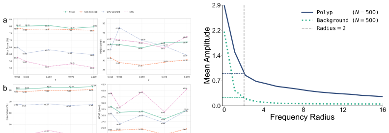
The load-bearing object is the learned one-dimensional frequency prior P̄: the EMA-aggregated radial amplitude profile of polyp edge regions. The mechanism that carries the argument is spectral shape alignment (Section 3.3): an unlabeled image's radial amplitude profile is split into total energy and a normalized shape, the shape is pulled toward the normalized template by the guidance factor γ = 0.05, re-scaled to the original energy, broadcast back to a two-dimensional spectrum, and recombined with the original phase via the inverse FFT. Everything that matters is in the separation of shape from energy and of amplitude from phase: the perturbation changes how spectral energy is distributed across frequencies, but not how much there is and not where structures sit. The paper's ablations show the learned template is the active ingredient—it beats a 1/f pink-noise prior and an ideal low-pass prior—and that it is specific to polyp edges: matched background profiles are flat and near zero, while the edge profile shows a rich, structured spectrum peaking at low radial frequencies.
What would settle it
Measure polyp-edge radial profiles in a dataset whose acquisition differs substantially from the six used here—for example, narrow-band imaging, a new scope vendor, or a different bowel-preparation protocol—and test whether the profile still matches the learned prior. Then run FPGM on that dataset's unlabeled images with the original prior and with a newly learned prior: if alignment to the original prior lowers Dice below the no-augmentation baseline while the new prior restores it, the domain-invariance claim is falsified.
Extended reading notes
Core claim
FPGM rests on the empirical discovery that the average radial profile of the FFT amplitude, computed at polyp boundary regions (Sobel gradient on the ground-truth mask, dilated into an edge band), is highly reproducible—between random subsets of the same dataset and across Kvasir, CVC-ClinicDB, CVC-ColonDB, and ETIS. The paper treats this profile as a domain-invariant stylistic prior: amplitude encodes texture, phase encodes structure, so aligning amplitude while preserving phase should strip domain-specific appearance without moving anatomy. Stage I aggregates this profile across labeled images with an exponential moving average (momentum 0.999), yielding a stable template P̄. Stage II takes each weakly augmented unlabeled image, decomposes its FFT into amplitude and phase, reduces the amplitude to a radial profile, factors it into total energy and normalized shape, interpolates the shape toward P̄ with strength γ = 0.05, rescales to the original energy, and reconstructs the image with the phase intact. The resulting consistency view forces the network to rely on structure rather than surface texture, giving the reported state-of-the-art results and the zero-shot gains on CVC-300 and BKAI.
Load-bearing premise
The load-bearing premise is that the average radial amplitude profile computed from ground-truth polyp edges in labeled images is a valid template for reshaping the full-image amplitude spectrum of any unlabeled colonoscopy image—that pulling each image's spectral profile toward this template removes domain-specific texture without disturbing the structure the segmentation model needs. If that transfer fails for some imaging domain or lesion subtype, the augmentation would distort unlabeled images and shrink, rather than grow, generalization.
Editorial extensions
If this is right
- At only 5% labeled data, zero-shot Dice on unseen CVC-300 reaches 82.46 versus 64.17 for UniMatch, a gap of 18.29 absolute points; on BKAI the gap is 10.22, so the value of spectral alignment grows as labels shrink.
- Because the frequency-guided loss alone beats conventional consistency augmentation on unseen domains by up to 6.89 Dice points (67.89 vs 61.00 on BKAI at 10%), the generalization gain is attributable to the learned prior, not to having more augmentations.
- The learned prior beats both the generic 1/f natural-image prior and an ideal low-pass prior, so task-specific spectral statistics—not frequency smoothing in general—are what carries the improvement.
- The method uses a standard U-Net backbone and an identical training protocol across all competing methods, so the reported margins trace to the augmentation strategy, and it should compose with stronger backbones or other SSL losses.
Reading between the lines
- A direct test of the domain-invariance claim is leave-one-dataset-out evaluation: train on three of the four datasets, compute the prior from those three, and test on the fourth; the paper evaluates generalization only on CVC-300 and BKAI, so a per-dataset held-out sweep would reveal whether the prior is equally stable for every source.
- The framework implies a recipe for other tasks: before applying FPGM to, say, tumor or lesion segmentation in MRI or CT, one should first measure whether the target structure's edge radial profile is as reproducible as it is for polyps; if it is not, the alignment step could actively damage unlabeled images.
- An adaptive guidance strength—scaling γ by the distance between the image's profile and the prior—could extend FPGM to domains far from the training distribution, where a fixed γ = 0.05 may be too weak to neutralize strong domain texture or too strong to preserve fine structure.
- The amplitude-as-style, phase-as-structure assumption could be probed quantitatively by swapping the roles: if perturbing phase (instead of amplitude) destroyed segmentation structure while amplitude perturbation did not, the mechanism behind FPGM would be confirmed as stated.
Editorial analysis
A structured set of objections, weighed in public.
Referee Report
Summary. The manuscript introduces FPGM, a two-stage semi-supervised augmentation framework for polyp segmentation. Stage I computes the average radial amplitude profile of ground-truth polyp edge regions in labeled images via FFT and aggregates it with an exponential moving average. Stage II transforms each unlabeled image into the Fourier domain, computes its full-image radial amplitude profile, interpolates the normalized profile by 5% toward the learned prior, energy-normalizes, broadcasts the result back to a 2D amplitude spectrum, and recombines it with the original phase. The model is trained with a supervised loss and two consistency losses, one against standard strong augmentation and one against the frequency-perturbed view. Experiments on four in-domain datasets under 5%, 10%, and 20% labeled data and two unseen datasets are reported against ten baselines, with claims of consistent state-of-the-art performance and large zero-shot gains.
Significance. The direction is practically useful and the experimental protocol is largely coherent: baselines are re-implemented under common settings, the code repository is released, and the paper includes ablations for loss components, hyperparameter sensitivity, and prior specificity. However, the central attribution of the gains to a domain-invariant polyp-edge frequency signature is not yet established. The prior computation is confounded by the spectrum of the binary edge mask and by the radialization operation in the reconstruction, and the reported numbers come from single runs with malformed table entries. If the mechanism is confirmed with appropriate controls, FPGM would be a solid contribution to semi-supervised medical image segmentation; in its current form, the causal claim is underdetermined.
major comments (5)
- [§4.1, Tables 2–3] All quantitative comparisons are reported as single numbers without standard deviations; because the headline is a state-of-the-art claim, the reader cannot tell which margins are meaningful. Please provide results over multiple random labeled/unlabeled splits and report mean±std for the main tables. In addition, Tables 2 and 3 contain malformed cells, e.g., '96.356.71' and '26.785.9863.03 54.66 35.96 11.88 38.98 33.8833.1313.82' in the 10% rows of Table 2, and similar concatenated numbers in Table 3; these must be fixed so that every cell is unambiguous.
- [§3.2, Eqs. (1)–(5); Figs. 1–2] The prior is computed from x_edge = x_gray ⊙ M_edge, where M_edge is a dilated binary boundary. The amplitude spectrum of this product is dominated by the spectrum of the binary mask itself, so the striking cross-dataset consistency in Figs. 1–2 may simply reflect that all ground-truth masks are thin binary contours rather than any property of polyp tissue. To support the key discovery claim, add a control in which the same pipeline is applied to random thin masks, to background-region masks, and to non-polyp foreground boundaries; if the profiles are equally consistent, the 'domain-invariant polyp-edge frequency signature' is an artifact of the masking operation.
- [§3.3, Eqs. (9)–(12)] Equation (11) broadcasts the 1D perturbed profile to every angle, making the reconstructed amplitude spectrum exactly radially symmetric. This discards all orientation-dependent amplitude structure, so the perturbation is far stronger than the γ=0.05 interpolation in shape space suggests; only the phase is preserved. The claim that the operation preserves structural integrity should be supported by quantifying how much each unlabeled image actually changes (e.g., mean absolute difference or structural similarity between x_w and x_freq) and by ablating radialization without any learned prior (e.g., using the image's own radial profile or a flat profile).
- [§5.2, Table 6] The ablation against a 1/f generic prior and a low-pass prior does not control for the two confounds identified above. A model trained with a prior learned from random binary masks, or with a radialization-only augmentation that never uses the learned template, would isolate the actual contribution of the learned polyp-edge prior. Without these controls, the reported gains in Table 6 could be produced by any edge-like template or even by radialization alone, so the paper's central attribution of state-of-the-art performance to the learned prior is not established.
- [§5.4, Fig. 8] The specificity analysis is also confounded by masking: the polyp-edge profile is computed on pixels multiplied by a thin boundary mask, while the background profile is computed on raw background pixels. If the same binary edge mask is applied to background control regions, the two profiles may become much more similar. Please rerun the comparison with matched masking and matched pixel counts for background regions.
minor comments (5)
- [Eq. (5)] Specify how the prior Pbar is initialized and over which samples the EMA is applied; currently it is unclear whether the update is per-image in arbitrary order or per-epoch.
- [§4.1] The sentence 'all experiments were performed under the same random seeds' is ambiguous; clarify the number of seeds and whether every baseline was run with identical data splits and initialization.
- [Eqs. (4) and (11)] Define the radius function r(u,v) and the discrete binning used in Eq. (4); the text uses both continuous radii and a sum over the set Omega_r.
- [§4.3, Table 1] CVC-300 contains only 60 test images; the boundary metrics HD95 and ASD on this set should be discussed with a caveat about the small test size.
- [Fig. 8] State the normalization of the amplitude axes; the text reports amplitudes near 1 at radius 2, which is hard to interpret without a normalization convention.
Circularity Check
No circularity: the frequency prior is learned from labeled polyp edges, the augmentation preserves phase, and all performance claims are evaluated on held-out test sets and completely unseen datasets, so the derivation chain is self-contained.
full rationale
FPGM's derivation chain is not circular. The frequency prior in Stage I (Eqs. 1-5) is estimated from ground-truth polyp edge regions of labeled images, and Stage II (Eqs. 6-12) perturbs unlabeled images by aligning their full-image radial amplitude profile toward that prior while preserving phase. The learned prior is a fitted template, but it is not used to 'predict' its own fitting data; the paper's performance claims are measured on unseen test splits of the four training datasets (Table 2) and on two completely held-out datasets, CVC-300 and BKAI (Table 3), which are excluded from training and validation. Hyperparameters such as gamma (Eq. 9) and the loss weights are set on validation, which is standard practice and does not constitute fitted-input-called-prediction. The two author self-citations (CUTS [16], DiffKillR [25]) appear only in the related-work discussion and are not load-bearing for the proposed method or its evaluation. The 'key discovery' of cross-dataset consistency in radial frequency profiles is an empirical observation presented with evidence in Figs. 1-2, not a derivation that assumes its conclusion. Critiques such as the radialization in Eq. 11 discarding orientation-dependent amplitude structure or the edge-mask spectrum dominating the prior are concerns about the mechanism or robustness of the augmentation, but they are not circularity: the method's gains are not entailed by its definition, and the ablations (Table 6) test alternative priors against the learned one. Overall, no step reduces by construction to its own inputs, so the paper receives a circularity score of 0.
Assumptions & free parameters
free parameters (6)
- gamma =
0.05
- mu =
0.999
- tau_c =
0.95
- lambda_unsup =
0.5
- lambda_freq =
0.5
- morphological dilation kernel size
assumptions (5)
- domain assumption Polyp edge regions have a stable radial frequency profile across datasets and clinical centers.
- domain assumption The amplitude spectrum encodes style and texture while the phase spectrum preserves spatial structure.
- domain assumption Aligning the full-image radial amplitude profile toward the edge-region prior normalizes domain-specific texture without corrupting polyp structure.
- domain assumption High-confidence pseudo-labels thresholded at 0.95 are reliable enough for consistency training.
- standard math Sobel edge detection with morphological dilation on ground-truth masks produces a precise polyp boundary mask.
Cite this review
Pith. "Pith review of Frequency Prior Guided Matching: A Data Augmentation Approach for Generalizable Semi-Supervised Polyp Segmentation." pith.science (2026). https://pith.science/paper/3PAUFIT7
@misc{pith2026250806517,
  author       = {Pith},
  title        = {Pith review of: Frequency Prior Guided Matching: A Data Augmentation Approach for Generalizable Semi-Supervised Polyp Segmentation},
  year         = {2026},
  howpublished = {\url{https://pith.science/paper/3PAUFIT7}},
  note         = {Machine review of arXiv:2508.06517}
}
read the original abstract
Automated polyp segmentation is essential for early diagnosis of colorectal cancer, yet developing robust models remains challenging due to limited annotated data and significant performance degradation under domain shift. Although semi-supervised learning (SSL) reduces annotation requirements, existing methods rely on generic augmentations that ignore polyp-specific structural properties, resulting in poor generalization to new imaging centers and devices. To address this, we introduce Frequency Prior Guided Matching (FPGM), a novel augmentation framework built on a key discovery: polyp edges exhibit a remarkably consistent frequency signature across diverse datasets. FPGM leverages this intrinsic regularity in a two-stage process. It first learns a domain-invariant frequency prior from the edge regions of labeled polyps. Then, it performs principled spectral perturbations on unlabeled images, aligning their amplitude spectra with this learned prior while preserving phase information to maintain structural integrity. This targeted alignment normalizes domain-specific textural variations, thereby compelling the model to learn the underlying, generalizable anatomical structure. Validated on six public datasets, FPGM establishes a new state-of-the-art against ten competing methods. It demonstrates exceptional zero-shot generalization capabilities, achieving over 10% absolute gain in Dice score in data-scarce scenarios. By significantly enhancing cross-domain robustness, FPGM presents a powerful solution for clinically deployable polyp segmentation under limited supervision.
Figures


Figures from the paper (3 more)


Reference graph
Works this paper leans on
-
[1]
Worldwide variations in colorectal cancer
Melissa M Center, Ahmedin Jemal, Robert A Smith, and Elizabeth Ward. Worldwide variations in colorectal cancer. CA: a cancer journal for clinicians, 59(6):366–378, 2009
work page 2009
-
[2]
J ¨urgen Weitz, Moritz Koch, J ¨urgen Debus, Thomas H ¨ohler, Peter R Galle, and Markus W B¨uchler. Colorectal cancer. The Lancet, 365(9454):153–165, 2005
work page 2005
-
[3]
Evelien Dekker, Pieter J Tanis, Jasper LA Vleugels, Pashtoon M Kasi, and Michael B Wallace. Colorectal cancer. The Lancet, 394(10207):1467–1480, 2019
work page 2019
-
[4]
Samir Jain, Rohan Atale, Anubhav Gupta, Utkarsh Mishra, Ayan Seal, Aparajita Ojha, Joanna Jaworek-Korjakowska, and Ondrej Krejcar. Coinnet: A convolution-involution network with a novel statistical attention for automatic polyp segmentation. IEEE Transactions on Medical Imaging, 42(12):3987–4000, 2023
work page 2023
-
[5]
Know your orientation: A viewpoint-aware framework for polyp segmentation
Linghan Cai, Lijiang Chen, Jianhao Huang, Yifeng Wang, and Yongbing Zhang. Know your orientation: A viewpoint-aware framework for polyp segmentation. Medical Image Analysis, 97:103288, 2024
work page 2024
-
[6]
Pranet: Parallel reverse attention network for polyp segmentation
Deng-Ping Fan, Ge-Peng Ji, Tao Zhou, Geng Chen, Huazhu Fu, Jianbing Shen, and Ling Shao. Pranet: Parallel reverse attention network for polyp segmentation. In International conference on medical image computing and computer-assisted intervention, pages 263–273. Springer, 2020
work page 2020
-
[7]
Adaptive context selection for polyp segmentation
Ruifei Zhang, Guanbin Li, Zhen Li, Shuguang Cui, Dahong Qian, and Yizhou Yu. Adaptive context selection for polyp segmentation. InMedical Image Computing and Computer Assisted Intervention–MICCAI 2020: 23rd International Conference, Lima, Peru, October 4–8, 2020, Proceedings, Part VI 23, pages 253–262. Springer, 2020
work page 2020
-
[8]
Huisi Wu, Guilian Chen, Zhenkun Wen, and Jing Qin. Collaborative and adversarial learning of focused and dispersive representations for semi-supervised polyp segmentation. In Proceedings of the IEEE/CVF International Conference on Computer Vision, pages 3489– 3498, 2021
work page 2021
Show all 59 references
-
[9]
Learning with local and global consistency
Dengyong Zhou, Olivier Bousquet, Thomas Lal, Jason Weston, and Bernhard Sch ¨olkopf. Learning with local and global consistency. Advances in neural information processing systems, 16, 2003
2003
-
[10]
Fixmatch: Simplifying semi-supervised learning with consistency and confidence
Kihyuk Sohn, David Berthelot, Nicholas Carlini, Zizhao Zhang, Han Zhang, Colin A Raffel, Ekin Dogus Cubuk, Alexey Kurakin, and Chun-Liang Li. Fixmatch: Simplifying semi-supervised learning with consistency and confidence. Advances in neural information processing systems, 33:5...
2020
-
[11]
Revisiting weak-to-strong consistency in semi-supervised semantic segmentation
Lihe Yang, Lei Qi, Litong Feng, Wayne Zhang, and Yinghuan Shi. Revisiting weak-to-strong consistency in semi-supervised semantic segmentation. In Proceedings of the IEEE/CVF conference on computer vision and pattern recognition, pages 7236–7246, 2023
2023
-
[12]
Dual student: Breaking the limits of the teacher in semi-supervised learning
Zhanghan Ke, Daoye Wang, Qiong Yan, Jimmy Ren, and Rynson WH Lau. Dual student: Breaking the limits of the teacher in semi-supervised learning. In Proceedings of the IEEE/CVF international conference on computer vision, pages 6728–6736, 2019
2019
-
[13]
Atso: Asynchronous teacher-student optimization for semi-supervised image segmentation
Xinyue Huo, Lingxi Xie, Jianzhong He, Zijie Yang, Wengang Zhou, Houqiang Li, and Qi Tian. Atso: Asynchronous teacher-student optimization for semi-supervised image segmentation. In Proceedings of the IEEE/CVF conference on computer vision and pattern recognition, pages 1235–1244, 2021
2021
-
[14]
Semi-supervised semantic segmentation with cross pseudo supervision
Xiaokang Chen, Yuhui Yuan, Gang Zeng, and Jingdong Wang. Semi-supervised semantic segmentation with cross pseudo supervision. In Proceedings of the IEEE/CVF conference on computer vision and pattern recognition, pages 2613–2622, 2021
2021
-
[15]
Mutual consistency learning for semi-supervised medical image segmentation
Yicheng Wu, Zongyuan Ge, Donghao Zhang, Minfeng Xu, Lei Zhang, Yong Xia, and Jianfei Cai. Mutual consistency learning for semi-supervised medical image segmentation. Medical Image Analysis, 81:102530, 2022. 16
2022
-
[16]
Cuts: A deep learning and topological framework for multigranular unsupervised medical image segmentation
Chen Liu, Matthew Amodio, Liangbo L Shen, Feng Gao, Arman Avesta, Sanjay Aneja, Jay C Wang, Lucian V Del Priore, and Smita Krishnaswamy. Cuts: A deep learning and topological framework for multigranular unsupervised medical image segmentation. In International Conference on Me...
-
[17]
Ambiguity-selective consistency regularization for mean-teacher semi-supervised medical image segmentation
Zhe Xu, Yixin Wang, Donghuan Lu, Xiangde Luo, Jiangpeng Yan, Yefeng Zheng, and Raymond Kai-yu Tong. Ambiguity-selective consistency regularization for mean-teacher semi-supervised medical image segmentation. Medical Image Analysis, 88:102880, 2023
2023
-
[18]
Semi-supervised medical image segmentation via uncertainty rectified pyramid consistency
Xiangde Luo, Guotai Wang, Wenjun Liao, Jieneng Chen, Tao Song, Yinan Chen, Shichuan Zhang, Dimitris N Metaxas, and Shaoting Zhang. Semi-supervised medical image segmentation via uncertainty rectified pyramid consistency. Medical Image Analysis, 80:102517, 2022
2022
-
[19]
Inconsistency-aware uncertainty estimation for semi-supervised medical image segmentation
Yinghuan Shi, Jian Zhang, Tong Ling, Jiwen Lu, Yefeng Zheng, Qian Yu, Lei Qi, and Yang Gao. Inconsistency-aware uncertainty estimation for semi-supervised medical image segmentation. IEEE transactions on medical imaging, 41(3):608–620, 2021
2021
-
[20]
Compete to win: Enhancing pseudo labels for barely-supervised medical image segmentation
Huimin Wu, Xiaomeng Li, Yiqun Lin, and Kwang-Ting Cheng. Compete to win: Enhancing pseudo labels for barely-supervised medical image segmentation. IEEE Transactions on Medical Imaging, 42(11):3244–3255, 2023
2023
-
[21]
Pseudo-label guided contrastive learning for semi- supervised medical image segmentation
Hritam Basak and Zhaozheng Yin. Pseudo-label guided contrastive learning for semi- supervised medical image segmentation. In Proceedings of the IEEE/CVF conference on computer vision and pattern recognition, pages 19786–19797, 2023
2023
-
[22]
Rcps: Rectified contrastive pseudo supervision for semi-supervised medical image segmentation
Xiangyu Zhao, Zengxin Qi, Sheng Wang, Qian Wang, Xuehai Wu, Ying Mao, and Lichi Zhang. Rcps: Rectified contrastive pseudo supervision for semi-supervised medical image segmentation. IEEE Journal of Biomedical and Health Informatics, 28(1):251–261, 2023
2023
-
[23]
Semi- supervised medical image segmentation using adversarial consistency learning and dynamic convolution network
Tao Lei, Dong Zhang, Xiaogang Du, Xuan Wang, Yong Wan, and Asoke K Nandi. Semi- supervised medical image segmentation using adversarial consistency learning and dynamic convolution network. IEEE transactions on medical imaging, 42(5):1265–1277, 2022
2022
-
[24]
Semi-supervised medical image segmentation through dual-task consistency
Xiangde Luo, Jieneng Chen, Tao Song, and Guotai Wang. Semi-supervised medical image segmentation through dual-task consistency. In Proceedings of the AAAI conference on artificial intelligence, volume 35, pages 8801–8809, 2021
2021
-
[25]
Diffkillr: Killing and recreating diffeomorphisms for cell annotation in dense microscopy images
Chen Liu, Danqi Liao, Alejandro Parada-Mayorga, Alejandro Ribeiro, Marcello DiStasio, and Smita Krishnaswamy. Diffkillr: Killing and recreating diffeomorphisms for cell annotation in dense microscopy images. In ICASSP 2025-2025 IEEE International Conference on Acoustics, Speec...
2025
-
[26]
Rethinking copy-paste for consistency learning in medical image segmentation
Senlong Huang, Yongxin Ge, Dongfang Liu, Mingjian Hong, Junhan Zhao, and Alexander C Loui. Rethinking copy-paste for consistency learning in medical image segmentation. IEEE Transactions on Image Processing, 2025
2025
-
[27]
Cutmix: Regularization strategy to train strong classifiers with localizable features
Sangdoo Yun, Dongyoon Han, Seong Joon Oh, Sanghyuk Chun, Junsuk Choe, and Youngjoon Yoo. Cutmix: Regularization strategy to train strong classifiers with localizable features. In Proceedings of the IEEE/CVF international conference on computer vision, pages 6023–6032, 2019
2019
-
[28]
Learn to threshold: Thresholdnet with confidence-guided manifold mixup for polyp segmentation.IEEE transactions on medical imaging, 40(4):1134–1146, 2020
Xiaoqing Guo, Chen Yang, Yajie Liu, and Yixuan Yuan. Learn to threshold: Thresholdnet with confidence-guided manifold mixup for polyp segmentation.IEEE transactions on medical imaging, 40(4):1134–1146, 2020
2020
-
[29]
Msrf-net: a multi-scale residual fusion network for biomedical image segmentation
Abhishek Srivastava, Debesh Jha, Sukalpa Chanda, Umapada Pal, H ˚avard D Johansen, Dag Johansen, Michael A Riegler, Sharib Ali, and P ˚al Halvorsen. Msrf-net: a multi-scale residual fusion network for biomedical image segmentation. IEEE Journal of Biomedical and Health Informa...
2021
-
[30]
Fuzzynet: A fuzzy attention module for polyp segmentation
Krushi Bharatbhai Patel, Fengjun Li, and Guanghui Wang. Fuzzynet: A fuzzy attention module for polyp segmentation. In NeurIPS’22 Workshop on All Things Attention: Bridging Different Perspectives on Attention, pages 0–0, 2022
2022
-
[31]
Cross- level feature aggregation network for polyp segmentation
Tao Zhou, Yi Zhou, Kelei He, Chen Gong, Jian Yang, Huazhu Fu, and Dinggang Shen. Cross- level feature aggregation network for polyp segmentation. Pattern Recognition, 140:109555, 2023
2023
-
[32]
Meganet: Multi-scale edge-guided attention network for weak boundary polyp segmentation
Nhat-Tan Bui, Dinh-Hieu Hoang, Quang-Thuc Nguyen, Minh-Triet Tran, and Ngan Le. Meganet: Multi-scale edge-guided attention network for weak boundary polyp segmentation. In Proceedings of the IEEE/CVF Winter Conference on Applications of Computer Vision, pages 7985–7994, 2024
2024
-
[33]
Uacanet: Uncertainty augmented context attention for polyp segmentation
Taehun Kim, Hyemin Lee, and Daijin Kim. Uacanet: Uncertainty augmented context attention for polyp segmentation. In Proceedings of the 29th ACM international conference on multimedia, pages 2167–2175, 2021
2021
-
[34]
Shallow attention network for polyp segmentation
Jun Wei, Yiwen Hu, Ruimao Zhang, Zhen Li, S Kevin Zhou, and Shuguang Cui. Shallow attention network for polyp segmentation. In Medical Image Computing and Computer Assisted Intervention–MICCAI 2021: 24th International Conference, Strasbourg, France, September 27–October 1, 202...
2021
-
[35]
Polyp-pvt: Polyp segmentation with pyramid vision transformers.arXiv preprint arXiv:2108.06932, 2021
Bo Dong, Wenhai Wang, Deng-Ping Fan, Jinpeng Li, Huazhu Fu, and Ling Shao. Polyp-pvt: Polyp segmentation with pyramid vision transformers.arXiv preprint arXiv:2108.06932, 2021
2021 arXiv
-
[36]
Ctnet: Contrastive transformer network for polyp segmentation
Bin Xiao, Jinwu Hu, Weisheng Li, Chi-Man Pun, and Xiuli Bi. Ctnet: Contrastive transformer network for polyp segmentation. IEEE Transactions on Cybernetics, 2024
2024
-
[37]
Fcn-transformer feature fusion for polyp segmentation
Edward Sanderson and Bogdan J Matuszewski. Fcn-transformer feature fusion for polyp segmentation. In Annual conference on medical image understanding and analysis, pages 892–907. Springer, 2022
2022
-
[38]
mixup: Beyond empirical risk minimization
Hongyi Zhang, Moustapha Cisse, Yann N Dauphin, and David Lopez-Paz. mixup: Beyond empirical risk minimization. arXiv preprint arXiv:1710.09412, 2017
2017 arXiv
-
[39]
Improved regularization of convolutional neural networks with cutout
Terrance DeVries and Graham W Taylor. Improved regularization of convolutional neural networks with cutout. arXiv preprint arXiv:1708.04552, 2017
2017 arXiv
-
[40]
Bidirectional copy-paste for semi-supervised medical image segmentation
Yunhao Bai, Duowen Chen, Qingli Li, Wei Shen, and Yan Wang. Bidirectional copy-paste for semi-supervised medical image segmentation. In Proceedings of the IEEE/CVF conference on computer vision and pattern recognition, pages 11514–11524, 2023
2023
-
[41]
Augmix: A simple data processing method to improve robustness and uncertainty
Dan Hendrycks, Norman Mu, Ekin D Cubuk, Barret Zoph, Justin Gilmer, and Balaji Lakshminarayanan. Augmix: A simple data processing method to improve robustness and uncertainty. arXiv preprint arXiv:1912.02781, 2019
1912 arXiv
-
[42]
Dfm-x: Augmentation by leveraging prior knowledge of shortcut learning
Shunxin Wang, Christoph Brune, Raymond Veldhuis, and Nicola Strisciuglio. Dfm-x: Augmentation by leveraging prior knowledge of shortcut learning. In Proceedings of the IEEE/CVF International Conference on Computer Vision, pages 129–138, 2023
2023
-
[43]
A fourier-based framework for domain generalization
Qinwei Xu, Ruipeng Zhang, Ya Zhang, Yanfeng Wang, and Qi Tian. A fourier-based framework for domain generalization. In Proceedings of the IEEE/CVF conference on computer vision and pattern recognition, pages 14383–14392, 2021
2021
-
[44]
Fsdr: Frequency space domain randomization for domain generalization
Jiaxing Huang, Dayan Guan, Aoran Xiao, and Shijian Lu. Fsdr: Frequency space domain randomization for domain generalization. In Proceedings of the IEEE/CVF conference on computer vision and pattern recognition, pages 6891–6902, 2021
2021
-
[45]
Design of an image edge detection filter using the sobel operator
Nick Kanopoulos, Nagesh Vasanthavada, and Robert L Baker. Design of an image edge detection filter using the sobel operator. IEEE Journal of solid-state circuits, 23(2):358–367, 1988. 18
1988
-
[46]
Combo loss: Handling input and output imbalance in multi-organ segmentation
Saeid Asgari Taghanaki, Yefeng Zheng, S Kevin Zhou, Bogdan Georgescu, Puneet Sharma, Daguang Xu, Dorin Comaniciu, and Ghassan Hamarneh. Combo loss: Handling input and output imbalance in multi-organ segmentation. Computerized Medical Imaging and Graphics, 75:24–33, 2019
2019
-
[47]
Kvasir-seg: A segmented polyp dataset
Debesh Jha, Pia H Smedsrud, Michael A Riegler, P ˚al Halvorsen, Thomas De Lange, Dag Johansen, and H ˚avard D Johansen. Kvasir-seg: A segmented polyp dataset. In International conference on multimedia modeling, pages 451–462. Springer, 2019
2019
-
[48]
Wm-dova maps for accurate polyp highlighting in colonoscopy: Validation vs
Jorge Bernal, F Javier S ´anchez, Gloria Fern´andez-Esparrach, Debora Gil, Cristina Rodr´ıguez, and Fernando Vilari ˜no. Wm-dova maps for accurate polyp highlighting in colonoscopy: Validation vs. saliency maps from physicians. Computerized medical imaging and graphics, 43:99–...
2015
-
[49]
Toward embedded detection of polyps in wce images for early diagnosis of colorectal cancer
Juan Silva, Aymeric Histace, Olivier Romain, Xavier Dray, and Bertrand Granado. Toward embedded detection of polyps in wce images for early diagnosis of colorectal cancer. International journal of computer assisted radiology and surgery, 9:283–293, 2014
2014
-
[50]
Towards automatic polyp detection with a polyp appearance model
Jorge Bernal, Javier S ´anchez, and Fernando Vilarino. Towards automatic polyp detection with a polyp appearance model. Pattern Recognition, 45(9):3166–3182, 2012
2012
-
[51]
Bkai-igh neopolyp
Dinh Sang. Bkai-igh neopolyp. https://kaggle.com/competitions/ bkai-igh-neopolyp, 2021. Kaggle
2021
-
[52]
Dynamic spectrum-driven hierarchical learning network for polyp segmentation
Haolin Wang, Kai-Ni Wang, Jie Hua, Yi Tang, Yang Chen, Guang-Quan Zhou, and Shuo Li. Dynamic spectrum-driven hierarchical learning network for polyp segmentation. Medical Image Analysis, 101:103449, 2025
2025
-
[53]
Bilateral supervision network for semi-supervised medical image segmentation
Along He, Tao Li, Juncheng Yan, Kai Wang, and Huazhu Fu. Bilateral supervision network for semi-supervised medical image segmentation. IEEE Transactions on Medical Imaging, 43(5):1715–1726, 2023
2023
-
[54]
Caussl: Causality- inspired semi-supervised learning for medical image segmentation
Juzheng Miao, Cheng Chen, Furui Liu, Hao Wei, and Pheng-Ann Heng. Caussl: Causality- inspired semi-supervised learning for medical image segmentation. In Proceedings of the IEEE/CVF international conference on computer vision, pages 21426–21437, 2023
2023
-
[55]
Adaptive bidirectional displacement for semi-supervised medical image segmentation
Hanyang Chi, Jian Pang, Bingfeng Zhang, and Weifeng Liu. Adaptive bidirectional displacement for semi-supervised medical image segmentation. In Proceedings of the IEEE/CVF conference on computer vision and pattern recognition, pages 4070–4080, 2024
2024
-
[56]
Dycon: Dynamic uncertainty-aware consistency and contrastive learning for semi-supervised medical image segmentation
Maregu Assefa, Muzammal Naseer, Iyyakutti Iyappan Ganapathi, Syed Sadaf Ali, Mohamed L Seghier, and Naoufel Werghi. Dycon: Dynamic uncertainty-aware consistency and contrastive learning for semi-supervised medical image segmentation. In Proceedings of the Computer Vision and P...
2025
-
[57]
Background matters: A cross-view bidirectional modeling framework for semi-supervised medical image segmentation
Luyang Cao, Jianwei Li, and Yinghuan Shi. Background matters: A cross-view bidirectional modeling framework for semi-supervised medical image segmentation. IEEE Transactions on Image Processing, 2025
2025
-
[58]
Pick: Predict and mask for semi-supervised medical image segmentation
Qingjie Zeng, Zilin Lu, Yutong Xie, and Yong Xia. Pick: Predict and mask for semi-supervised medical image segmentation. International Journal of Computer Vision, pages 1–16, 2025
2025
-
[59]
U-net: Convolutional networks for biomedical image segmentation
Olaf Ronneberger, Philipp Fischer, and Thomas Brox. U-net: Convolutional networks for biomedical image segmentation. Springer, Cham, 2015. 19
2015
Reviewed August 6, 2026 · model on record in the stance chip above.
Discussion (0). Sign in to comment.