Recognition: unknown
A Specialized Importance-Aware Quantum Convolutional Neural Network with Ring-Topology (IA-QCNN) for MGMT Promoter Methylation Prediction in Glioblastoma
Pith reviewed 2026-05-08 12:17 UTC · model grok-4.3
The pith
A ring-topology quantum CNN predicts MGMT promoter methylation in glioblastoma patients with high accuracy using few parameters and less overfitting than classical models.
A machine-rendered reading of the paper's core claim, the machinery that carries it, and where it could break.
Core claim
The IA-QCNN model integrates energy-based slice selection, importance-aware weighting, ring-topology quantum convolution, and folding-based pooling to predict MGMT promoter methylation status from mpMRI and T1Gd images, achieving high accuracy with a low number of trainable parameters while reducing the overfitting typical of classical models and showing robustness when noise is present in the input data.
What carries the argument
The importance-aware quantum convolutional neural network with ring topology (IA-QCNN), which applies quantum convolution operations arranged in a ring structure together with importance weighting to perform feature extraction directly in high-dimensional Hilbert space.
If this is right
- T1Gd images provide stronger discriminative information for MGMT status than standard multi-parametric MRI sequences.
- Noise in the imaging data can function as a beneficial regularizer that improves predictive performance rather than degrading it.
- Quantum convolutional layers with ring topology enable accurate biomarker prediction while using far fewer trainable parameters than classical equivalents.
- The framework establishes a direct methodological connection between radiogenomic MRI analysis and quantum deep learning techniques.
Where Pith is reading between the lines
- Architectures of this form could be adapted to predict other molecular markers from imaging data in additional cancer types.
- Lower parameter counts may allow such models to run on standard clinical workstations without specialized hardware.
- Validation on larger, multi-institutional datasets would be required to assess whether the reported robustness holds across different scanners and patient populations.
- Hybrid quantum-classical pipelines might further scale the approach for real-time clinical decision support.
Load-bearing premise
The specific combination of energy-based slice selection, importance-aware weighting, ring-topology quantum convolution, and folding pooling produces more efficient feature learning than classical neural networks when applied to correlated, high-dimensional radiogenomic MRI data.
What would settle it
A classical convolutional neural network trained and tested on the identical mpMRI and T1Gd dataset achieving equal or higher accuracy with a comparable or lower parameter count would falsify the claimed efficiency advantage of the quantum architecture.
Figures


read the original abstract
GBM is a highly aggressive primary malignancy in adults, necessitating personalized therapeutic strategies due to its inherent molecular heterogeneity. MGMT promoter methylation is a pivotal prognostic biomarker for anticipating response to temozolomide-based chemotherapy. Although various AI frameworks have been developed for non-invasive MGMT prediction, spatial heterogeneity of methylation status and the high-dimensional and correlated nature of MRI data frequently constrain discriminative feature learning and generalizability of classical models. To circumvent these limitations, a specialized IA-QCNN architecture is proposed, based on the principles of quantum mechanics, including superposition and entanglement, and enabling more efficient representation learning in high-dimensional Hilbert space. The framework establishes a methodological bridge between GBM radiogenomics and quantum deep learning by integrating energy-based slice selection, importance-aware weighting, ring-topology quantum convolution, and folding-based pooling layers. When the model predicts MGMT promoter methylation status using both mpMRI and T1Gd images, experimental results demonstrate that the IA-QCNN achieves high accuracy despite its low number of trainable parameters while effectively minimizing the overfitting problem observed in classical models. Quantitative analyses reveal that the T1Gd modality possesses higher discriminative power than mpMRI, establishing a clinically significant sequence preference. Furthermore, the model exhibits exceptional robustness in hybrid noise environments, effectively utilizing noise as a regularization mechanism to enhance predictive performance. Consequently, the specialized IA-QCNN architecture provides a robust and computationally efficient alternative to classical approaches in the analysis of heterogeneous radiogenomic data.
Editorial analysis
A structured set of objections, weighed in public.
Referee Report
Summary. The manuscript introduces an Importance-Aware Quantum Convolutional Neural Network with Ring-Topology (IA-QCNN) for non-invasive prediction of MGMT promoter methylation status in glioblastoma multiforme (GBM) from multi-parametric MRI (mpMRI) and T1Gd images. The architecture combines energy-based slice selection, importance-aware weighting, ring-topology quantum convolution, and folding-based pooling layers to exploit superposition and entanglement for representation learning in high-dimensional Hilbert space. The central claims are that IA-QCNN achieves high accuracy with a low number of trainable parameters, reduces overfitting relative to classical CNNs, demonstrates superior discriminative power for the T1Gd modality, and exhibits robustness to hybrid noise that can act as regularization.
Significance. If the performance and generalization claims are substantiated with rigorous validation, the work would represent a concrete demonstration of quantum machine learning advantages in radiogenomics, particularly for handling spatial heterogeneity and high-dimensional correlated MRI data with reduced parameter count and overfitting. The noise-robustness finding and modality preference for T1Gd could have direct clinical implications for efficient biomarker prediction.
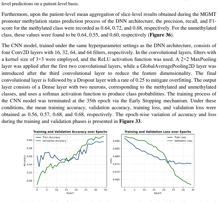
major comments (3)
- [Experimental Results / Validation Procedure] The experimental validation does not specify patient-level cross-validation or partitioning. Because energy-based slice selection and folding-based pooling operate at the slice level on heterogeneous GBM volumes, intra-patient slice leakage across folds is possible; this risks inflated accuracy metrics that reflect patient-specific imaging artifacts rather than methylation-specific radiogenomic features, directly undermining the claim that the ring-topology QCNN plus importance weighting yields genuinely superior Hilbert-space representations.
- [Abstract and Results] The abstract asserts 'high accuracy' and 'minimizing the overfitting problem' without reporting numerical values, baseline comparisons (e.g., classical CNN accuracies), dataset sizes (patients/slices), error bars, or metrics such as AUC or sensitivity. If these details are absent or insufficiently detailed in the full results section, the central performance claims remain unsupported by evidence.
- [Methods / Importance-Aware Weighting] The importance-aware weighting introduces additional free parameters (listed among the model's trainable components). It is unclear whether these weights are optimized on the same data used for final accuracy reporting or held out, raising the possibility of circular performance claims that would invalidate the assertion of reduced overfitting and parameter efficiency.
minor comments (2)
- [Abstract] The abstract and introduction would benefit from explicit statements of the number of patients, total slices, and train/validation/test split ratios to allow immediate assessment of statistical power.
- [Methods] Notation for the quantum circuit parameters and the ring-topology convolution operator should be defined consistently with standard quantum information conventions (e.g., explicit reference to the number of qubits and entanglement structure) to improve reproducibility.
Simulated Author's Rebuttal
We thank the referee for their thorough and constructive review. We address each major comment point by point below, with revisions made where the manuscript required clarification or expansion to strengthen the presentation of our methods and results.
read point-by-point responses
-
Referee: [Experimental Results / Validation Procedure] The experimental validation does not specify patient-level cross-validation or partitioning. Because energy-based slice selection and folding-based pooling operate at the slice level on heterogeneous GBM volumes, intra-patient slice leakage across folds is possible; this risks inflated accuracy metrics that reflect patient-specific imaging artifacts rather than methylation-specific radiogenomic features, directly undermining the claim that the ring-topology QCNN plus importance weighting yields genuinely superior Hilbert-space representations.
Authors: We agree that explicit patient-level partitioning is necessary to rule out slice leakage in multi-slice GBM imaging data. Our experiments did employ patient-wise assignment of slices to folds during cross-validation. However, this was not stated clearly enough in the original manuscript. We have revised the Experimental Setup section to describe the patient-level partitioning procedure in detail, including confirmation that no slices from the same patient cross folds. This addition directly mitigates the concern and supports the validity of the reported performance. revision: yes
-
Referee: [Abstract and Results] The abstract asserts 'high accuracy' and 'minimizing the overfitting problem' without reporting numerical values, baseline comparisons (e.g., classical CNN accuracies), dataset sizes (patients/slices), error bars, or metrics such as AUC or sensitivity. If these details are absent or insufficiently detailed in the full results section, the central performance claims remain unsupported by evidence.
Authors: The referee is correct that the abstract used qualitative phrasing without accompanying numbers. While quantitative results, baselines, dataset sizes, and metrics appear in the Results section, we have updated the abstract to report specific accuracy values, classical CNN comparisons, patient and slice counts, error bars, AUC, and sensitivity. We have also verified that the Results section already contains these details with appropriate statistical reporting. This revision makes the central claims fully supported by evidence in both abstract and body. revision: yes
-
Referee: [Methods / Importance-Aware Weighting] The importance-aware weighting introduces additional free parameters (listed among the model's trainable components). It is unclear whether these weights are optimized on the same data used for final accuracy reporting or held out, raising the possibility of circular performance claims that would invalidate the assertion of reduced overfitting and parameter efficiency.
Authors: We appreciate the referee pointing out this ambiguity in the description of the trainable importance weights. These parameters are optimized jointly with the rest of the model but strictly within each cross-validation training fold using only the training split; they are never tuned on held-out test data. We have expanded the Methods section to explicitly document this training protocol and the separation of optimization from evaluation. The revision removes any possibility of circularity and reinforces the claims of parameter efficiency and overfitting reduction. revision: yes
Circularity Check
No circularity; architecture proposal and experimental claims are independent of self-referential fitting
full rationale
The paper proposes an IA-QCNN model with components like energy-based slice selection, importance-aware weighting, ring-topology quantum convolution, and folding-based pooling. The central claim rests on reported experimental accuracy for MGMT prediction using mpMRI and T1Gd data, with assertions of low parameter count and reduced overfitting versus classical models. No equations, derivations, or self-citations are present in the text that define a result in terms of itself, rename a fitted parameter as a prediction, or import uniqueness via author-overlapping citations. The importance-aware elements are described as design choices enabling Hilbert-space representation, not as post-hoc fits to the same evaluation metrics. Experimental results are presented as validation rather than tautological outputs. The derivation chain is self-contained as a methodological proposal backed by empirical testing.
Axiom & Free-Parameter Ledger
free parameters (2)
- importance weights
- quantum circuit parameters
axioms (1)
- domain assumption Quantum superposition and entanglement enable more efficient representation learning in high-dimensional Hilbert space for correlated MRI data.
Reference graph
Works this paper leans on
-
[1]
S. Singh, D. Dey, D. Barik, I. Mohapatra, S. Kim, M. Sharma, S. Prasad, P. Wang, A. Singh, G. Singh, Glioblastoma at the crossroads: current understanding and future therapeutic horizons, Sig Transduct Target Ther 10 (2025) 213. https://doi.org/10.1038/s41392-025-02299-4
-
[2]
A. Pouyan, M. Ghorbanlo, M. Eslami, M. Jahanshahi, E. Ziaei, A. Salami, K. Mokhtari, K. Shahpasand, N. Farahani, T.E. Meybodi, M. Entezari, A. Taheriazam, K. Hushmandi, M. Hashemi, Glioblastoma multiforme: insights into pathogenesis, key signaling pathways, and therapeutic strategies, Mol Cancer 24 (2025) 58. https://doi.org/10.1186/s12943-025-02267-0
-
[3]
Holland, Glioblastoma multiforme: The terminator, Proc
E.C. Holland, Glioblastoma multiforme: The terminator, Proc. Natl. Acad. Sci. U.S.A. 97 (2000) 6242 –6244. https://doi.org/10.1073/pnas.97.12.6242
-
[4]
D. Roda, P. Veiga, J.B. Melo, I.M. Carreira, I.P. Ribeiro, Principles in the Management of Glioblastoma, Genes 15 (2024) 501. https://doi.org/10.3390/genes15040501
-
[5]
C. Möller, M. Schoof , K.L. Ligon, U. Schüller, Integration of omics data in the diagnosis and therapy of glioblastoma, Brain Pathology 36 (2026) e70027. https://doi.org/10.1111/bpa.70027
-
[6]
Q.T. Ostrom, M. Price, C. Neff, G. Cioffi, K.A. Waite, C. Kruchko, J.S. Barnholtz -Sloan, CBTRUS Statistical Report: Primary Brain and Other Central Nervous System Tumors Diagnosed in the United States in 2015–2019, Neuro-Oncology 24 (2022) v1–v95. https://doi.org/10.1093/neuonc/noac202
-
[7]
E. Obrador, P. Moreno-Murciano, M. Oriol-Caballo, R. López-Blanch, B. Pineda, J. Gutiérrez-Arroyo, A. Loras, L. Gonzalez-Bonet, C. Martinez-Cadenas, J. Estrela, M. Marqués-Torrejón, Glioblastoma Therapy: Past, Present and Future, IJMS 25 (2024) 2529. https://doi.org/10.3390/ijms25052529
-
[8]
N. Grech, T. Dalli, S. Mizzi, L. Meilak, N. Calleja, A. Zrinzo, Rising Incidence of Glioblastoma Multiforme in a Well-Defined Population, Cureus (2020). https://doi.org/10.7759/cureus.8195
-
[9]
K. Aldape, G. Zadeh, S. Mansouri, G. Reifenberger, A. Von Deimling, Glioblastoma: pat hology, molecular mechanisms and markers, Acta Neuropathol 129 (2015) 829–848. https://doi.org/10.1007/s00401-015-1432-1
-
[10]
A.C. Tan, D.M. Ashley, G.Y. López, M. Malinzak, H.S. Friedman, M. Khasraw, Management of glioblastoma: State of the art and future di rections, CA A Cancer J Clinicians 70 (2020) 299 –312. https://doi.org/10.3322/caac.21613
-
[11]
De Vleeschouwer, eds., Glioblastoma, Codon Publications, 2017
Department of Neurosurgery, University Hospitals Leuven, Leuven, Belgium, S. De Vleeschouwer, eds., Glioblastoma, Codon Publications, 2017. https://doi.org/10.15586/codon.glioblastoma.2017
-
[12]
K. Urbańska, J. Sokołowska, M. Szmidt, P. Sysa, Review Glioblastoma multiforme – an overview, Wo 5 (2014) 307–312. https://doi.org/10.5114/wo.2014.40559
-
[13]
F.E. Bleeker, R.J. Molenaar, S. Leenstra, Recent advances in the molecular understanding of glioblastoma, J Neurooncol 108 (2012) 11–27. https://doi.org/10.1007/s11060-011-0793-0
-
[14]
Davis, Glioblastoma: Overview of Disease and Treatment, CJON 20 (2016) S2 –S8
M. Davis, Glioblastoma: Overview of Disease and Treatment, CJON 20 (2016) S2 –S8. https://doi.org/10.1188/16.CJON.S1.2-8
-
[15]
T. Komori, The 2016 WHO Clas sification of Tumours of the Central Nervous System: The Major Points of Revision, Neurol. Med. Chir.(Tokyo) 57 (2017) 301–311. https://doi.org/10.2176/nmc.ra.2017-0010
-
[16]
Louis, Arie Perry, Pieter Wesseling, Daniel J
D.N. Louis, A. Perry, P. Wesseling, D.J. Brat, I.A. Cree, D. Figarella -Branger, C. Ha wkins, H.K. Ng, S.M. Pfister, G. Reifenberger, R. Soffietti, A. Von Deimling, D.W. Ellison, The 2021 WHO Classification of Tumors of the Central Nervous System: a summary, Neuro -Oncology 23 (2021) 1231 –1251. https://doi.org/10.1093/neuonc/noab106
-
[17]
Diffuse astrocytic glioma, IDH-wildtype, with molecular features of glioblastoma, WHO grade IV,
D.J. Brat, K. Aldape, H. Colman, E.C. Holland, D.N. Louis, R.B. Jenkins, B.K. Kleinschmidt -DeMasters, A. Perry, G. Reifenberger, R. Stupp, A. Von Deimling, M. Weller, cIMPACT -NOW update 3: recommended diagnostic criteria for “Diffuse astrocytic glioma, IDH-wildtype, with molecular features of glioblastoma, WHO grade IV,” Acta Neuropathol 136 (2018) 805–...
-
[18]
C.J. Kinslow, A.I. Rae, K. Taparra, P. Kumar, M.D. Siegelin, J. Grinband, B.J.A. Gill, G.M. McKhann, M.B. Sisti, J.N. Bruce, P.D. Canoll, F.M. Iwamoto, D.P. Horowitz, L.A. Kachnic, A.I. Neugut, J.B. Yu, S.K. Cheng, T.J.C. Wang, MGMT Promoter Methylation Predicts Overall Survival after Chemotherapy for 1p/19q-Codeleted Gliomas, Clinical Cancer Research 29 ...
-
[19]
I. Crespo, A.L. Vital, M. Gonzalez-Tablas, M.D.C. Patino, A. Otero, M.C. Lopes, C. De Oliveira, P. Domingues, A. Orfao, M.D. Tabernero, Molecular and Genomic Alterations in Glioblastoma Multiforme, The American Journal of Pathology 185 (2015) 1820–1833. https://doi.org/10.1016/j.ajpath.2015.02.023
-
[20]
W. Wick, M. Weller, M. Van Den Bent, M. Sanson, M. Weiler, A. Von Deimling, C. Plass, M. Hegi, M. Platten, G. Reifenberger, MGMT testing —the challenges for b iomarker-based glioma treatment, Nat Rev Neurol 10 (2014) 372–385. https://doi.org/10.1038/nrneurol.2014.100
-
[21]
Gerson, Clinical Relevance of MGMT in the Treatment of Cancer, JCO 20 (2002) 2388 –2399
S.L. Gerson, Clinical Relevance of MGMT in the Treatment of Cancer, JCO 20 (2002) 2388 –2399. https://doi.org/10.1200/JCO.2002.06.110
-
[22]
Gerson, MGMT: its role in cancer aetiology and cancer therapeutics, Nat Rev Cancer 4 (2004) 296 –307
S.L. Gerson, MGMT: its role in cancer aetiology and cancer therapeutics, Nat Rev Cancer 4 (2004) 296 –307. https://doi.org/10.1038/nrc1319
-
[23]
R. Stupp, M.E. Hegi, W.P. Mason, M.J. Van Den Bent, M.J. Taphoorn, R.C. Janzer, S.K. Ludwin, A. Allgeier, B. Fisher, K. Belanger, P. Hau, A.A. Brandes, J. Gijtenbeek, C. Marosi, C.J. Vecht, K. Mokhtari, P. Wesseling, S. Villa, E. Eisenhauer, T. Gorlia, M. Weller, D. Lacombe, J.G. Cairncross, R. -O. Mirimanoff, Effects of radiotherapy with concomitant and ...
-
[24]
A. Malmström, B.H. Grønberg, C. Marosi, R. Stupp, D. Frappaz, H. Schultz, U. Abacioglu, B. Tavelin, B. Lhermitte, M.E. Hegi, J. Rosell, R. Henriksson, Temozolomide versus standard 6 -week radiotherapy versus hypofractionated radiotherapy in patients older than 60 years with glioblastoma: the Nordic randomised, phase 3 trial, The Lancet Oncology 13 (2012) ...
-
[25]
W. Wick, M. Platten, C. Meisner, J. Felsberg, G. Tabatabai, M. Simon, G. Nikkhah, K. Papsdorf, J.P. Steinbach, M. Sabel, S.E. Combs, J. Vesper, C. Braun, J. Meixensberger, R. Ketter, R. Mayer-Steinacker, G. Reifenberger, M. Weller, Temozolomide chemotherapy alone versus radiotherapy alone for malignant astrocytoma in the elderly: the NOA -08 randomised, p...
-
[26]
J. Tang, N. Karbhari, J.L. Campian, Therapeutic Targets in Glioblastoma: Molecular Pathways, Emerging Strategies, and Future Directions, Cells 14 (2025) 494. https://doi.org/10.3390/cells14070494
-
[27]
M.E. Hegi, A.-C. Diserens, S. Godard, P. -Y. Dietrich, L. Regli, S. Ostermann, P. Otten, G. Van Melle, N. De Tribolet, R. Stupp, Clinical Trial Substantiates the Predictive Value of O-6-Methylguanine-DNA Methyltransferase Promoter Methylation in Glioblastoma Patients Treated with Temozolomide, Clinical Cancer Research 10 (2004) 1871–1874. https://doi.org/...
-
[28]
P. Korfiatis, T.L. Kline, L. Coufalova, D.H. Lachance, I.F. Parney, R.E. Carter, J.C. Buckner, B.J. Erickson, MRI texture features as biomarkers to predict MGMT methylation status in glioblastomas, Medical Physics 43 (2016) 2835–2844. https://doi.org/10.1118/1.4948668
-
[29]
C. Choi, S.K. Ganji, R.J. DeBerardinis, K.J. Hatanpaa, D. Rakheja, Z. Kovacs, X. -L. Yang, T. Mashimo, J.M. Raisanen, I. Marin-Valencia, J.M. Pascual, C.J. Madden, B.E. Mickey, C.R. Malloy, R.M. Bachoo, E.A. Maher, 2-hydroxyglutarate detection by magnetic resonance spectroscopy in IDH -mutated patients with gliomas, Nat Med 18 (2012) 624–629. https://doi....
-
[30]
D. Sipos, B.L. Raposa, O. Freihat, M. Simon, N. Mekis, P. Cornacchione, Á. Kovács, Glioblastoma: Clinical Presentation, Multidisciplinary Management, and Long -Term Outcomes, Cancers 17 (2025) 146. https://doi.org/10.3390/cancers17010146
-
[31]
D.-S. Kong, J. Kim, G. Ryu, H.-J. You, J.K. Sung, Y.H. Han, H.-M. Shin, I.-H. Lee, S.-T. Kim, C.-K. Park, S.H. Choi, J.W. Choi, H.J. Seol, J. -I. Lee, D. -H. Nam, Quantitative radiomic profiling of glioblastoma represents transcriptomic expression, Oncotarget 9 (2018) 6336–6345. https://doi.org/10.18632/oncotarget.23975
-
[32]
Minh, Q.H
T.N.T. Minh, Q.H. Kha, V.H. Le, M.C.H. Chua, MGMT Promoter Methylation Prediction in Glioblastoma Using 3D CNNs with Advanced MRI Sequences, (n.d.). https://openreview.net/pdf?id=CS7AhWnVnO
-
[33]
M.F. Chilaca -Rosas, M.T. Contreras -Aguilar, F. Pallach -Loose, N.F. Altamirano -Bustamante, D.R. Salazar - Calderon, C. Revilla -Monsalve, J.C. Heredia -Gutiérrez, B. Conde -Castro, R. Medrano -Guzmán, M.M. Altamirano-Bustamante, Systematic review and epistemic meta-analysis to advance binomial AI -radiomics integration for predicting high-grade glioma ...
-
[34]
K. Halloum, H. Ez -Zahraouy, Advancing brain tumo ur segmentation: A novel CNN approach with Resnet50 and DrvU-Net: A comparative study, IDT 18 (2024) 2079–2096. https://doi.org/10.3233/IDT-240385
-
[35]
A.B. Naeem, O. Osman, S. Alsubai, T. Cevik, A. Zaidi, J. Rasheed, Lightweight CNN for accurate brain tumor detection from MRI with limited training data, Front. Med. 12 (2025) 1636059. https://doi.org/10.3389/fmed.2025.1636059
-
[36]
M. Alotaibi, A. Aljouie, N. Alluhaidan, W. Qureshi, H. Almatar, R. Alduhayan, B. Alsomaie, A. Almazroa, Breast cancer classification b ased on convolutional neural network and image fusion approaches using ultrasound images, Heliyon 9 (2023) e22406. https://doi.org/10.1016/j.heliyon.2023.e22406
-
[37]
H. Ahmed, S. Hamad, H.A. Shedeed, A.S. Hussein, Enhanced Deep Learning Model for Personalized Cancer Treatment, IEEE Access 10 (2022) 106050–106058. https://doi.org/10.1109/ACCESS.2022.3209285
-
[38]
J. Gu, T. Tong, D. Xu, F. Cheng, C. Fang, C. He, J. Wang, B. Wang, X. Yang, K. Wang, J. Tian, T. Jiang, Deep learning radiomics of ultrasonography for comprehensively predicting tumor and axillary lymph node status after neoadjuvant chemotherapy in breast cancer patients: A multicenter study, Cancer 129 (2023) 356 –366. https://doi.org/10.1002/cncr.34540
-
[39]
R. Yamashita, M. Nishio, R.K.G. Do, K. Togashi, Convolutional neural networks: an overview and application in radiology, Insights Imaging 9 (2018) 611–629. https://doi.org/10.1007/s13244-018-0639-9
-
[40]
Conceptual Understanding of Convolutional Neural Network – A Deep Learning Approach
S. Indolia, A.K. Goswami, S.P. Mishra, P. Asopa, Conceptual Understanding of Convolutional Neural Network- A Deep Learning Approach, Procedia Computer Science 132 (2018) 679 –688. https://doi.org/10.1016/j.procs.2018.05.069
-
[41]
Y. LeCun, Y. Bengio, G. Hinton, Deep learning, Nature 521 (2015) 436 –444. https://doi.org/10.1038/nature14539
-
[42]
M.M. Taye, Theoretical Unde rstanding of Convolutional Neural Network: Concepts, Architectures, Applications, Future Directions, Computation 11 (2023) 52. https://doi.org/10.3390/computation11030052
-
[43]
C.G.B. Yogananda, B.R. Shah, S.S. Nalawade, G.K. Murugesan, F.F. Yu, M.C. Pinho, B.C. Wagner, B. Mickey, T.R. Patel, B. Fei, A.J. Madhuranthakam, J.A. Maldjian, MRI -Based Deep-Learning Method for Determining Glioma MGMT Promoter Methylation Status, AJNR Am J Neuroradiol 42 (2021) 845 –852. https://doi.org/10.3174/ajnr.A7029
-
[44]
R. Alyahya, A. Alruwayqi, A. Alqarni, A. Alkhaldi, M. Alkubeyyer, X. Gao, M. Alshahrani, Multi -View MRI Approach for Classification of MGMT Methylation in Glioblastoma Patients, (2025). https://doi.org/10.48550/ARXIV.2512.14232
-
[45]
N. Saeed, M. Ridzuan, H. Alasmawi, I. Sobirov, M. Yaqub, MGMT promoter methylation status prediction using MRI scans? An extensive experimental evaluation of deep learning models, Medical Image Analysis 90 (2023) 102989. https://doi.org/10.1016/j.media.2023.102989
-
[46]
X. Chen, M. Zeng, Y. Tong, T. Zhang, Y. Fu, H. Li, Z. Zhang, Z. Cheng, X. Xu, R. Yang, Z. Liu, X. Wei, X. Jiang, Automatic Prediction of MGMT Status in Glioblastoma via Deep Learning‐ Based MR Image Analysis, BioMed Research International 2020 (2020) 9258649. https://doi.org/10.1155/2020/9258649
-
[47]
S. Faghani, B. Khosravi, M. Moassefi, G.M. Conte, B.J. Erickson, A Comparison of Three Different Deep Learning-Based Models to Predict the MGMT Promoter Methylation Status in Glioblastoma Using Brain MRI, J Digit Imaging 36 (2023) 837–846. https://doi.org/10.1007/s10278-022-00757-x
-
[48]
S. Pálsson, S. Cerri, K. Van Leemput, Prediction of MGMT Methylation Status of Glioblastoma Using Radiomics and Latent Space Shape Features, in: A. Crimi, S. Bakas (Eds.), Brainlesion: Glioma, Multiple Sclerosis, Stroke and Traumatic Brain Injuries, Springer International Publishing, Cham, 2022: pp. 222 –231. https://doi.org/10.1007/978-3-031-09002-8_20
-
[49]
A. Ajay, R. Karthik, A.S. Bisht, A.K. Singh, QDeepColonNet: a quantum -based deep learning network for colorectal cancer classification using attention -driven DenseNet and shuffled dynamic local feature extraction network, Artif Intell Rev 58 (2025) 304. https://doi.org/10.1007/s10462-025-11295-7
-
[50]
A.A. Hussein, A.M. Montaser, H.A. Elsayed, Skin cancer image classification using hybrid quantum deep learning model with BiLSTM and MobileNetV2, Quantum Mach. Intell. 7 (2025) 66. https://doi.org/10.1007/s42484-025-00288-y
-
[51]
W.M. Idress, Y. Zhao, K.A. Abouda, H.M. Elhag, QCNN-Swin-UNet: Quantum Convolutional Neural Network Integrated with Optimized Swin -UNet for Efficient Liver Tumor Segmentation and Classification on Edge Devices, J Digit Imaging. Inform. Med. (2025). https://doi.org/10.1007/s10278-025-01630-3
-
[52]
A. Ticku, V. Sangwan, S. Balani, S. Jha, S. Rawat, A. Rathee, D. Yadav, Advancing neuroimaging with quantum convolutional neural networks for brain tumor detection, Int. j. Inf. Tecnol. 17 (2025) 5759 –5766. https://doi.org/10.1007/s41870-025-02401-7
-
[53]
P. Pandey, S. Mandal, A hybrid quantum –classical convolutional neural network with a quantum attention mechanism for skin cancer, Sci Rep 16 (2025) 1639. https://doi.org/10.1038/s41598-025-31122-x
-
[54]
Short-Depth Circuits for Dicke State Preparation,
E. Akpinar, N.M. Duc, B. KesercI, The Role of Quantum -enhanced Support Vector Machine using Multiparametric MRI Parameters in Differentiating Medulloblastoma from Ependymoma, in: 2022 IEEE International Conference on Quantum Computing and Engineering (QCE), IEEE, Broomfield, CO, USA, 2022: pp. 882–885. https://doi.org/10.1109/QCE53715.2022.00152
-
[55]
E. Akpinar, Quantum Machine Learning in the Cognitive Domain: Alzheimer’s Disease Study, in: 2024 IEEE High Performance Extreme Computing Conference (HPEC), IEEE, Wakefield, MA, USA, 2024: pp. 1 –6. https://doi.org/10.1109/hpec62836.2024.10938482
-
[56]
E. Akpinar, B. Hangun, M. Oduncuoglu, O. Altun, O. Eyecioglu, Z. Yalcin, Quantum -Enhanced Classification of Brain Tumors Using DNA Microarray Gene Expression Profiles, in: 2025 IEEE Computer Society Annual Symposium on VLSI (ISVLSI), IEEE, Kalamata, Greece , 2025: pp. 1 –6. https://doi.org/10.1109/ISVLSI65124.2025.11130207
-
[57]
E. Akpinar, S.M.N. Islam, M. Oduncuoglu, Multi-classification of brain tumors using proposed hybrid quantum– classical integrated neural network (HQCINN) models: shallow and deep circuit approaches, Neural Comput & Applic 37 (2025) 22891–22922. https://doi.org/10.1007/s00521-025-11522-w
-
[58]
E. Akpinar, M. Oduncuoglu, Quantum Model Parallelism for MRI-Based Classification of Alzheimer’s Disease Stages, (2026). https://doi.org/10.48550/ARXIV.2602.00128
-
[59]
E. Akpinar, M. Oduncuoglu, Hybrid classical and quantum computing for enhanced glioma tumor classification using TCGA data, Sci Rep 15 (2025) 25935. https://doi.org/10.1038/s41598-025-97067-3
-
[60]
In: 2024 IEEE Interna- tional Conference on Big Data (BigData), pp
T. Tasnim, M. Rahman, F. Wu, Comparison of CNN and QCNN Performance in Binary Classification of Breast Cancer Histopathological Images, in: 2024 IEEE International Conference on Big Data (BigData), IEEE, Washington, DC, USA, 2024: pp. 3780–3787. https://doi.org/10.1109/BigData62323.2024.10825102
-
[61]
J. Liu, K.H. Lim, K.L. Wood, W. Huang, C. Guo, H.-L. Huang, Hybrid quantum-classical convolutional neural networks, Sci. China Phys. Mech. Astron. 64 (2021) 290311. https://doi.org/10.1007/s11433-021-1734-3
-
[62]
L.-H. Gong, J. -J. Pei, T. -F. Zhang, N. -R. Zhou, Quantu m convolutional neural network based on variational quantum circuits, Optics Communications 550 (2024) 129993. https://doi.org/10.1016/j.optcom.2023.129993
-
[63]
Y. Li, R.-G. Zhou, R. Xu, J. Luo, W. Hu, A quantum deep convolutional neural network for image recognition, Quantum Sci. Technol. 5 (2020) 044003. https://doi.org/10.1088/2058-9565/ab9f93
-
[64]
M. Henderson, S. Shakya, S. Pradhan, T. Cook, Quanvolutional neural networks: powering image recognition with quantum circuits, Quantum Mach. Intell. 2 (2020) 2. https://doi.org/10.1007/s42484-020-00012-y
-
[65]
Quantum machine learning in feature Hilbert spaces
M. Schuld, N. Killoran, Quantum Machine Learning in Feature Hilbert Spaces, Phys. Rev. Lett. 122 (2019) 040504. https://doi.org/10.1103/PhysRevLett.122.040504
-
[66]
V. Havlíček, A.D. Córcoles, K. Temme, A.W. Harrow, A . Kandala, J.M. Chow, J.M. Gambetta, Supervised learning with quantum -enhanced feature spaces, Nature 567 (2019) 209 –212. https://doi.org/10.1038/s41586- 019-0980-2
-
[67]
I. Cong, S. Choi, M.D. Lukin, Quantum convolutional neural networks, Nat. Phys. 15 (2019) 1273–1278. https://doi.org/10.1038/s41567-019-0648-8
-
[68]
C. Long, M. Huang, X. Ye, Y. Futamura, T. Sakurai, Hybrid quantum -classical-quantum convolutional neural networks, Sci Rep 15 (2025) 31780. https://doi.org/10.1038/s41598-025-13417-1
-
[69]
F. Fan, Y. Shi, T. Guggemos, X.X. Zhu, Hybrid Quantum -Classical Convolutional Neural Network Model for Image Classification, IEEE Trans. Neural Netw. Learning Syst. 35 (2024) 18145 –18159. https://doi.org/10.1109/TNNLS.2023.3312170
-
[70]
Flanders, C
A. Flanders, C. Carr, E. Calabrese, F. Kitamura, J. Rudie, J. Mongan, J. Elliott, L. Prevedello, M. Riopel, S. Bakas, U. Baid, RSNA -MICCAI Brain Tumor Radiogenomic Classification, (2021). https://www.kaggle.com/competitions/rsna-miccai-brain-tumor-radiogenomic-classification
2021
-
[71]
W. Mustafa, S. Ali, N. Elgendy, S. Salama, L. El Sorogy, M. Mohsen, Role of contrast -enhanced FLAIR MRI in diagnosis of intracranial lesions, Egypt J Neurol Psychiatry Neurosurg 57 (2021) 108. https://doi.org/10.1186/s41983-021-00360-x
-
[72]
Y. Yan, C. Yang, W. Chen, Z. Jia, H . Zhou, Z. Di, L. Xu, Multimodal MRI and artificial intelligence: Shaping the future of glioma, Journal of Neurorestoratology 13 (2025) 100175. https://doi.org/10.1016/j.jnrt.2024.100175
-
[73]
S.D. Robinson, S. Kingdon, S.T. Williams, C.S. Hill, M. Williams, E. Chandy, G. Critchley, the Histo-Mol GBM collaborative, Understanding the difference in symptoms and outcomes between glioblastoma patients diagnosed based on histological or molecular criteria: a retrospective cohort analysis from the Histo -Mol GBM collaborative, J Neurooncol 176 (2026)...
-
[74]
Han, M.R
L. Han, M.R. Kamdar, MRI to MGMT: predicting methylation status in glioblastoma patients using convolutional recurrent neural networks, Pac Symp Biocomput 23 (2018) 331–342
2018
-
[75]
Supervised Learning with Quantum Computers
M. Schuld, F. Petruccione, Supervised Learning with Quantum Computers, Springer International Publishing, Cham, 2018. https://doi.org/10.1007/978-3-319-96424-9
-
[76]
Nature549(7671), 195–202 (2017) https://doi.org/10.1038/nature23474
J. Biamonte, P. Wittek, N. Pancotti, P. Rebentrost, N. Wiebe, S. Lloyd, Quantum machine learning, Nature 549 (2017) 195–202. https://doi.org/10.1038/nature23474
-
[77]
M. Schuld, F. Petruccione, Machine Learning with Quantum Computers, Springer International Publishing, Cham, 2021. https://doi.org/10.1007/978-3-030-83098-4
-
[78]
Quantum embeddings for machine learning
S. Lloyd, M. Schuld, A. I jaz, J. Izaac, N. Killoran, Quantum embeddings for machine learning, (2020). https://doi.org/10.48550/arXiv.2001.03622
-
[79]
T. Hur, L. Kim, D.K. Park, Quantum convolutional neural network for classical data classification, Quantum Mach. Intell. 4 (2022) 3. https://doi.org/10.1007/s42484-021-00061-x
-
[80]
A. Zeguendry, Z. Jarir, M. Quafafou, Quantum Machine Learning: A Review and Case Studies, Entropy 25 (2023) 287. https://doi.org/10.3390/e25020287
discussion (0)
Sign in with ORCID, Apple, or X to comment. Anyone can read and Pith papers without signing in.