Recognition: unknown
Physics-Guided Deep Learning For High Resolution X-ray Imaging
Pith reviewed 2026-05-09 18:02 UTC · model grok-4.3
The pith
Modeling X-ray artifacts as a separable feature layer lets a U-Net estimate and remove them directly from single-shot data.
A machine-rendered reading of the paper's core claim, the machinery that carries it, and where it could break.
Core claim
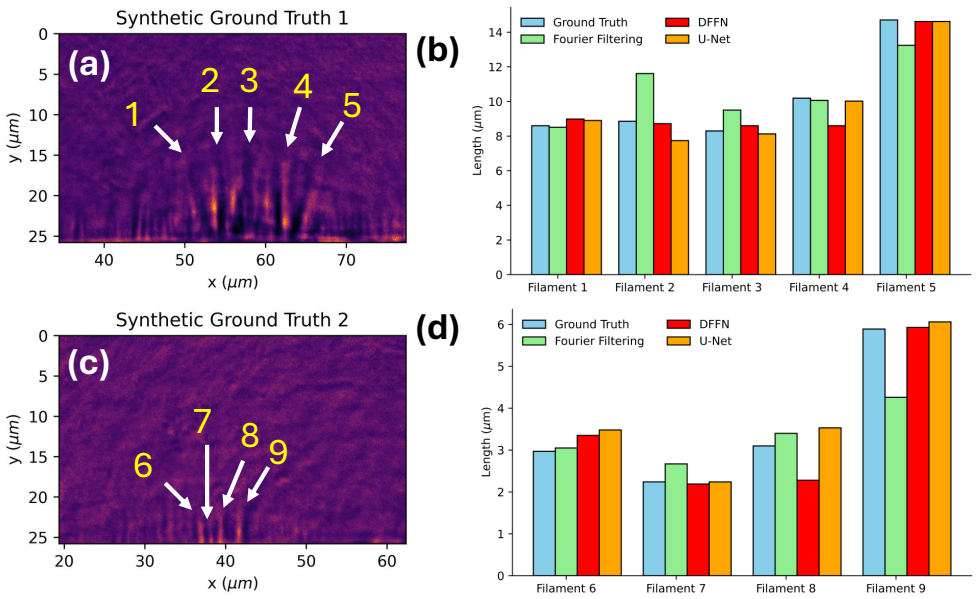
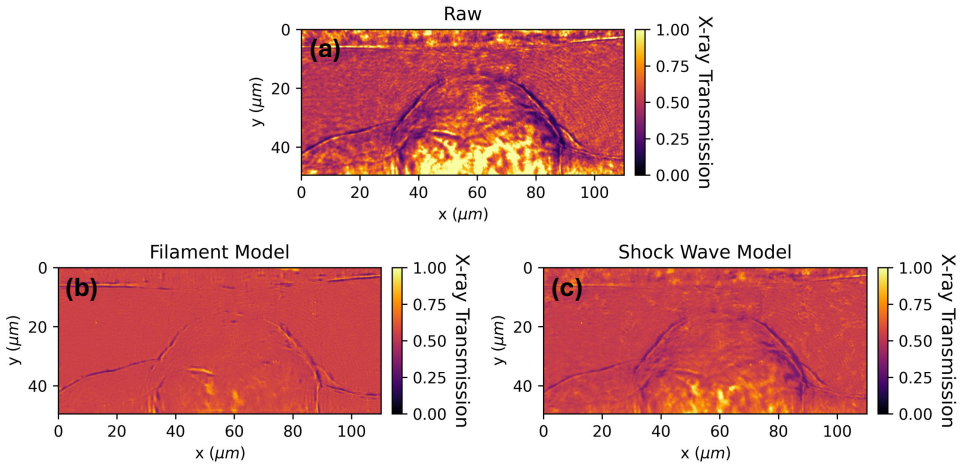
By treating artifacts as a separable feature layer and training a U-Net to estimate that layer from experimental radiographs, the method reconstructs transmission maps that preserve filament signals and yield higher structural similarity scores than conventional normalization techniques.
What carries the argument
U-Net architecture trained to estimate the artifact feature layer from raw measurements, using physics-guided synthetic injections for supervision.
If this is right
- Transmission maps with higher structural fidelity improve downstream calculations of electron density, velocity, and feature size.
- Filament profiles remain intact during artifact removal, reducing systematic bias in plasma diagnostics.
- Epistemic uncertainty estimates from deep ensembles flag shots where the reconstruction may be unreliable.
- The approach reduces reliance on repeated reference exposures that are often unavailable in single-shot experiments.
Where Pith is reading between the lines
- If the method holds on real data it could shorten the time needed to extract quantitative results from each expensive shot.
- The separable-layer premise may transfer to other single-exposure imaging modalities that suffer structured detector artifacts.
- Uncertainty maps could be used to weight or discard individual pixels in downstream physics analysis.
Load-bearing premise
X-ray artifacts can be treated as a separable feature layer independent of the true signal and that a U-Net trained only on synthetic examples will generalize to real experimental images without introducing new biases or signal loss.
What would settle it
Direct application of the trained U-Net to real HED X-ray radiographs that shows either lower SSIM than dynamic flat-field normalization or measurable attenuation of known filament features.
Figures


read the original abstract
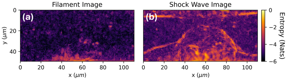
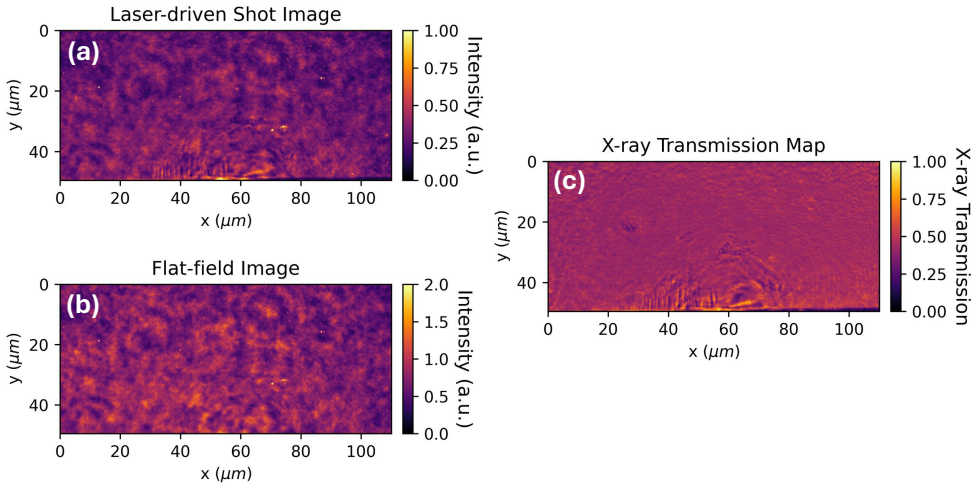
Imperfections in X-ray imaging systems can limit their performance, especially in High Energy Density (HED) or Inertial Fusion Energy (IFE)-relevant experiments that are typically single shot, by introducing structured, non-stationary features that overlap with the signal of interest. When the X-ray transmission is reconstructed by typical flat-field normalization, even small shot-to-shot drift of structured features imprints residual patterns onto transmission maps, degrading signal visibility and biasing measurements such as electron density, velocity and feature sizes. We investigate this limitation by modeling the artifacts as a separable feature layer and training a U-Net architecture to estimate and infer them directly from the experimental data. We compare our method against Fourier filtering and more advanced procedures like Dynamic Flat-Field Normalization (DFFN) to evaluate artifact suppression capability and signal preservation in the reconstructed transmission maps. In multiple synthetic injection tests, our Physics-Guided Deep Learning approach is able to obtain an improvement in mean Structural Similarity Index (SSIM) from 0.345 to 0.906 and from 0.0679 to 0.945, while better preserving filament profiles and reducing degradation of the filament signal during artifact suppression. Additionally, we utilize deep ensembles to obtain predictive epistemic uncertainty estimates for the U-Net based reconstruction, to ensure Out Of Distribution (OOD) robustness for this procedure.
Editorial analysis
A structured set of objections, weighed in public.
Referee Report
Summary. The paper proposes modeling X-ray imaging artifacts in HED/IFE experiments as a separable additive feature layer and training a U-Net to estimate and subtract them directly from experimental radiographs. The method is compared to Fourier filtering and Dynamic Flat-Field Normalization (DFFN) on synthetic artifact-injection tests, reporting mean SSIM improvements from 0.345 to 0.906 and from 0.0679 to 0.945 while claiming better filament-profile preservation; deep ensembles are used to provide epistemic uncertainty estimates for OOD robustness.
Significance. If the separability assumption and synthetic-to-real generalization hold, the approach could meaningfully improve single-shot transmission measurements by reducing residual artifact bias without excessive signal loss, with the deep-ensemble uncertainty providing a practical safeguard. The independent SSIM evaluation on held-out synthetics and the explicit comparison to DFFN are strengths that support the quantitative claims within the synthetic regime.
major comments (3)
- [Abstract] Abstract: The headline SSIM gains (0.345→0.906 and 0.0679→0.945) and the claim of “better preserving filament profiles and reducing degradation of the filament signal” are demonstrated exclusively on synthetic injections that presuppose artifacts form an additive, separable layer independent of the transmission signal. No real experimental radiographs are shown, leaving the central generalization claim untested against the structured, shot-to-shot, non-stationary artifacts actually present in HED/IFE data.
- [Abstract] Abstract: The manuscript provides no description of how the synthetic artifacts were generated (e.g., spatial-frequency content, amplitude distribution, or correlation with the underlying filament signal). Without these details it is impossible to judge whether the training distribution adequately covers the real artifact statistics that the U-Net is intended to handle.
- [Abstract] Abstract: No error bars, statistical significance tests, or cross-validation results accompany the reported mean SSIM values, making it difficult to assess the robustness of the claimed improvement over DFFN and Fourier filtering.
Simulated Author's Rebuttal
We thank the referee for their constructive and detailed comments on our manuscript. We have reviewed each major comment carefully and provide point-by-point responses below, indicating where revisions will be made to strengthen the paper.
read point-by-point responses
-
Referee: [Abstract] Abstract: The headline SSIM gains (0.345→0.906 and 0.0679→0.945) and the claim of “better preserving filament profiles and reducing degradation of the filament signal” are demonstrated exclusively on synthetic injections that presuppose artifacts form an additive, separable layer independent of the transmission signal. No real experimental radiographs are shown, leaving the central generalization claim untested against the structured, shot-to-shot, non-stationary artifacts actually present in HED/IFE data.
Authors: We agree that all quantitative results, including the reported SSIM improvements and filament profile preservation, are obtained exclusively from controlled synthetic artifact-injection experiments that assume an additive, separable artifact layer. This modeling choice reflects the physical characteristics of X-ray system imperfections in single-shot HED/IFE setups, where shot-to-shot drifts produce structured features largely independent of the underlying transmission. Because real single-shot experiments lack pixel-wise ground truth, synthetic tests enable rigorous quantitative evaluation that would otherwise be impossible. We will revise the abstract to explicitly note that the SSIM gains and profile comparisons are demonstrated on synthetic data, and we will expand the discussion section to elaborate on the separability assumption, its physical basis, and the challenges of direct validation on real radiographs. This will clarify the scope of the current claims while highlighting the method's potential for real-world application. revision: partial
-
Referee: [Abstract] Abstract: The manuscript provides no description of how the synthetic artifacts were generated (e.g., spatial-frequency content, amplitude distribution, or correlation with the underlying filament signal). Without these details it is impossible to judge whether the training distribution adequately covers the real artifact statistics that the U-Net is intended to handle.
Authors: We thank the referee for highlighting this omission. Although the full manuscript describes the artifact synthesis procedure in the Methods section (including generation of non-stationary features via spatially varying low-frequency modulations with controlled amplitudes and minimal correlation to the filament signal), these details were not summarized in the abstract. In the revised version we will add a concise description of the synthetic artifact generation process to the abstract, specifying the spatial-frequency content, amplitude ranges, and correlation properties used. This will allow readers to assess how well the training distribution matches expected real-world artifact statistics. revision: yes
-
Referee: [Abstract] Abstract: No error bars, statistical significance tests, or cross-validation results accompany the reported mean SSIM values, making it difficult to assess the robustness of the claimed improvement over DFFN and Fourier filtering.
Authors: We acknowledge that the abstract reports only mean SSIM values without accompanying variability measures or statistical analysis. The means were computed across multiple independent synthetic injection trials, and the underlying experiments incorporated cross-validation folds, yet these details were not included in the abstract. In the revised manuscript we will add error bars (standard deviation across trials) to the SSIM results, report the outcomes of cross-validation, and include statistical significance tests (e.g., paired Wilcoxon signed-rank tests) comparing our method to Fourier filtering and DFFN. These additions will be placed both in the abstract and in the results section to demonstrate the robustness of the observed improvements. revision: yes
Circularity Check
No significant circularity in derivation or claims
full rationale
The paper presents an empirical ML method: artifacts are modeled as a separable layer, a U-Net is trained on synthetic injections, and performance is measured with independent SSIM on held-out synthetics. No equations, derivations, or self-citations are shown that reduce the reported SSIM gains (0.345→0.906, 0.0679→0.945) or filament preservation claims to a fitted parameter, self-definition, or tautological input by construction. The evaluation metrics are external benchmarks, not internal to the training or modeling assumptions.
Axiom & Free-Parameter Ledger
axioms (1)
- domain assumption X-ray imaging artifacts can be modeled as a separable feature layer independent of the underlying transmission signal.
Reference graph
Works this paper leans on
-
[1]
Reports15, 7588, DOI: 10.1038/s41598-025-91989-8 (2025)
Galtier, E.et al.X-ray microscopy and talbot imaging with the matter in extreme conditions x-ray imager at lcls.Sci. Reports15, 7588, DOI: 10.1038/s41598-025-91989-8 (2025)
-
[2]
Bradley, D. K., Landen, O. L., Bullock, A. B., Glendinning, S. G. & Turner, R. E. Efficient, 1–100-kev x-ray radiography with high spatial and temporal resolution.Opt. Lett.27, 134–136, DOI: 10.1364/OL.27.000134 (2002)
-
[3]
Plasmas15, 072705, DOI: 10.1063/1.2957918 (2008)
Park, H.-S.et al.High-resolution 17–75 kev backlighters for high energy density experiments.Phys. Plasmas15, 072705, DOI: 10.1063/1.2957918 (2008). 11/13 4.Momose, A.et al.Demonstration of x-ray talbot interferometry.Jpn. J. Appl. Phys.42, L866–L868, DOI: 10.1143/JJAP. 42.L866 (2003)
-
[4]
Buakor, K.et al.Shot-to-shot flat-field correction at x-ray free-electron lasers.Opt. Express30, 10633, DOI: 10.1364/OE. 451914 (2022)
work page doi:10.1364/oe 2022
-
[5]
Synchrotron Radiat.28, 52–63, DOI: 10.1107/S160057752001557X (2021)
Hagemann, J.et al.Single-pulse phase-contrast imaging at free-electron lasers in the hard x-ray regime.J. Synchrotron Radiat.28, 52–63, DOI: 10.1107/S160057752001557X (2021)
-
[6]
Express23, 27975–27989, DOI: 10.1364/OE.23.027975 (2015)
Van Nieuwenhove, V .et al.Dynamic intensity normalization using eigen flat fields in x-ray imaging.Opt. Express23, 27975–27989, DOI: 10.1364/OE.23.027975 (2015)
-
[7]
Dover, N. P.et al.Optical imaging of laser-driven fast electron weibel-like filamentation in overcritical density plasma. Phys. Rev. Lett.134, 025102, DOI: 10.1103/PhysRevLett.134.025102 (2025)
-
[8]
Commun.15, 7528, DOI: 10.1038/s41467-024-51084-4 (2024)
Sawada, H.et al.Spatiotemporal dynamics of fast electron heating in solid-density matter via xfel.Nat. Commun.15, 7528, DOI: 10.1038/s41467-024-51084-4 (2024)
-
[9]
S.et al.Observations of the filamentation of high-intensity laser-produced electron beams.Phys
Wei, M. S.et al.Observations of the filamentation of high-intensity laser-produced electron beams.Phys. Rev. E70, 056412, DOI: 10.1103/PhysRevE.70.056412 (2004)
-
[10]
Romagnani, L.et al.Dynamics of the electromagnetic fields induced by fast electron propagation in near-solid-density media.Phys. Rev. Lett.122, 025001, DOI: 10.1103/PhysRevLett.122.025001 (2019)
-
[11]
Kodama, R.et al.Fast heating of ultrahigh-density plasma as a step towards laser fusion ignition.Nature412, 798–802, DOI: 10.1038/35090525 (2001)
-
[12]
Robinson, A. P. L.et al.Theory of fast electron transport for fast ignition.Nucl. Fusion54, 054003, DOI: 10.1088/ 0029-5515/54/5/054003 (2014)
2014
-
[13]
Plasmas1, 1626–1634, DOI: 10.1063/1.870664 (1994)
Tabak, M.et al.Ignition and high gain with ultrapowerful lasers.Phys. Plasmas1, 1626–1634, DOI: 10.1063/1.870664 (1994)
-
[14]
Commun.17, 467, DOI: 10.1038/s41467-025-67160-2 (2026)
Schoenwaelder, C.et al.Time-resolved x-ray imaging of the current filamentation instability in solid-density plasmas.Nat. Commun.17, 467, DOI: 10.1038/s41467-025-67160-2 (2026)
-
[15]
V ., Schafer, R
Oppenheim, A. V ., Schafer, R. W. & Stockham, T. G. Nonlinear filtering of multiplied and convolved signals.IEEE Transactions on Audio Electroacoustics16, 437–466 (1968)
1968
-
[16]
& Brox, T
Ronneberger, O., Fischer, P. & Brox, T. U-net: Convolutional networks for biomedical image segmentation. InMedical Image Computing and Computer-Assisted Intervention–MICCAI 2015: 18th International Conference, Munich, Germany, October 5-9, 2015, Proceedings, Part III 18, 234–241 (Springer, 2015)
2015
-
[17]
Siddique, N., Paheding, S., Elkin, C. P. & Devabhaktuni, V . U-net and its variants for medical image segmentation: A review of theory and applications.IEEE Access9, 82031, DOI: 10.1109/ACCESS.2021.3086020 (2021)
-
[18]
Intell.46, 10076, DOI: 10.1109/TPAMI.2024.3435571 (2024)
Azad, R.et al.Medical image segmentation review: The success of u-net.IEEE Transactions on Pattern Analysis Mach. Intell.46, 10076, DOI: 10.1109/TPAMI.2024.3435571 (2024)
-
[19]
Fornek, T. E. Advanced photon source upgrade project final design report. Tech. Rep., Argonne National Laboratory (ANL), Argonne, IL (United States) (2019)
2019
-
[20]
W.et al.LCLS-II High Energy (LCLS-II-HE): A transformative x-ray laser for science
Schoenlein, R. W.et al.LCLS-II High Energy (LCLS-II-HE): A transformative x-ray laser for science. Tech. Rep., SLAC National Accelerator Laboratory (SLAC), Menlo Park, CA (United States) (2016)
2016
-
[21]
Synchrotron Radiat
Mishra, A.et al.A start to end machine learning approach to maximize scientific throughput from the LCLS-II-HE. Synchrotron Radiat. News38, 10–17 (2025)
2025
-
[22]
& Ratner, D
Edelen, A., Neveu, N., Mayes, C., Emma, C. & Ratner, D. Machine learning models for optimization and control of x-ray free electron lasers. InNeurIPS Machine Learning for the Physical Sciences Workshop(2019)
2019
-
[23]
Münch, B., Trtik, P., Marone, F. & Stampanoni, M. Stripe and ring artifact removal with combined wavelet - fourier filtering.Opt. Express17, 8567–8591, DOI: 10.1364/OE.17.008567 (2009)
-
[24]
Wang, Z., Bovik, A. C., Sheikh, H. R. & Simoncelli, E. P. Image quality assessment: from error visibility to structural similarity.IEEE Transactions on Image Process.13, 600–612, DOI: 10.1109/TIP.2003.819861 (2004)
-
[25]
& Blundell, C
Lakshminarayanan, B., Pritzel, A. & Blundell, C. Simple and scalable predictive uncertainty estimation using deep ensembles. InAdvances in Neural Information Processing Systems, vol. 30 (2017). 12/13
2017
-
[26]
Sepúlveda-Fontaine, S. A. & Amigó, J. M. Applications of entropy in data analysis and machine learning: A review. Entropy26, 1126, DOI: 10.3390/e26121126 (2024)
-
[27]
L., Gullikson, E
Henke, B. L., Gullikson, E. M. & Davis, J. C. X-ray interactions: photoabsorption, scattering, transmission, and reflection atE=50−30,000 eV ,Z=1−92.At. Data Nucl. Data Tables54, 181–342 (1993)
1993
-
[28]
Ghiasi, G.et al.Simple copy-paste is a strong data augmentation method for instance segmentation. InProceedings of the IEEE/CVF Conference on Computer Vision and Pattern Recognition (CVPR), DOI: 10.1109/CVPR46437.2021.00294 (2021). 30.Hendrycks, D. & Gimpel, K. Gaussian error linear units (gelus).arXiv preprint arXiv:1606.08415(2016). 31.Kingma, D. P. & B...
discussion (0)
Sign in with ORCID, Apple, or X to comment. Anyone can read and Pith papers without signing in.