Recognition: 2 theorem links
· Lean TheoremEpitaxial growth of beta-bismuthene on Sb2Te3
Pith reviewed 2026-05-11 02:31 UTC · model grok-4.3
The pith
Beta-bismuthene forms an epitaxial interface on the topological insulator Sb2Te3 when bismuth is deposited under controlled coverage and temperature.
A machine-rendered reading of the paper's core claim, the machinery that carries it, and where it could break.
Core claim
The authors report the successful epitaxial growth of beta-bismuthene on Sb2Te3. They show that bismuth coverage controls nucleation density and island size while substrate temperature affects the quality of the atomic ordering inside the islands. Throughout the bismuthene lattice, defects induced by the substrate are visible in the STM images.
What carries the argument
The epitaxial heterointerface between beta-bismuthene and Sb2Te3, whose formation is tuned by bismuth coverage and substrate temperature and imaged at atomic resolution with scanning tunneling microscopy.
If this is right
- Increasing bismuth coverage increases the density and size of bismuth islands on the surface.
- Raising substrate temperature improves the atomic ordering inside the bismuthene islands.
- Defects from the Sb2Te3 substrate appear uniformly across the bismuthene layer and limit its perfection.
- The interface forms without detectable intermixing when growth parameters are chosen correctly.
Where Pith is reading between the lines
- The same deposition approach could be tested on other topological insulators to produce families of similar 2D interfaces.
- The substrate-induced defects could be mapped against electronic measurements to see how they scatter carriers in the bismuthene.
- Because Sb2Te3 already supports topological surface states, the completed stack offers a platform for studying proximity effects between 2D bismuthene and those states.
Load-bearing premise
The atomic lattices and island shapes seen in the microscope truly belong to beta-bismuthene and the layer sits in registry with the Sb2Te3 surface without intermixing or other phases.
What would settle it
An STM image that reveals an atomic spacing or symmetry different from the expected beta-bismuthene structure, or that shows no consistent alignment with the Sb2Te3 lattice, would show the growth is not epitaxial beta-bismuthene.
Figures


read the original abstract
Over the past decades, two-dimensional crystals have attracted considerable interest as promising materials for electronic and optoelectronic applications. Among them, graphene analogs composed of heavy atoms occupy a particularly distinctive niche due to their enhanced spin-orbit interaction. Here, we present an epitaxial heterointerface formed by beta-bismuthene on Sb2Te3, a well-known three-dimensional topological insulator. Using scanning tunneling microscopy, we systematically investigated the effects of Bi coverage and substrate temperature on nucleation processes, island morphology, and atomic structure. In addition, substrate-induced defects were identified throughout the bismuthene lattice.
Editorial analysis
A structured set of objections, weighed in public.
Referee Report
Summary. The manuscript reports the epitaxial growth of beta-bismuthene on the topological insulator Sb2Te3. Using scanning tunneling microscopy, the authors systematically vary bismuth coverage and substrate temperature to examine effects on nucleation processes, island morphology, and atomic structure, while also identifying substrate-induced defects throughout the bismuthene lattice.
Significance. If the phase identification and epitaxial character are confirmed, the work would establish a new 2D/3D heterointerface combining a buckled honeycomb bismuthene layer (with strong spin-orbit coupling) and a well-characterized topological insulator substrate. This could enable controlled studies of interface-induced phenomena relevant to spintronics and topological electronics. The systematic exploration of growth parameters is a positive feature that provides a practical route toward reproducible synthesis.
major comments (1)
- The central claim of an epitaxial beta-bismuthene/Sb2Te3 heterointerface rests on the assignment of observed atomic lattices to the buckled honeycomb beta phase rather than alpha-bismuthene, gamma phases, reconstructions, or Bi-Sb intermixing. No quantitative lattice constants, Fourier-transform analysis, symmetry identification, or bias-dependent imaging are supplied to exclude these alternatives, leaving the structural interpretation unsupported by the presented evidence.
minor comments (1)
- The abstract states that systematic STM studies were performed, yet the manuscript would benefit from explicit inclusion of representative images, error bars on coverage/temperature trends, and raw data metrics in the main text or supplementary information to allow independent assessment of the growth conclusions.
Simulated Author's Rebuttal
We thank the referee for the constructive review and positive assessment of the work's potential significance. We address the single major comment below and have revised the manuscript to incorporate additional supporting analysis.
read point-by-point responses
-
Referee: The central claim of an epitaxial beta-bismuthene/Sb2Te3 heterointerface rests on the assignment of observed atomic lattices to the buckled honeycomb beta phase rather than alpha-bismuthene, gamma phases, reconstructions, or Bi-Sb intermixing. No quantitative lattice constants, Fourier-transform analysis, symmetry identification, or bias-dependent imaging are supplied to exclude these alternatives, leaving the structural interpretation unsupported by the presented evidence.
Authors: We agree that the original manuscript would benefit from more quantitative structural characterization to firmly support the beta-bismuthene assignment. In the revised version, we have added high-resolution STM data with measured lattice constants (consistent with the known beta phase), Fourier-transform images confirming hexagonal symmetry, explicit symmetry analysis, and bias-dependent imaging across a range of voltages. These additions allow direct comparison to expected features of beta-bismuthene while addressing why alpha, gamma, or intermixing phases are inconsistent with the observed nucleation, morphology, and atomic contrast under our growth conditions. The new analysis appears in the revised results section and an expanded supplementary figure. revision: yes
Circularity Check
No circularity: experimental STM study with direct observations and no derivations or fitted predictions
full rationale
The paper is a purely experimental report on epitaxial growth and STM imaging of bismuthene islands on Sb2Te3. It contains no equations, no parameter fitting, no derivations, and no modeling steps that could reduce to self-referential inputs. Central claims rest on direct imaging of nucleation, morphology, atomic structure, and defects under varied coverage and temperature conditions. No self-citation chains, uniqueness theorems, or ansatzes are invoked to support any derivation. The structural assignment to beta-bismuthene is an empirical interpretation of observed lattices, not a circular reduction of a claimed prediction to its own inputs. This is the normal case of a self-contained experimental study.
Axiom & Free-Parameter Ledger
axioms (2)
- domain assumption Beta-bismuthene denotes the buckled honeycomb monolayer structure of bismuth.
- domain assumption Sb2Te3 surface supports epitaxial growth of bismuth without intermixing under the stated conditions.
Lean theorems connected to this paper
-
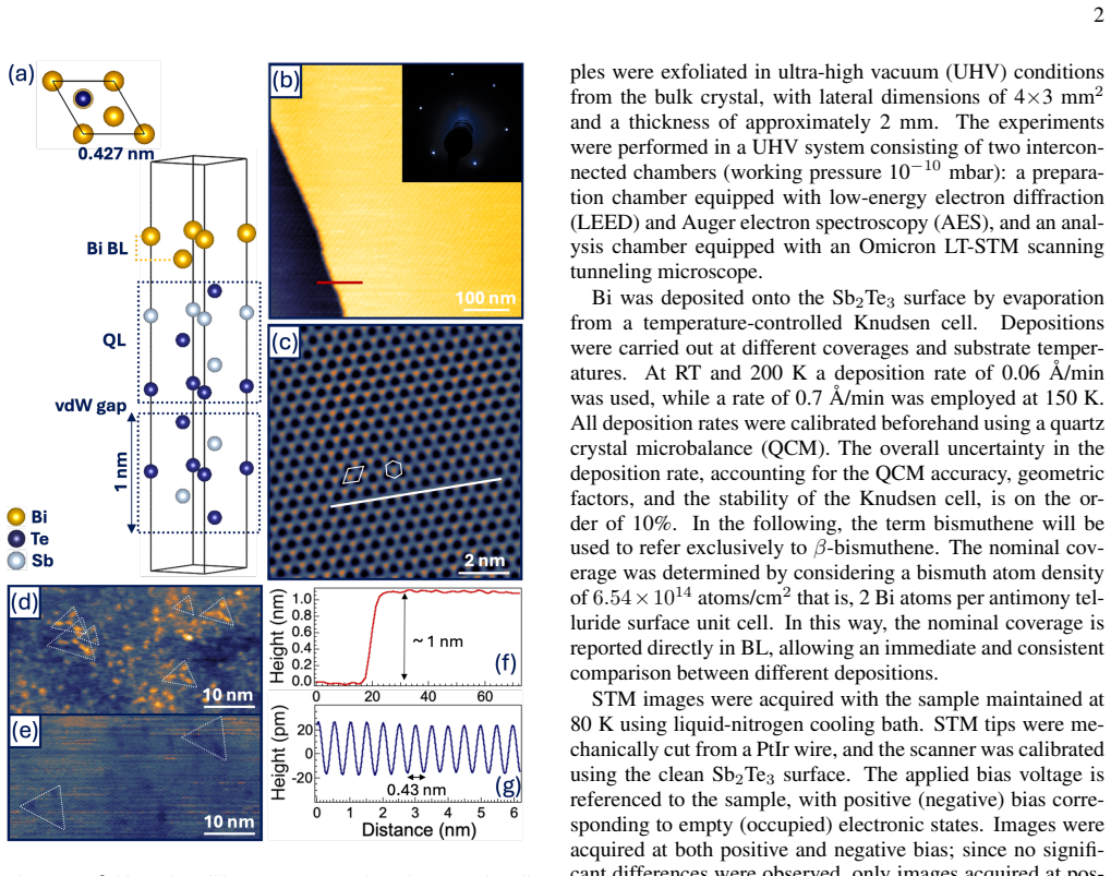
IndisputableMonolith/Foundation/AlexanderDuality.leanalexander_duality_circle_linking unclearAtomic-resolution images reveal indeed a hexagonal structure... period of nearly 0.43 nm... in perfect registry with the Te layer underneath... apparent height of approximately 0.5 nm... compatible with a bismuthene BL
-
IndisputableMonolith/Foundation/ArithmeticFromLogic.leanembed_strictMono_of_one_lt unclearthe formation of bismuthene was confirmed in all cases... hexagonal honeycomb structure... triangular morphology... epitaxial relationship with the Sb2Te3 substrate
Reference graph
Works this paper leans on
-
[1]
M. Z. Hasan and C. L. Kane, Rev. Mod. Phys.82, 3045 (2010)
work page 2010
-
[2]
F. Schindler, Z. Wang, M. G. Vergniory, A. M. Cook, A. Mu- rani, S. Sengupta, A. Y . Kasumov, R. Deblock, S. Jeon, I. Droz- dov, H. Bouchiat, S. Guéron, A. Yazdani, B. A. Bernevig, and T. Neupert, Nature Physics14, 918 (2018)
work page 2018
-
[3]
Y . M. Koroteev, G. Bihlmayer, J. E. Gayone, E. V . Chulkov, S. Blügel, P. M. Echenique, and P. Hofmann, Phys. Rev. Lett. 93, 046403 (2004)
work page 2004
-
[4]
B. Q. Lv, T. Qian, and H. Ding, Rev. Mod. Phys.93, 025002 (2021)
work page 2021
- [5]
- [6]
-
[7]
Y . Lu, W. Xu, M. Zeng, G. Yao, L. Shen, M. Yang, Z. Luo, F. Pan, K. Wu, T. Das, P. He, J. Jiang, J. Martin, Y . P. Feng, H. Lin, and X.-s. Wang, Nano Letters15, 80 (2015), pMID: 25495154, https://doi.org/10.1021/nl502997v
-
[8]
Y . Lyu, S. Daneshmandi, S. Huyan, and C.-W. Chu, Materials Today Physics18, 100380 (2021)
work page 2021
-
[9]
J. Gou, H. Bai, X. Zhang, Y . L. Huang, S. Duan, A. Ariando, S. A. Yang, L. Chen, Y . Lu, and A. T. S. Wee, Nature617, 67
-
[10]
F. Reis, G. Li, L. Dudy, M. Bauernfeind, S. Glass, W. Hanke, R. Thomale, J. Schaefer, and R. Claessen, Science357(2016)
work page 2016
- [11]
-
[12]
M. Wada, S. Murakami, F. Freimuth, and G. Bihlmayer, Phys. Rev. B83, 121310 (2011)
work page 2011
- [13]
-
[14]
P. Kowalczyk, O. Mahapatra, D. McCarthy, W. Kozlowski, Z. Klusek, and S. Brown, Surface Science605, 659 (2011)
work page 2011
-
[15]
F. Song, J. W. Wells, Z. Jiang, M. Saxegaard, and E. Wahlström, ACS Applied Materials & Interfaces7, 8525 (2015), pMID: 25849866, https://doi.org/10.1021/acsami.5b00264
-
[16]
N. Tilgner, S. Wolff, S. Soubatch, T.-L. Lee, A. D. Peña Uni- garro, S. Gemming, F. S. Tautz, T. Seyller, C. Kumpf, F. Göhler, and P. Schädlich, Nature Communications16, 6171 (2025)
work page 2025
-
[17]
Hofmann, Progress in Surface Science81, 191 (2006)
P. Hofmann, Progress in Surface Science81, 191 (2006)
work page 2006
- [18]
-
[19]
T. Hirahara, T. Nagao, I. Matsuda, G. Bihlmayer, E. V . Chulkov, Y . M. Koroteev, P. M. Echenique, M. Saito, and S. Hasegawa, Phys. Rev. Lett.97, 146803 (2006)
work page 2006
-
[20]
S.-H. Bae, H. Kum, W. Kong, Y . Kim, C. Choi, B. Lee, P. Lin, Y . Park, and J. Kim, Nature Materials18, 550 (2019)
work page 2019
- [21]
-
[22]
T. Hirahara, G. Bihlmayer, Y . Sakamoto, M. Yamada, H. Miyazaki, S.-i. Kimura, S. Blügel, and S. Hasegawa, Physi- cal Review Letters107, 166801 (2011)
work page 2011
-
[23]
M. Chen, J.-P. Peng, H. Zhang, l. Jun, K. He, X.-C. Ma, and Q.-K. Xue, Applied Physics Letters101(2012)
work page 2012
-
[24]
T. Hirahara, N. Fukui, T. Shirasawa, M. Yamada, M. Ai- tani, H. Miyazaki, M. Matsunami, S. Kimura, T. Takahashi, S. Hasegawa, and K. Kobayashi, Phys. Rev. Lett.109, 227401 (2012)
work page 2012
-
[25]
F. Yang, L. Miao, Z. F. Wang, M.-Y . Yao, F. Zhu, Y . R. Song, M.-X. Wang, J.-P. Xu, A. V . Fedorov, Z. Sun, G. B. Zhang, C. Liu, F. Liu, D. Qian, C. L. Gao, and J.-F. Jia, Physical Review Letters109, 016801 (2012)
work page 2012
-
[26]
L. Miao, M.-Y . Yao, W. Ming, F. Zhu, C. Q. Han, Z. F. Wang, D. D. Guan, C. L. Gao, C. Liu, F. Liu, D. Qian, and J.-F. Jia, Physical Review B91, 205414 (2015)
work page 2015
-
[27]
A. Eich, M. Michiardi, G. Bihlmayer, X.-G. Zhu, J.-L. Mi, B. B. Iversen, R. Wiesendanger, P. Hofmann, A. A. Khajetoorians, and J. Wiebe, Phys. Rev. B90, 155414 (2014)
work page 2014
- [28]
-
[29]
S. H. Su, P. Y . Chuang, S. W. Chen, H. Y . Chen, Y . Tung, W.-C. Chen, C.-H. Wang, Y .-W. Yang, J. C. A. Huang, T.-R. Chang, H. Lin, H.-T. Jeng, C.-M. Cheng, K.-D. Tsuei, H. L. Su, and Y . C. Wu, Chemistry of Materials29, 8992 (2017)
work page 2017
-
[30]
S. H. Kim, K.-H. Jin, J. Park, J. S. Kim, S.-H. Jhi, T.-H. Kim, and H. W. Yeom, Physical Review B89, 155436 (2014)
work page 2014
-
[31]
I. I. Klimovskikh, S. V . Eremeev, D. A. Estyunin, S. O. Filnov, K. Shimada, V . A. Golyashov, N. Solovova, O. E. Tereshchenko, K. A. Kokh, A. S. Frolov, A. I. Sergeev, V . S. Stolyarov, V . M. Trontl, L. Petaccia, G. Di Santo, M. Tallar- ida, J. Dai, S. Blanco-Canosa, T. Valla, A. M. Shikin, and E. V . Chulkov, Materials Today Advances23, 100511 (2024)
work page 2024
-
[32]
F. Yang, L. Miao, Z. F. Wang, M.-Y . Yao, F. Zhu, Y . R. Song, M.-X. Wang, J.-P. Xu, A. V . Fedorov, Z. Sun, G. B. Zhang, C. Liu, F. Liu, D. Qian, C. L. Gao, and J.-F. Jia, Phys. Rev. Lett. 109, 016801 (2012)
work page 2012
-
[33]
G. Chun-Lei, Q. Dong, L. Can-Hua, J. Jin-Feng, and L. Feng, Chinese Physics B22, 067304 (2013)
work page 2013
-
[34]
H. Zhang, C.-X. Liu, X.-L. Qi, X. Dai, Z. Fang, and S.-C. Zhang, Nature Physics5, 438 (2009)
work page 2009
-
[35]
J. A. Hutasoit and T. D. Stanescu, Phys. Rev. B84, 085103 (2011)
work page 2011
-
[36]
T. V . Menshchikova, M. M. Otrokov, S. S. Tsirkin, D. A. Samorokov, V . V . Bebneva, A. Ernst, V . M. Kuznetsov, and E. V . Chulkov, Nano Letters13, 6064 (2013), pMID: 24274792, https://doi.org/10.1021/nl403312y
- [37]
-
[38]
K.-H. Jin, H. W. Yeom, and S.-H. Jhi, Phys. Rev. B93, 075308 (2016)
work page 2016
-
[39]
K. Holtgrewe, S. Mahatha, P. Sheverdyaeva, P. Moras, R. Flam- mini, S. Colonna, F. Ronci, M. Papagno, A. Barla, L. Petaccia, Z. Aliev, M. Babanly, E. Chulkov, S. Sanna, C. Hogan, and C. Carbone, Scientific Reports10, 14619 (2020)
work page 2020
-
[40]
I. A. Nechaev, I. Aguilera, V . De Renzi, A. di Bona, A. Lodi Rizzini, A. M. Mio, G. Nicotra, A. Politano, S. Scalese, Z. S. Aliev, M. B. Babanly, C. Friedrich, S. Blügel, and E. V . Chulkov, Phys. Rev. B91, 245123 (2015)
work page 2015
-
[41]
D. Ne ˇcas and P. Klapetek, Central European Journal of Physics 10(2011)
work page 2011
-
[42]
N. Abrikosov, L. Ivanova, and T. Fetisova, Izv. Akad. Nauk SSSR, Neorg. Mater.12, 810 (1976)
work page 1976
-
[43]
T. L. Anderson and H. B. Krause, Acta Crystallographica Sec- tion B30, 1307 (1974)
work page 1974
-
[44]
G. Wang, X. Zhu, J. Wen, X. Chen, K. He, L. Wang, X. Ma, Y . Liu, X. Dai, Z. Fang, J. Jia, and Q. Xue, Nano Research3, 874 (2010)
work page 2010
-
[45]
S. Urazhdin, D. Bilc, S. H. Tessmer, S. D. Mahanti, T. Kyratsi, and M. G. Kanatzidis, Phys. Rev. B66, 161306 (2002)
work page 2002
- [46]
- [47]
- [48]
-
[49]
L. Plucinski, A. Herdt, S. Fahrendorf, G. Bihlmayer, G. Mus- sler, S. Döring, J. Kampmeier, F. Matthes, D. Bürgler, D. Grutz- macher, S. Blügel, and C. Schneider, Journal of Applied Physics 113, 053706 (2013)
work page 2013
- [50]
-
[51]
J. Dai, D. West, X. Wang, Y . Wang, D. Kwok, S.-W. Cheong, S. B. Zhang, and W. Wu, Phys. Rev. Lett.117, 106401 (2016)
work page 2016
- [52]
-
[53]
G. Jnawali, T. Wagner, H. Hattab, R. Möller, and M. Horn-von Hoegen, Phys. Rev. B79, 193306 (2009). 7
work page 2009
-
[54]
J. Dong, L. Zhang, X. Dai, and F. Ding, Nature Communica- tions11(2020)
work page 2020
-
[55]
C. Rodríguez-Fernández, K. Akius, M. Morais de Lima, A. Cantarero, J. M. van Ruitenbeek, and C. Sabater, Materials Science and Engineering: B270, 115240 (2021)
work page 2021
- [56]
-
[57]
V . Ivaništšev, R. R. Nazmutdinov, and E. Lust, Surface Science 604, 1919 (2010)
work page 1919
-
[58]
R. Flammini, S. Colonna, C. Hogan, S. K. Mahatha, M. Pa- pagno, A. Barla, P. M. Sheverdyaeva, P. Moras, Z. S. Aliev, M. B. Babanly, E. V . Chulkov, C. Carbone, and F. Ronci, Nan- otechnology29, 065704 (2018)
work page 2018
-
[59]
C. Hogan, K. Holtgrewe, F. Ronci, S. Colonna, S. Sanna, P. Moras, P. M. Sheverdyaeva, S. Mahatha, M. Papagno, Z. S. Aliev, M. Babanly, E. V . Chulkov, C. Carbone, and R. Flammini, ACS Nano13, 10481 (2019), pMID: 31469534, https://doi.org/10.1021/acsnano.9b04377
-
[60]
R. Flammini, C. Hogan, S. Colonna, F. Ronci, M. Satta, M. Pa- pagno, Z. S. Aliev, S. V . Eremeev, E. V . Chulkov, Z. R. Benher, S. Gardonio, L. Petaccia, G. Di Santo, C. Carbone, P. Moras, and P. M. Sheverdyaeva, Applied Physics Reviews12, 011336 (2025)
work page 2025
discussion (0)
Sign in with ORCID, Apple, or X to comment. Anyone can read and Pith papers without signing in.