Oxygen-Pressure-Limited Recovery of the Hematite {α}-Fe₂O₃(0001) Surface from a Reduced Fe₃O₄(111)-Like Layer
Pith reviewed 2026-05-21 03:47 UTC · model grok-4.3
The pith
Recovery of the hematite surface from a reduced layer occurs through nucleation and lateral growth of a two-dimensional honeycomb phase and is limited by oxygen supply.
A machine-rendered reading of the paper's core claim, the machinery that carries it, and where it could break.
Core claim
The authors report that complete recovery of the hematite surface termination is closely linked to the nucleation and lateral growth of a two-dimensional honeycomb (H) phase. While higher temperatures accelerate nucleation, they slow lateral growth at constant oxygen pressure, indicating that oxygen supply limits the oxidation rate. Below an oxygen partial pressure threshold of approximately 2×10^{-6} mbar, growth dramatically slows.
What carries the argument
nucleation and lateral growth of the two-dimensional honeycomb (H) phase
If this is right
- Full surface recovery requires both successful nucleation and sufficient lateral expansion of the honeycomb phase.
- Higher temperatures promote faster nucleation but reduce the speed of lateral growth when oxygen pressure is kept constant.
- Oxygen partial pressure below approximately 2×10^{-6} mbar causes a sharp rise in the time required for recovery.
- The oxidation process becomes supply-limited by oxygen availability rather than by temperature once pressure drops below the identified threshold.
Where Pith is reading between the lines
- Adjusting oxygen pressure upward during processing could offset the slowing of lateral growth that occurs at higher temperatures.
- The same oxygen-supply limitation may govern recovery kinetics on related iron-oxide or metal-oxide surfaces used in catalysis.
- Surface termination in industrial catalysts could be tuned by holding oxygen pressure above the threshold while selecting appropriate temperatures.
Load-bearing premise
The LEEM and LEED observations under the reported conditions directly capture the intrinsic oxidation kinetics without artifacts from electron-beam exposure, sample preparation, or vacuum effects.
What would settle it
Observation of the honeycomb phase expanding at comparable rates when oxygen pressure is varied across the 2×10^{-6} mbar threshold, or when temperature is raised at fixed pressure without slowing the lateral growth.
Figures


read the original abstract
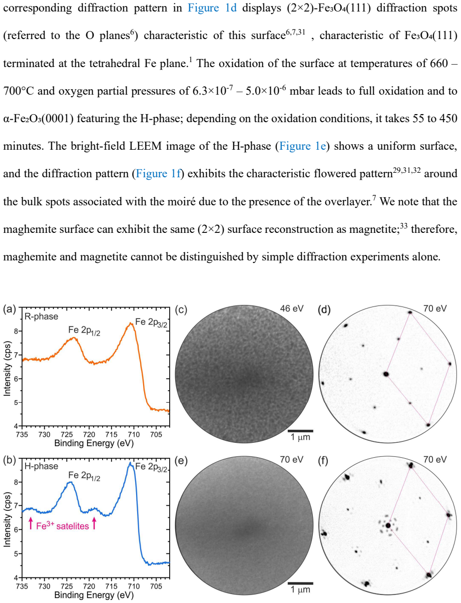
The oxidation kinetics of hematite {\alpha}-Fe$_2$O$_3$(0001) surfaces are vital for its applications in catalysis, environmental remediation, and industrial processes. Despite prior studies, the roles of temperature, oxygen partial pressure, and oxygen chemical potential in controlling nucleation and growth kinetics are not fully understood. Using real-time Low Energy Electron Microscopy/Diffraction (LEEM/LEED), we systematically investigate the oxidation of a reduced Fe$_3$O$_4$(111)-like surface layer to hematite under controlled conditions. We show that complete recovery of the hematite surface termination is closely linked to the nucleation and lateral growth of a two-dimensional honeycomb (H) phase. While higher temperatures accelerate nucleation, they slow lateral growth at constant oxygen pressure, indicating that oxygen supply limits the oxidation rate. Below an oxygen partial pressure threshold (~2$\times$10$^{-6}$ mbar), growth dramatically slows, underscoring the critical role of oxygen availability. Below a certain oxygen pressure threshold, the growth time rapidly increases. Our study elucidates the interplay between thermodynamics and kinetics in hematite surface oxidation, informing strategies to optimize surface properties for catalytic and industrial processes.
Editorial analysis
A structured set of objections, weighed in public.
Referee Report
Summary. The manuscript reports an experimental investigation of the oxidation kinetics of the α-Fe₂O₃(0001) surface starting from a reduced Fe₃O₄(111)-like layer, using real-time LEEM/LEED. The central claim is that complete recovery of the hematite surface termination is directly linked to the nucleation and lateral growth of a two-dimensional honeycomb (H) phase. Higher temperatures are reported to accelerate nucleation while slowing lateral growth at fixed oxygen pressure, which the authors interpret as evidence that oxygen supply limits the oxidation rate. A threshold oxygen partial pressure of ~2×10^{-6} mbar is identified below which growth slows dramatically.
Significance. If the LEEM/LEED observations prove free of probe-induced artifacts, the work supplies valuable real-time microscopic data on the interplay between nucleation, lateral growth, temperature, and oxygen pressure during hematite surface recovery. The direct correlation with the honeycomb phase and the reported pressure threshold could help guide optimization of iron-oxide surfaces for catalysis and related applications. The in-situ microscopy approach is a methodological strength for capturing dynamic phase evolution.
major comments (2)
- Abstract: The claim that oxygen supply limits the oxidation rate (inferred from faster nucleation but slower growth at higher temperature and the ~2×10^{-6} mbar threshold) rests entirely on LEEM/LEED contrast and diffraction changes. The manuscript provides no description of controls for low-energy electron beam effects (e.g., beam-current variation, intermittent imaging, or post-exposure verification), which are known to induce O₂ dissociation, reduction, or desorption on iron oxides and could therefore alter the very nucleation/growth kinetics being measured.
- Abstract: The temperature dependence and pressure-threshold conclusions are presented without quantitative growth-rate data, error bars, or statistical analysis of multiple runs. Without these, it is difficult to assess whether the observed trends robustly support the oxygen-limited interpretation or could arise from other factors such as surface preparation variability.
minor comments (1)
- Abstract: The final two sentences are nearly redundant ('Below an oxygen partial pressure threshold (~2×10^{-6} mbar), growth dramatically slows...' and 'Below a certain oxygen pressure threshold, the growth time rapidly increases'). Consolidating them would improve clarity.
Simulated Author's Rebuttal
We thank the referee for the careful reading and constructive comments on our manuscript. We address each major point below and indicate the revisions made to strengthen the presentation of controls and quantitative analysis.
read point-by-point responses
-
Referee: [—] Abstract: The claim that oxygen supply limits the oxidation rate (inferred from faster nucleation but slower growth at higher temperature and the ~2×10^{-6} mbar threshold) rests entirely on LEEM/LEED contrast and diffraction changes. The manuscript provides no description of controls for low-energy electron beam effects (e.g., beam-current variation, intermittent imaging, or post-exposure verification), which are known to induce O₂ dissociation, reduction, or desorption on iron oxides and could therefore alter the very nucleation/growth kinetics being measured.
Authors: We agree that the original manuscript lacked an explicit description of beam-effect controls. In the revised version we have added a new paragraph to the Experimental section that specifies the typical beam current (1–5 nA), the protocol of intermittent imaging with beam blanking between frames, and dedicated control runs in which the beam was extinguished for the entire oxidation period and only briefly turned on for final imaging. These controls reproduce the same nucleation densities and growth velocities, confirming that the reported temperature and pressure trends are not dominated by probe-induced artifacts. revision: yes
-
Referee: [—] Abstract: The temperature dependence and pressure-threshold conclusions are presented without quantitative growth-rate data, error bars, or statistical analysis of multiple runs. Without these, it is difficult to assess whether the observed trends robustly support the oxygen-limited interpretation or could arise from other factors such as surface preparation variability.
Authors: We accept that the original submission presented the trends qualitatively. The revised manuscript now contains a new figure that reports lateral growth velocities extracted from LEEM image sequences as a function of temperature (at fixed pO₂) and as a function of pO₂ (at fixed temperature). Each data point includes error bars representing the standard deviation across at least three independent runs performed on separately prepared surfaces. Linear fits with associated R² values and confidence intervals are provided in the text to quantify the opposing temperature effects on nucleation versus growth and to support the identification of the ~2×10^{-6} mbar threshold. revision: yes
Circularity Check
No circularity: purely observational experimental study
full rationale
The paper reports direct LEEM/LEED microscopy and diffraction observations of surface phase recovery under controlled temperature and oxygen pressure. No equations, fitted models, derivations, or predictions appear in the text. Claims about nucleation rates, lateral growth, and the ~2×10^{-6} mbar threshold rest on empirical contrast changes and time-resolved imaging rather than any reduction to prior results or self-citations by construction. The derivation chain is therefore self-contained against external benchmarks with no load-bearing steps that collapse to inputs.
Axiom & Free-Parameter Ledger
axioms (1)
- domain assumption LEEM/LEED diffraction patterns can be reliably assigned to specific surface phases such as the honeycomb (H) phase based on established literature interpretations.
Reference graph
Works this paper leans on
-
[1]
(1) Parkinson, G. S. Iron Oxide Surfaces. Surf. Sci. Rep. 2016 , 71 (1), 272–365. https://doi.org/10.1016/j.surfrep.2016.02.001. (2) Sivula, K.; Le Formal, F.; Grätzel, M. Solar Water S plitting: Progress Using Hematite (Α‐Fe 2 O 3 ) Photoelectrodes. ChemSusChem 2011 , 4 (4), 432–449. https://doi.org/10.1002/cssc.201000416. (3) Tamirat, A. G.; Rick, J.; D...
-
[2]
Defects and Cation Diffusi on in Magnetite (II)
(22) Dieckmann, R.; Schmalzried, H. Defects and Cation Diffusi on in Magnetite (II). Ber. Bunsenges. Phys. Chem. 1977 , 81 ,
work page 1977
-
[3]
Fabric and Structura l Characteristics of the Martitization Process
(23) Davis, B.; Rapp, G.; Walawender, M. Fabric and Structura l Characteristics of the Martitization Process. Am. J. Sci. 1968 , 266 , 482–496. (24) Cornell, R. M.; Schwertmann, U. The Iron Oxides ; Wiley,
work page 1968
-
[4]
25 (25) Freindl, K.; Wojas, J.; Kwiatek, N.; Korecki, J.; Spi ridis, N
https://doi.org/10.1002/3527602097. 25 (25) Freindl, K.; Wojas, J.; Kwiatek, N.; Korecki, J.; Spi ridis, N. Reversible Oxidation–Reduction of Epitaxial Iron Oxide Films on Pt(111): Magnetite–Hematite Interconversion. J. Chem. Phys. 2020 , 152 (5). https://doi.org/10.1063/1.5136322. (26) Dieckmann, R. Defects and Cation Diffusion in Magnetite (IV ): Nonsto...
-
[5]
(28) McIntyre, N. S.; Zetaruk, D. G. X-Ray Photoelectron Spectr oscopic Studies of Iron Oxides. Anal. Chem. 1977 , 49 (11), 1521–1529. https://doi.org/10.1021/ac50019a016. (29) Lad, R. J.; Henrich, V. E. Structure of α-Fe2O3 Single Crys tal Surfaces Following Ar+ Ion Bombardment and Annealing in O2. Surf. Sci. 1988 , 193 (1–2), 81–93. https://doi.org/10.1...
-
[6]
(32) Condon, N. G.; Leibsle, F. M.; Lennie, A. R.; Murray, P. W.; Vaughan, D. J.; Thornton, G. Biphase Ordering of Iron Oxide Surfaces. Phys. Rev. Lett. 1995 , 75 (10), 1961–1964. https://doi.org/10.1103/PhysRevLett.75.1961. (33) Bowker, M.; Hutchings, G.; Davies, P. R.; Edwards, D.; Da vies, R.; Shaikhutdinov, S.; Freund, H.-J. Surface Structure of γ-Fe2...
discussion (0)
Sign in with ORCID, Apple, or X to comment. Anyone can read and Pith papers without signing in.