Resilience of the physicochemical properties of graphene-based materials for applications in harsh radiation environments
Pith reviewed 2026-06-29 21:07 UTC · model grok-4.3
The pith
Ion irradiation disorders highly oriented graphite while partially reordering multilayer reduced graphene oxide at higher fluences.
A machine-rendered reading of the paper's core claim, the machinery that carries it, and where it could break.
Core claim
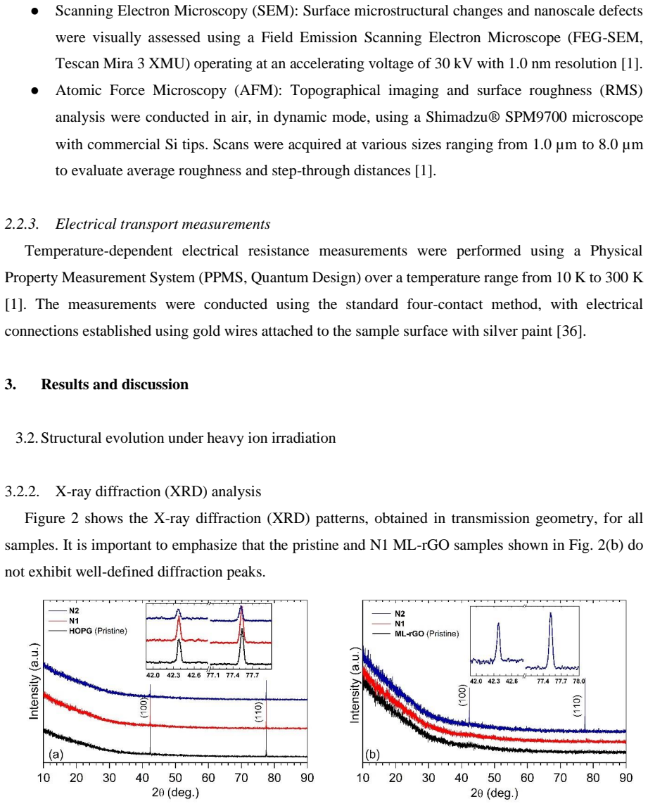
The irradiation response is strongly influenced by the initial structural organization of the material. In HOPG, ion exposure leads to a progressive loss of crystalline order, evidenced by XRD peak broadening and an increase in the Raman ID/IG ratio, accompanied by a reduction in electrical transport performance. In contrast, ML-rGO exhibits distinct behavior at higher fluences, suggesting partial structural reorganization. The appearance of more defined graphitic features in XRD and Raman analyses, along with changes in surface morphology and electrical response, suggests the formation of more ordered sp2 domains.
What carries the argument
Initial structural organization of the carbon lattice, which determines whether 60 MeV 35Cl ions cause net disordering (HOPG) or partial reordering (ML-rGO), tracked through XRD peak width, Raman ID/IG ratio, SEM/AFM morphology, and electrical transport.
If this is right
- HOPG loses crystalline order and electrical performance under the tested ion fluences.
- ML-rGO develops sharper graphitic XRD and Raman signatures at the higher fluence, consistent with formation of ordered sp2 domains.
- Surface morphology and electrical response shift in parallel with the structural changes in both materials.
- Material choice for radiation environments must take account of the starting degree of crystalline order.
Where Pith is reading between the lines
- Disordered carbon forms could tolerate moderate radiation exposure through ion-driven reorganization rather than pure damage.
- The same reordering tendency might appear in other defective carbon structures under comparable ion bombardment.
- Device layers in radiation settings might benefit from using reduced graphene oxide instead of perfect graphite sheets.
- Mapping the fluence threshold that separates damage from reorganization across different ions would be a direct next measurement.
Load-bearing premise
The reported changes in XRD, Raman, morphology, and transport are produced by the ion irradiation rather than by sample-to-sample differences or post-exposure handling.
What would settle it
If identical unirradiated HOPG control samples, measured under the same conditions and sequence, show the same XRD peak broadening and ID/IG increase as the irradiated specimens, the claim that irradiation drives the loss of order would be falsified.
Figures


read the original abstract
The development of radiation-tolerant materials capable of maintaining structural, electrical, and thermal stability in extreme, radiation-rich environments remains a critical challenge in materials science. In this work, the effects of 60 MeV 35Cl ion irradiation on highly oriented pyrolytic graphite (HOPG) and multilayer reduced graphene oxide (ML-rGO) were investigated. The samples were exposed to fluences of 5.11 x 10^9 and 1.3 x 10^10 ions/cm^2 and characterized by X-ray diffraction (XRD), Raman spectroscopy, scanning electron microscopy (SEM), atomic force microscopy (AFM), and electrical transport measurements. The results show that the irradiation response is strongly influenced by the initial structural organization of the material. In HOPG, ion exposure leads to a progressive loss of crystalline order, evidenced by XRD peak broadening and an increase in the Raman ID/IG ratio, accompanied by a reduction in electrical transport performance. In contrast, ML-rGO exhibits distinct behavior at higher fluences, suggesting partial structural reorganization. The appearance of more defined graphitic features in XRD and Raman analyses, along with changes in surface morphology and electrical response, suggests the formation of more ordered sp2 domains. These findings indicate that irradiation effects vary with the initial degree of order, providing useful insights for selecting carbon-based materials for devices operating under severe radiation conditions.
Editorial analysis
A structured set of objections, weighed in public.
Referee Report
Summary. The manuscript reports an experimental study of 60 MeV 35Cl ion irradiation at fluences of 5.11×10^9 and 1.3×10^10 ions/cm² on HOPG and ML-rGO. Characterization by XRD, Raman, SEM, AFM, and electrical transport is used to claim that response depends on initial structural organization: HOPG exhibits progressive loss of crystalline order while ML-rGO shows partial reorganization into more ordered sp2 domains at higher fluence.
Significance. If the differential response is shown to be irradiation-driven rather than an artifact of sample variation, the findings would provide practical guidance for selecting carbon-based materials in radiation environments. The experimental approach is standard for the field, but the absence of statistical controls limits the strength of the conclusions.
major comments (2)
- [Abstract] Abstract and results: Changes in XRD peak width, Raman I_D/I_G ratio, morphology, and transport are presented as irradiation-induced without reported error bars, standard deviations, replicate counts, or statistical tests. This prevents evaluation of whether the reported differences between HOPG and ML-rGO exceed sample-to-sample variation.
- [Methods] Experimental section: No description of pre-irradiation measurements on the identical specimens, multiple independent samples per fluence, or unirradiated controls is provided. Without these, the central claim that initial structural organization dictates the irradiation response cannot be isolated from batch variation or handling effects.
minor comments (1)
- [Abstract] The fluence values are given with scientific notation but without explicit units in the abstract; ensure consistent reporting of units and ion species throughout.
Simulated Author's Rebuttal
We thank the referee for the constructive comments on statistical reporting and experimental controls. We have revised the manuscript to address these points as described below.
read point-by-point responses
-
Referee: [Abstract] Abstract and results: Changes in XRD peak width, Raman I_D/I_G ratio, morphology, and transport are presented as irradiation-induced without reported error bars, standard deviations, replicate counts, or statistical tests. This prevents evaluation of whether the reported differences between HOPG and ML-rGO exceed sample-to-sample variation.
Authors: We agree that the absence of error bars and replicate information limits the ability to assess variability. In the revised manuscript we have added error bars (standard deviations) to all relevant plots in the results section, along with a statement specifying the number of measurements per condition and observed sample-to-sample variation. These additions allow direct evaluation of whether the reported differences between HOPG and ML-rGO exceed typical variation. revision: yes
-
Referee: [Methods] Experimental section: No description of pre-irradiation measurements on the identical specimens, multiple independent samples per fluence, or unirradiated controls is provided. Without these, the central claim that initial structural organization dictates the irradiation response cannot be isolated from batch variation or handling effects.
Authors: We have expanded the experimental section to describe the use of unirradiated control samples from the same preparation batches and to note that multiple specimens were measured per fluence. Pre-irradiation characterization was performed on representative samples from each batch. We acknowledge that pre-irradiation data on the exact same specimen locations were not obtained for every technique in every case. The revised text now includes an explicit discussion of this limitation and explains how the distinctly different initial structures of HOPG versus ML-rGO still permit attribution of the differential response to irradiation. This constitutes a partial revision because full isolation from all possible batch effects would require additional replicate experiments that were not part of the original study. revision: partial
Circularity Check
No circularity: purely experimental reporting with no models or derivations
full rationale
The paper consists entirely of experimental irradiation of HOPG and ML-rGO samples followed by direct characterization via XRD, Raman, SEM, AFM, and transport measurements. No equations, fitted parameters, predictive models, or derivation steps appear in the abstract or described content. Claims about differential response to irradiation are presented as observations, not as outputs of any chain that could reduce to inputs by construction. This matches the default case of a self-contained experimental report with score 0.
Axiom & Free-Parameter Ledger
axioms (1)
- domain assumption XRD peak broadening and increased Raman ID/IG ratio reliably indicate loss of crystalline order and increased defects
Reference graph
Works this paper leans on
-
[1]
Guazzelli MA, et al., Effects of neutron radiation on the thermal conductivity of multilayer, Diam. Relat. Mater. 151 (2025) 111803. https://doi.org/10.1016/j.diamond.2024.111803
-
[2]
https://doi.org/10.1007/978-1-4939-3438-6
Was GS, Fundamentals of Radiation Materials Science, Springer, New York, NY (2017). https://doi.org/10.1007/978-1-4939-3438-6
-
[3]
https://doi.org/10.1016/j.carbon.2020.05.027
Al-Qasir II, et al., Vacancy-driven variations in the phonon density of states of fast neutron irradiated nuclear graphite, Carbon 168 (2020) 42. https://doi.org/10.1016/j.carbon.2020.05.027
-
[4]
Khandaker MU, et al., Defects and structural changes of graphite-rich media subjected to low-level neutron doses for radiation dosimetry, Radiat. Phys. Chem. 201 (2022) 110498. https://doi.org/10.1016/j.radphyschem.2022.110498
-
[5]
Avanzi LH, et al., Using TRIM-SRIM code simulations to determine defect density produced in HOPG irradiated with high energy heavy ions, J. Phys. Conf. Ser. 2340 (2022) 012002. https://dx.doi.org/10.1088/1742-6596/2340/1/012002
-
[6]
Cappuzzello F, et al., The NUMEN Technical Design Report, Int. J. Mod. Phys. A 36 (2021) 2130018. https://doi.org/10.1142/S0217751X21300180
-
[7]
Lo Presti D, et al., Neutron radiation effects on an electronic system on module, Rev. Sci. Instrum. 91 (2020) 083301. https://doi.org/10.1063/5.0010968
-
[8]
F. Pinna, V. Capirossi et al., Tests of a cooling system for thin targets submitted to intense ion beams for the NUMEN experiment, Acta Phys. Pol. B 51, 655 (2020). https://doi.org/10.5506/APhysPolB.51.655
-
[9]
Pinna F, et al., Project of thin targets for the NUMEN experiment, J. Phys. Conf. Ser. 1056 (2018) 012046. https://iopscience.iop.org/article/10.1088/1742-6596/1056/1/012046
-
[10]
Balandin AA, Thermal properties of graphene and nanostructured carbon materials, Nat. Mater. 10 (2011) 569. https://doi.org/10.1038/nmat3064
-
[11]
https://doi.org/10.1126/science.1157996
Lee C, Wei X, Kysar JW, Hone J, Measurement of the Elastic Properties and Intrinsic Strength of Monolayer Graphene, Science 321 (2008) 385–388. https://doi.org/10.1126/science.1157996
-
[12]
https://doi.org/10.1016/j.cartre.2024.100429
Kaftelen-Odabaşı Ü, Evaluation of morphological, structural, thermal, electrical, and chemical composition properties of graphene oxide and reduced graphene oxide obtained by sequential reduction methods, Carbon Trends 17 (2024) 100429. https://doi.org/10.1016/j.cartre.2024.100429
-
[13]
https://doi.org/10.1016/j.cartre.2026.100619
Tjenreng JSZ, Sukma FOR, Santjojo DJDH, Harun SW, Masruroh, From graphene oxide (GO) to reduced graphene oxide (rGO) films: A hybrid approach combining film transfer and vapor reduction for enhanced structural and optical properties, Carbon Trends (2026) 100619. https://doi.org/10.1016/j.cartre.2026.100619
-
[14]
https://doi.org/10.1016/j.carbon.2013.07.017
Mortazavi B, Ahzi S, Thermal conductivity and tensile response of defective graphene: A molecular dynamics study, Carbon 63 (2013) 460. https://doi.org/10.1016/j.carbon.2013.07.017
-
[15]
Patil S, Kolekar S, Deshpande A, Revisiting HOPG superlattices: Structure and conductance properties, Surf. Sci. 658 (2017) 55–60. https://doi.org/10.1016/j.susc.2016.12.002
-
[16]
2026, Journal of High Energy Astrophysics, 50, 100490, doi: 10.1016/j.jheap.2025.100490
Gupta S, Kumar R, Kumar A, Devi KD, Structural and electrical changes in multilayer graphene induced by negative oxygen ion bombardment, Results Surf. Interfaces 19 (2025) 100490. https://doi.org/10.1016/j.rsurfi.2025.100490
-
[17]
Fok T, Janulewicz KA, Wachulak P, et al., Electronic structure of multi-layered graphene oxide membrane moderately reduced in vacuum, J. Phys. Chem. Solids 164 (2022) 110623. https://doi.org/10.1016/j.jpcs.2022.110623
-
[18]
Gupta A, Chen G, Joshi P, Tadigadapa S, Eklund PC, Raman scattering from high-frequency phonons in supported n-graphene layer films, Nano Lett. 6 (2006) 2667–2673. https://doi.org/10.1021/nl061420a
-
[19]
Ávila M, Venosta L, Bajales N, Bercoff P, Structural and magnetic changes induced by electron and ion irradiation on HOPG, Procedia Mater. Sci. 9 (2015) 62–68. https://doi.org/10.1016/j.mspro.2015.04.008
-
[20]
Ziang Z, et al., Ultra-low threshold optically pumped random laser emission behaviour of highly oriented pyrolytic graphite, Mater. Lett. 115 (2014) 261. https://doi.org/10.1016/j.matlet.2013.10.045
-
[21]
Ruddy FH, et al., The fast neutron response of 4H silicon carbide semiconductor radiation detectors, IEEE Trans. Nucl. Sci. 53 (2006). https://doi.org/10.1109/TNS.2006.875151
-
[22]
https://doi.org/10.1016/j.carbon.2023.04.004
Jimenez-Rioboo RJ, et al., Boron-doped diamond by 9 MeV microbeam implantation: Damage and recovery, Carbon 208 (2023) 421. https://doi.org/10.1016/j.carbon.2023.04.004
-
[23]
https://doi.org/10.1016/j.carbon.2020.10.086
Liu D, et al., A macro-scale ruck and tuck mechanism for deformation in ion-irradiated polycrystalline graphite, Carbon 173 (2021) 215. https://doi.org/10.1016/j.carbon.2020.10.086
-
[24]
Luhmann T, et al., Investigation of the graphitization process of ion-beam irradiated diamond using ellipsometry, Raman spectroscopy and electrical transport measurements, Carbon 121 (2017)
2017
-
[25]
https://doi.org/10.1016/j.carbon.2017.05.093
-
[26]
Da Rocha MS, et al., Applying the inelastic thermal spike model to the investigation of damage induced by high-energy ions in polymers, Macromol. Chem. Phys. 224 (2023) 2200339. https://doi.org/10.1002/macp.202200339
-
[27]
Abdol MA, Sadeghzadeh S, Jalaly M, et al., Constructing a three-dimensional graphene structure via bonding layers by ion beam irradiation, Sci. Rep. 9 (2019) 8127. https://doi.org/10.1038/s41598- 019-44697-z
-
[28]
Kolawole FO, Mitma Pillaca EJD, Martins GV, Kolawole SK, Corat EJ, Trava-Airoldi VJ, Deposition of graphene incorporated diamond-like carbon coatings using pulsed-DC PECVD with an additional cathode for space applications, Diam. Relat. Mater. 156 (2025) 112422. https://doi.org/10.1016/j.diamond.2025.112422
-
[29]
Moon I, Lee J, Ruoff R, et al., Reduced graphene oxide by chemical graphitization, Nat. Commun. 1 (2010) 73. https://doi.org/10.1038/ncomms1067
-
[30]
Wei Y, Pastuovic Ž, Murphy T, Gore DB, Precise tuning chemistry and tailoring defects of graphene oxide films by low energy ion beam irradiation, Appl. Surf. Sci. 505 (2020) 144651. https://doi.org/10.1016/j.apsusc.2019.144651
-
[31]
Tyagi C, Khan SA, Sulania I, Meena R, Avasthi DK, Tripathi A, Evidence of ion-beam-induced annealing in graphene oxide films using in situ X-ray diffraction and spectroscopy techniques, J. Phys. Chem. C 122 (2018) 9632–9640. https://doi.org/10.1021/acs.jpcc.7b10699
-
[32]
Capan I, Bernat R, Makino T, Knežević T, 4H-SiC Schottky barrier diodes as radiation detectors: A role of Schottky contact area, Diam. Relat. Mater. 137 (2023) 110072. https://doi.org/10.1016/j.diamond.2023.110072
-
[33]
Babu KV, Sree GNJ, Das S, Algarni AD, Ghzaoui MEL, Devana VNKR, A graphene material- based wideband metamaterial absorber exploiting slotted substrate integrated waveguide (SIW) for multifarious terahertz applications, Diam. Relat. Mater. 157 (2025) 112585. https://doi.org/10.1016/j.diamond.2025.112585
-
[34]
https://doi.org/10.1016/j.carbon.2015.12.101
Zeng J, et al., Comparative study of irradiation effects in graphite and graphene induced by swift heavy ions and highly charged ions, Carbon 100 (2016) 16–26. https://doi.org/10.1016/j.carbon.2015.12.101
-
[35]
Inagaki M, et al., Advanced materials science and engineering of carbon, Elsevier (2014)
2014
-
[36]
Aguiar VAP, et al., SAFIIRA: A heavy-ion multi-purpose irradiation facility in Brazil, Rev. Sci. Instrum. 91 (2020) 053301. https://doi.org/10.1063/1.5138644
-
[37]
https://doi.org/10.1016/0008-6223(92)90047-Z
Hishiyama Y, Kaburagi Y, Electrical resistivity of highly crystallized kish graphite, Carbon 30 (1992) 483–486. https://doi.org/10.1016/0008-6223(92)90047-Z
-
[38]
Blanton TN, Majumdar D, X-ray diffraction characterization of polymer intercalated graphite oxide, Powder Diffr. 27 (2012) 104. https://doi.org/10.1017/S0885715612000292
-
[39]
Ferreira EHM, et al., Evolution of the Raman spectra from single-, few-, and many-layer graphene with increasing disorder, Phys. Rev. B 82 (2010) 125429. https://doi.org/10.1103/PhysRevB.82.125429
-
[40]
Livneh T, Haslett TL, Moskovits M, Distinguishing disorder-induced bands from allowed Raman bands in graphite, Phys. Rev. B 66 (2002) 195110. https://doi.org/10.1103/PhysRevB.66.195110
-
[41]
Nakamura K, Kitajima M, Ion-irradiation effects on the phonon correlation length of graphite studied by Raman spectroscopy, Phys. Rev. B 45 (1992) 78. https://doi.org/10.1103/PhysRevB.45.78
-
[42]
https://doi.org/10.1016/0008-6223(94)90096-5
Klemens PG, Pedraza DF, Thermal conductivity of graphite in the basal plane, Carbon 32 (1994) 735–741. https://doi.org/10.1016/0008-6223(94)90096-5
-
[43]
Zhao Y, et al., Experimental measurement of thermal conductivity along different crystallographic planes in graphite, J. Appl. Phys. 128 (2020). https://doi.org/10.1063/5.0013474
-
[44]
https://doi.org/10.1007/978-0-387-09579-0
Pecharsky VK, Zavalij PY, Fundamentals of Powder Diffraction and Structural Characterization of Materials, 2nd ed., Springer (2009). https://doi.org/10.1007/978-0-387-09579-0
discussion (0)
Sign in with ORCID, Apple, or X to comment. Anyone can read and Pith papers without signing in.