Polariton spectroscopy at the diamond K-edge via X-ray parametric down-conversion
Pith reviewed 2026-06-29 08:59 UTC · model grok-4.3
The pith
X-ray parametric down-conversion at the diamond K-edge produces polariton hybridization that enters the strong-coupling regime.
A machine-rendered reading of the paper's core claim, the machinery that carries it, and where it could break.
Core claim
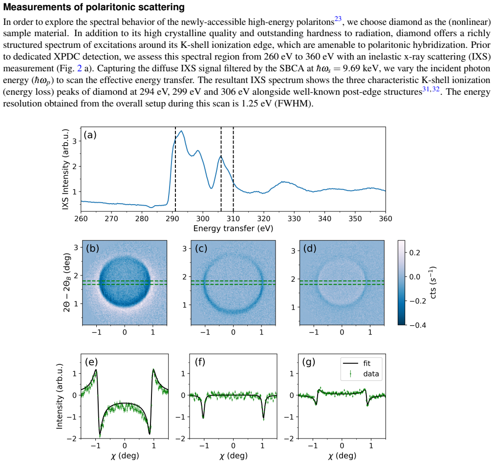
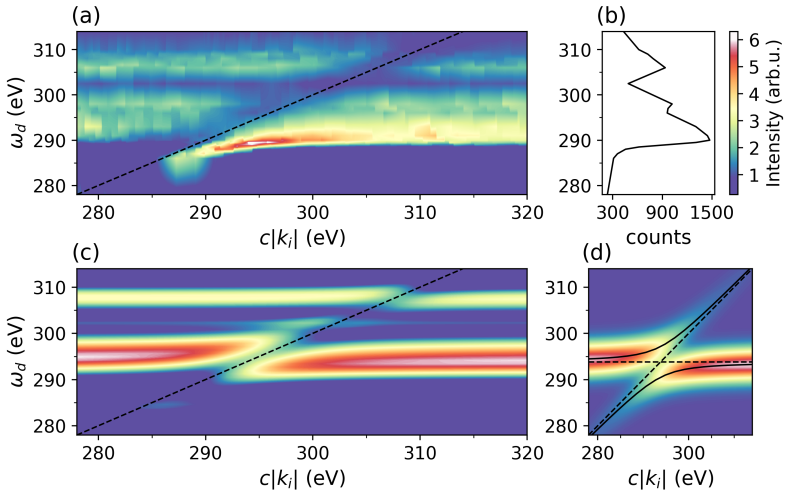
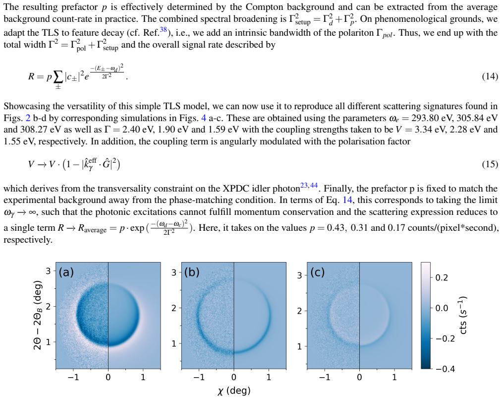
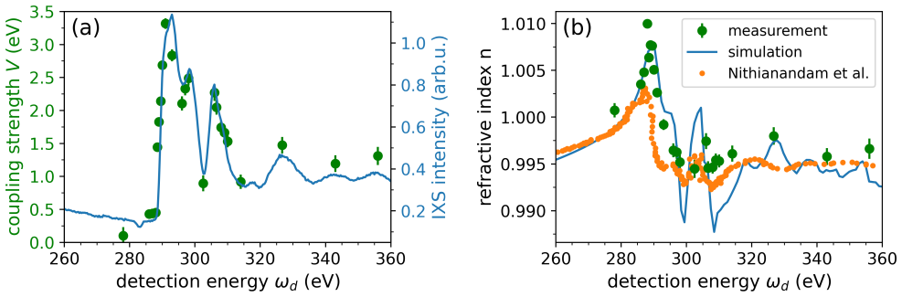
XPDC provides access to high-energy polaritons resulting from hybridization of down-converted photons with electronic excitations in a nonlinear medium. Around the K-shell absorption edge in diamond the measurements display pronounced signatures of this hybridization, visualized through a polariton spectral map and analyzed with theoretical modeling. The hybridization produces substantially higher coupling strength than previously reported for a non-resonant case and reaches well into the strong-coupling regime. The same polaritonic XPDC measurements allow extraction of the refractive index for bulk diamond at high spectral resolution around the carbon K-edge.
What carries the argument
Polariton spectral map together with theoretical modeling of the XPDC process, which isolates the hybridization signatures between down-converted photons and K-edge excitations.
Load-bearing premise
The observed spectral features arise primarily from polaritonic hybridization rather than competing nonlinear processes or absorption artifacts, and the fitting model does not reproduce the data without the polariton mechanism.
What would settle it
An experiment or calculation in which the same spectral features appear when the data are fitted with a model containing only linear absorption and no polariton term, or a measurement showing no increase in coupling strength when tuned across the K-edge.
Figures

read the original abstract
It has recently been shown that x-ray parametric down-conversion (XPDC) provides access to high-energy polaritons, resulting from the hybridization of down-converted photons with electronic excitations in a nonlinear medium. Here, we present a spectrally resolved study of this effect around the K-shell absorption edge in diamond. Our results exhibit pronounced signatures of polaritonic hybridization, which we visualize by introducing a polariton spectral map and analyze by help of theoretical modelling. We find that the hybridization at this absorption edge results in substantially higher coupling strength than previously reported for a non-resonant case and reaches well into the strong-coupling regime. In addition, we demonstrate how our measurements of polaritonic XPDC allow us to extract the refractive index for bulk diamond at high spectral resolution around the carbon K-edge.
Editorial analysis
A structured set of objections, weighed in public.
Referee Report
Summary. The manuscript reports a spectrally resolved X-ray parametric down-conversion (XPDC) experiment at the carbon K-edge in diamond. It introduces a polariton spectral map to visualize hybridization signatures between down-converted photons and electronic excitations, analyzes the data with theoretical modeling, claims substantially higher coupling strengths than prior non-resonant cases that reach the strong-coupling regime, and shows that the measurements enable high-resolution extraction of the bulk refractive index around the K-edge.
Significance. If the central claims are substantiated, the work would establish XPDC-based polariton spectroscopy as a practical route to strong-coupling studies at core-level edges, offering a new experimental handle on X-ray polaritonics and a method for precise refractive-index determination that complements existing techniques.
major comments (2)
- [Theoretical modelling] Theoretical modelling section: the description of the model used to fit the polariton spectral map and extract the coupling strength does not include the explicit form of the interaction Hamiltonian, the fitting procedure, or quantitative metrics (e.g., χ² or residual comparison) showing that the polariton term is required; without this, it remains possible that phenomenological absorption or competing χ^(3) processes could reproduce the reported line shapes and apparent splitting.
- [Results and discussion] Results and discussion: the manuscript presents no raw spectra, error bars on the extracted coupling strengths, or tabulated fit parameters with uncertainties, so the claim that the hybridization 'reaches well into the strong-coupling regime' and exceeds prior non-resonant values cannot be quantitatively assessed from the provided data.
minor comments (2)
- [Figures] Figure captions for the polariton spectral map should explicitly state the normalization, energy resolution, and any background subtraction applied.
- [Abstract and refractive-index extraction] The abstract states that the refractive index is extracted 'at high spectral resolution,' but the main text does not specify the achieved resolution or compare it to tabulated values near the K-edge.
Simulated Author's Rebuttal
We thank the referee for their careful reading of the manuscript and for the constructive comments. We address each major point below and will revise the manuscript to incorporate the requested details.
read point-by-point responses
-
Referee: [Theoretical modelling] Theoretical modelling section: the description of the model used to fit the polariton spectral map and extract the coupling strength does not include the explicit form of the interaction Hamiltonian, the fitting procedure, or quantitative metrics (e.g., χ² or residual comparison) showing that the polariton term is required; without this, it remains possible that phenomenological absorption or competing χ^(3) processes could reproduce the reported line shapes and apparent splitting.
Authors: We agree that the theoretical modelling section requires additional detail to fully substantiate the analysis. In the revised manuscript we will explicitly state the interaction Hamiltonian employed for the polariton spectral map, describe the fitting procedure (including initial parameters and constraints), and supply quantitative goodness-of-fit metrics such as χ² values together with residual plots. These additions will demonstrate that the polariton term is necessary and that simpler phenomenological absorption models or competing χ^(3) processes do not reproduce the observed line shapes and apparent splitting. revision: yes
-
Referee: [Results and discussion] Results and discussion: the manuscript presents no raw spectra, error bars on the extracted coupling strengths, or tabulated fit parameters with uncertainties, so the claim that the hybridization 'reaches well into the strong-coupling regime' and exceeds prior non-resonant values cannot be quantitatively assessed from the provided data.
Authors: We acknowledge that the current presentation omits raw spectra, error bars, and tabulated parameters with uncertainties. In the revision we will add the raw spectra (as supplementary material if space is limited), include error bars on all extracted coupling strengths, and provide a table of fit parameters together with their uncertainties. These changes will enable quantitative evaluation of the strong-coupling claim and direct comparison with prior non-resonant results. revision: yes
Circularity Check
No significant circularity; experimental data and modeling remain independent.
full rationale
The paper reports direct experimental XPDC spectra around the diamond K-edge, introduces a polariton spectral map as a visualization tool, and applies separate theoretical modeling to extract coupling strengths and refractive index values. No quoted derivation step reduces the reported hybridization signatures, coupling strength, or extracted index to a fitted input or self-citation by construction; the central claims rest on measured spectral features analyzed against an external model rather than tautological redefinition. This is the expected outcome for an experimental spectroscopy study with independent fitting.
Axiom & Free-Parameter Ledger
Reference graph
Works this paper leans on
-
[1]
A., Fadeev, V
Akhmanov, S. A., Fadeev, V . V ., Khokhlov, R. V . & Chunaev, O. N. Quantum Noise in Parametric Light Amplifiers. ZhETF Pisma Redaktsiiu6, 575 (1967)
1967
-
[2]
Harris, S. E., Oshman, M. K. & Byer, R. L. Observation of Tunable Optical Parametric Fluorescence.Phys. Rev. Lett.18, 732–734, DOI: 10.1103/PhysRevLett.18.732 (1967)
-
[3]
& Chunaev, O
Akhmanov, S., Fadeev, V ., Khokhlov, R. & Chunaev, O. Quantum noise in parametric light amplifiers.JETP Lett6(1967)
1967
-
[4]
Magde, D. & Mahr, H. Study in Ammonium Dihydrogen Phosphate of Spontaneous Parametric Interaction Tunable from 4400 to 16 000 Å.Phys. Rev. Lett.18, 905–907, DOI: 10.1103/PhysRevLett.18.905 (1967)
-
[5]
Giordmaine, J. A. & Miller, R. C. Tunable Coherent Parametric Oscillation in LiNb O 3 at Optical Frequencies.Phys. Rev. Lett.14, 973–976, DOI: 10.1103/PhysRevLett.14.973 (1965)
-
[6]
A., Kovrigin, A., Piskarskas, A., Fadeev, V
Akhmanov, S. A., Kovrigin, A., Piskarskas, A., Fadeev, V . & Khokhlov, R. Observation of parametric amplification in the optical range.JETP Lett2, 191 (1965). 9.Menzel, R.Photonics: linear and nonlinear interactions of laser light and matter(Springer, Berlin, 2007), 2nd ed edn
1965
-
[7]
Li, B.et al.Down-converted photon pairs in a high-Q silicon nitride microresonator.Nature639, 922–927, DOI: 10.1038/s41586-025-08662-3 (2025)
-
[8]
J.et al.NIF final optics system: frequency conversion and beam conditioning
Wegner, P. J.et al.NIF final optics system: frequency conversion and beam conditioning. 180, DOI: 10.1117/12.538481 (San Jose, Ca, 2004)
-
[9]
Zhang, S.et al.Recent advances in nonlinear optics for bio-imaging applications.Opto-Electronic Adv.3, 200003–200003, DOI: 10.29026/oea.2020.200003 (2020)
-
[10]
M., Peacock, A
Dudley, J. M., Peacock, A. C., Stiller, B. & Tissoni, G. Nonlinear optics and its applications 2024. InProc. of SPIE V ol, vol. 13004, 1300401–1 (2024)
2024
-
[11]
G.et al.New High-Intensity Source of Polarization-Entangled Photon Pairs.Phys
Kwiat, P. G.et al.New High-Intensity Source of Polarization-Entangled Photon Pairs.Phys. Rev. Lett.75, 4337–4341, DOI: 10.1103/PhysRevLett.75.4337 (1995)
-
[12]
Rubin, M. H., Klyshko, D. N., Shih, Y . H. & Sergienko, A. V . Theory of two-photon entanglement in type-II optical parametric down-conversion.Phys. Rev. A50, 5122–5133, DOI: 10.1103/PhysRevA.50.5122 (1994)
-
[13]
H.et al.A large-scale reconfigurable multiplexed quantum photonic network.Nat
Valencia, N. H.et al.A large-scale reconfigurable multiplexed quantum photonic network.Nat. PhotonicsDOI: 10.1038/s41566-025-01806-x (2025)
-
[14]
Commun.16, 9616, DOI: 10.1038/s41467-025-64620-7 (2025)
Lu, Z.et al.Counter-propagating entangled photon pairs from monolayer GaSe.Nat. Commun.16, 9616, DOI: 10.1038/s41467-025-64620-7 (2025)
-
[15]
Chakraborty, T.et al.Towards a spectrally multiplexed quantum repeater.npj Quantum Inf.11, 3, DOI: 10.1038/ s41534-024-00946-2 (2025)
2025
-
[16]
Commun.16, 1899, DOI: 10.1038/s41467-025-56436-2 (2025)
Lyu, X.et al.A tunable entangled photon-pair source based on a Van der Waals insulator.Nat. Commun.16, 1899, DOI: 10.1038/s41467-025-56436-2 (2025)
-
[17]
Hopfield, J. J. Theory of the Contribution of Excitons to the Complex Dielectric Constant of Crystals.Phys. Rev.112, 1555–1567, DOI: 10.1103/PhysRev.112.1555 (1958)
-
[18]
N., Asenjo-Garcia, A., Schuck, P
Basov, D. N., Asenjo-Garcia, A., Schuck, P. J., Zhu, X. & Rubio, A. Polariton panorama.Nanophotonics10, 549–577, DOI: 10.1515/nanoph-2020-0449 (2020). 22.Klyshko, D. N. Scattering of light in a medium with nonlinear polarizability.JETP28(1968)
-
[19]
Commun.16, DOI: 10.1038/ s41467-025-60845-8 (2025)
Krebs, D.et al.X-ray parametric down-conversion reveals EUV-polariton.Nat. Commun.16, DOI: 10.1038/ s41467-025-60845-8 (2025). Publisher: Springer Science and Business Media LLC. 10/12
2025
-
[20]
Coffinet, J. P. & De Martini, F. Coherent Excitation of Polaritons in Gallium Phosphide.Phys. Rev. Lett.22, 60–64, DOI: 10.1103/PhysRevLett.22.60 (1969)
-
[21]
Kulevsky, L. A., Polivanov, Y . N. & Poluektov, S. N. Light scattering by polaritons in LiIO3.J. Raman Spectrosc.3, 239–254, DOI: 10.1002/jrs.1250030213 (1975)
-
[22]
& Panin, A
Aktsipetrov, A., Ivanov, V . & Panin, A. Frequency-angle spectrum of light scattering by polaritons and interference of susceptibilities of different orders.Zh. Eksp. Teor . Fiz78, 2309–2315 (1980)
1980
-
[23]
Chekhova, M. V . & Penin, A. N. Study of second-order excitations in α-iodic acid crystal by means of polaritonk -spectroscopy.J. Raman Spectrosc.24, 581–584, DOI: 10.1002/jrs.1250240904 (1993)
-
[24]
Synchrotron Radiat.24, 521–530, DOI: 10.1107/S1600577516020579 (2017)
Huotari, S.et al.A large-solid-angle X-ray Raman scattering spectrometer at ID20 of the European Synchrotron Radiation Facility.J. Synchrotron Radiat.24, 521–530, DOI: 10.1107/S1600577516020579 (2017)
-
[25]
Henry, C. H. & Hopfield, J. J. Raman Scattering by Polaritons.Phys. Rev. Lett.15, 964–966, DOI: 10.1103/PhysRevLett. 15.964 (1965)
-
[26]
J.et al.Planning, performing and analyzing X-ray Raman scattering experiments.J
Sahle, C. J.et al.Planning, performing and analyzing X-ray Raman scattering experiments.J. Synchrotron Radiat.22, 400–409, DOI: 10.1107/S1600577514027581 (2015)
-
[27]
Galambosi, S., Soininen, J. A., Nygård, K., Huotari, S. & Hämäläinen, K. Symmetry of the 1 s core exciton in diamond studied using x-ray Raman scattering.Phys. Rev. B76, 195112, DOI: 10.1103/PhysRevB.76.195112 (2007)
-
[28]
Morar, J. F., Himpsel, F. J., Hollinger, G., Hughes, G. & Jordan, J. L. Observation of a C- 1 s Core Exciton in Diamond. Phys. Rev. Lett.54, 1960–1963, DOI: 10.1103/PhysRevLett.54.1960 (1985)
-
[29]
Effects of Configuration Interaction on Intensities and Phase Shifts.Phys
Fano, U. Effects of Configuration Interaction on Intensities and Phase Shifts.Phys. Rev.124, 1866–1878, DOI: 10.1103/PhysRev.124.1866 (1961)
-
[30]
Weinhardt, L.et al.Resonant inelastic soft x-ray scattering of CdS: A two-dimensional electronic structure map approach. Phys. Rev. B79, 165305, DOI: 10.1103/PhysRevB.79.165305 (2009)
-
[31]
Noda, I. Generalized two-dimensional correlation method applicable to infrared, raman, and other types of spectroscopy. Appl. Spectrosc.47, 1329–1336, DOI: 10.1366/0003702934067694 (1993). https://doi.org/10.1366/0003702934067694
-
[32]
Huang, K. Lattice Vibrations and Optical Waves in Ionic Crystals.Nature167, 779–780, DOI: 10.1038/167779b0 (1951)
-
[33]
Rev.123, 9786–9879, DOI: 10.1021/acs.chemrev.2c00855 (2023)
Mandal, A.et al.Theoretical Advances in Polariton Chemistry and Molecular Cavity Quantum Electrodynamics.Chem. Rev.123, 9786–9879, DOI: 10.1021/acs.chemrev.2c00855 (2023). 38.Törmä, P. & Barnes, W. L. Strong coupling between surface plasmon polaritons and emitters: a review.Reports on Prog. Phys.78, 013901, DOI: 10.1088/0034-4885/78/1/013901 (2015)
-
[34]
Ebbesen, T. W. Hybrid Light–Matter States in a Molecular and Material Science Perspective.Accounts Chem. Res.49, 2403–2412, DOI: 10.1021/acs.accounts.6b00295 (2016)
-
[35]
Toffoletti, F. & Collini, E. Coherent phenomena in exciton–polariton systems.J. Physics: Mater.8, 022002, DOI: 10.1088/2515-7639/adcbd6 (2025)
-
[36]
G., Wersäll, M., Cuadra, J., Antosiewicz, T
Baranov, D. G., Wersäll, M., Cuadra, J., Antosiewicz, T. J. & Shegai, T. Novel Nanostructures and Materials for Strong Light–Matter Interactions.ACS Photonics5, 24–42, DOI: 10.1021/acsphotonics.7b00674 (2018)
-
[37]
Blaha, M., Johnson, A., Rauschenbeutel, A. & V olz, J. Beyond the Tavis-Cummings model: Revisiting cavity QED with ensembles of quantum emitters.Phys. Rev. A105, 013719, DOI: 10.1103/PhysRevA.105.013719 (2022)
-
[38]
Schülke, W.Electron dynamics by inelastic X-ray scattering. No. 7 in Oxford series on synchrotron radiation (Oxford University Press, Oxford ; New York, 2007). OCLC: ocm85862430
2007
-
[39]
Boemer, C.et al.Towards novel probes for valence chargesviaX-ray optical wave mixing.Faraday Discuss.228, 451–469, DOI: 10.1039/D0FD00130A (2021)
-
[40]
Painter, G. S., Ellis, D. E. & Lubinsky, A. R. Ab initio calculation of the electronic structure and optical properties of diamond using the discrete variational method.Phys. Rev. B4, 3610–3622, DOI: 10.1103/PhysRevB.4.3610 (1971)
-
[41]
Tamasaku, K., Sawada, K. & Ishikawa, T. Determining X-Ray Nonlinear Susceptibility of Diamond by the Optical Fano Effect.Phys. Rev. Lett.103, 254801, DOI: 10.1103/PhysRevLett.103.254801 (2009)
-
[42]
Garcia-Vidal, F. J., Ciuti, C. & Ebbesen, T. W. Manipulating matter by strong coupling to vacuum fields.Science373, eabd0336, DOI: 10.1126/science.abd0336 (2021). 11/12
-
[43]
Sánchez-Barquilla, M., Fernández-Domínguez, A. I., Feist, J. & García-Vidal, F. J. A Theoretical Perspective on Molecular Polaritonics.ACS Photonics9, 1830–1841, DOI: 10.1021/acsphotonics.2c00048 (2022)
-
[44]
Li, T. E., Cui, B., Subotnik, J. E. & Nitzan, A. Molecular Polaritonics: Chemical Dynamics Under Strong Light–Matter Coupling.Annu. Rev. Phys. Chem.73, 43–71, DOI: 10.1146/annurev-physchem-090519-042621 (2022)
-
[45]
Nithianandam, J. & Rife, J. C. Synchrotron x-ray optical properties of natural diamond.Phys. Rev. B47, 3517–3521, DOI: 10.1103/PhysRevB.47.3517 (1993)
-
[46]
Blum, V .et al.Ab initio molecular simulations with numeric atom-centered orbitals.Comput. Phys. Commun.180, 2175–2196, DOI: 10.1016/j.cpc.2009.06.022 (2009)
-
[47]
Phys.14, 053020, DOI: 10.1088/1367-2630/14/5/053020 (2012)
Ren, X.et al.Resolution-of-identity approach to Hartree–Fock, hybrid density functionals, RPA, MP2 andGWwith numeric atom-centered orbital basis functions.New J. Phys.14, 053020, DOI: 10.1088/1367-2630/14/5/053020 (2012)
-
[48]
Heyd, J., Scuseria, G. E. & Ernzerhof, M. Hybrid functionals based on a screened Coulomb potential.The J. Chem. Phys. 118, 8207–8215, DOI: 10.1063/1.1564060 (2003)
-
[49]
Schülke, W., Bonse, U., Nagasawa, H., Kaprolat, A. & Berthold, A. Interband transitions and core excitation in highly oriented pyrolytic graphite studied by inelastic synchrotron x-ray scattering: Band-structure information.Phys. Rev. B38, 2112–2123, DOI: 10.1103/PhysRevB.38.2112 (1988)
-
[50]
McDougall, N. L., Nicholls, R. J., Partridge, J. G. & McCulloch, D. G. The Near Edge Structure of Hexagonal Boron Nitride.Microsc. Microanal.20, 1053–1059, DOI: 10.1017/S1431927614000737 (2014)
-
[51]
Soufli, R. & Gullikson, E. M. Reflectance measurements on clean surfaces for the determination of optical constants of silicon in the extreme ultraviolet–soft-x-ray region.Appl. Opt.36, 5499, DOI: 10.1364/AO.36.005499 (1997)
-
[52]
Dorney, K. M.et al.Actinic inspection of the extreme ultraviolet optical parameters of lithographic materials enabled by a table-top, coherent extreme ultraviolet source.J. Micro/Nanopatterning, Materials, Metrol.23, DOI: 10.1117/1.JMM.23.4. 041406 (2024)
-
[53]
Tallents, G., Wagenaars, E. & Pert, G. Lithography at EUV wavelengths.Nat. Photonics4, 809–811, DOI: 10.1038/ nphoton.2010.277 (2010). 59.Wagner, C. & Harned, N. Lithography gets extreme.Nat. Photonics4, 24–26, DOI: 10.1038/nphoton.2009.251 (2010)
-
[54]
Ciesielski, R.et al.Determination of optical constants of thin films in the EUV.Appl. Opt.61, 2060, DOI: 10.1364/AO. 447152 (2022)
work page doi:10.1364/ao 2060
-
[55]
Tarrio, C.et al.Improved measurement capabilities at the NIST EUV reflectometry facility. 90481I, DOI: 10.1117/12. 2046290 (San Jose, California, United States, 2014)
work page doi:10.1117/12 2014
-
[56]
Sokolov, A.et al.At-wavelength metrology facility for soft X-ray reflection optics.Rev. Sci. Instruments87, 052005, DOI: 10.1063/1.4950731 (2016)
-
[57]
Laubis, C., Haase, A., Soltwisch, V . & Scholze, F. Characterization of optical material parameters for EUV Lithography applications at PTB. 96610W, DOI: 10.1117/12.2195009 (Eindhoven, Netherlands, 2015). Acknowledgements We acknowledge the European Synchrotron Radiation Facility (ESRF) for provision of synchrotron radiation facilities under proposal numb...
discussion (0)
Sign in with ORCID, Apple, or X to comment. Anyone can read and Pith papers without signing in.