Lattice genome: representation and analysis of heterogeneous crystalline microstructures
Pith reviewed 2026-06-27 15:24 UTC · model grok-4.3
The pith
A variational autoencoder turns Kikuchi diffraction patterns into compact lattice genes that reconstruct originals and support heterogeneity metrics.
A machine-rendered reading of the paper's core claim, the machinery that carries it, and where it could break.
Core claim
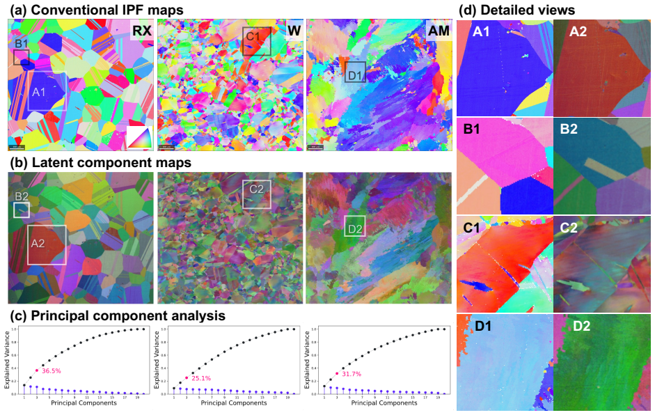
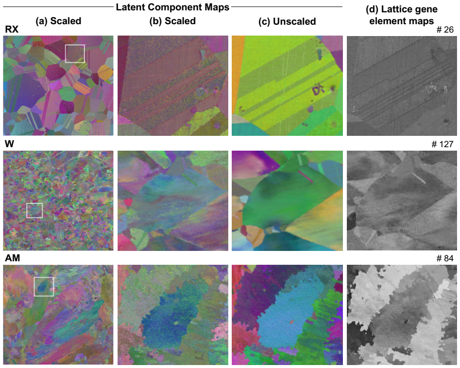
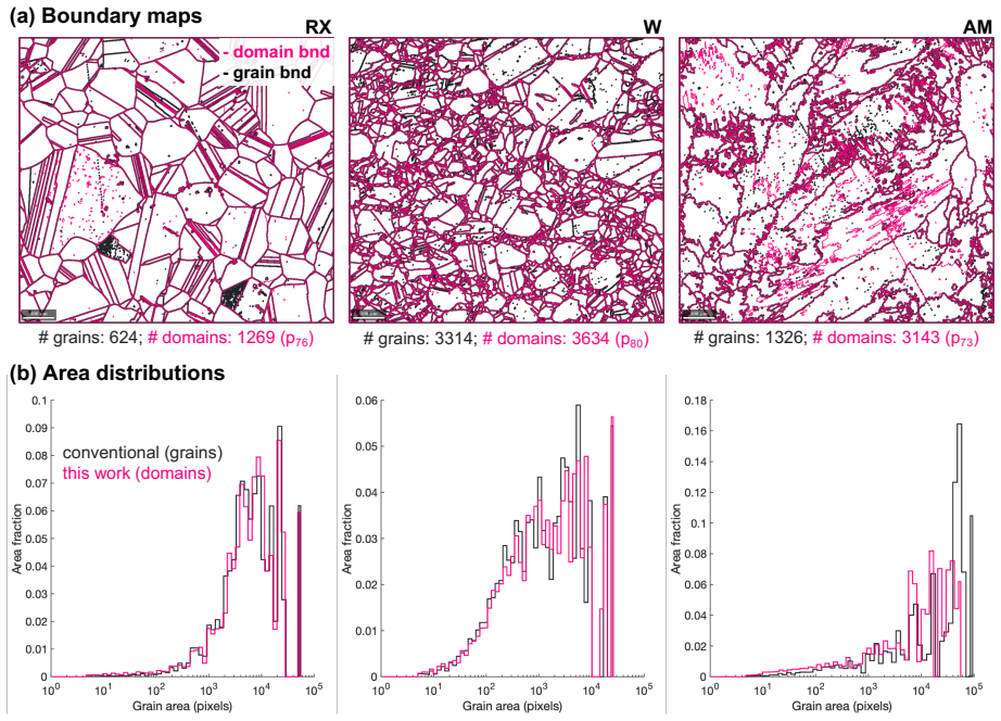
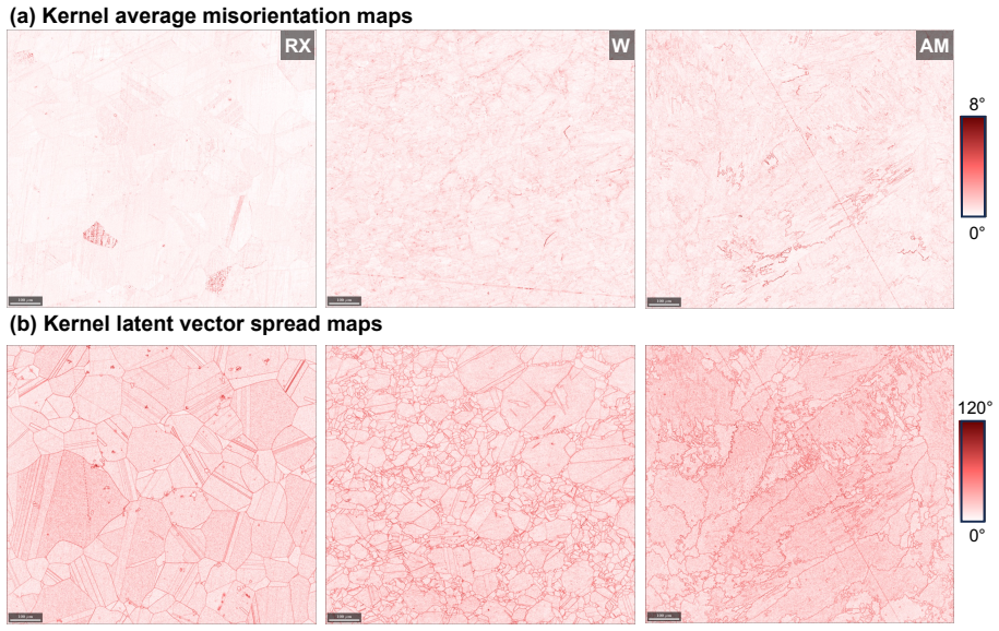
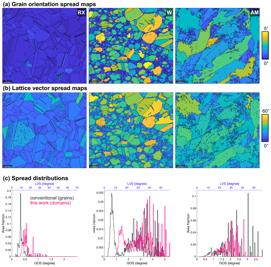
The lattice gene is a compact variational-autoencoder encoding of Kikuchi diffraction patterns that is experimentally accessible, admits a distance metric reflecting structural similarity, and retains enough information to reconstruct the original patterns. The lattice genome is the spatially resolved collection of lattice genes across an EBSD-mapped area; latent-component maps, distance- and angle-based segmentation, and kernel/domain latent-vector spreads derived from it quantify grain-scale and intragranular heterogeneity in additively manufactured and wrought Ni-base superalloys.
What carries the argument
Variational autoencoder latent encoding of Kikuchi diffraction patterns, which produces a low-dimensional vector space used for distance calculations, reconstruction, and spatial mapping.
If this is right
- Latent component maps visualize both grain-scale boundaries and intra-grain orientation variations without explicit indexing.
- Domain segmentation partitions the microstructure using only latent-space distance and angle thresholds.
- Kernel and domain latent-vector spreads serve as high-dimensional generalizations of kernel average misorientation and grain orientation spread.
- All three quantities are validated on additively manufactured and recrystallized Ni-base superalloy samples.
Where Pith is reading between the lines
- The same latent representation could be applied to other diffraction modalities such as transmission Kikuchi diffraction or X-ray microdiffraction to produce comparable genomes.
- If the latent distance correlates with local property variations, the genome could serve as input for spatially resolved property prediction without separate crystal-plasticity simulations.
- The compactness of the gene may allow storage and comparison of large EBSD datasets across processing conditions or alloy variants at lower computational cost than raw pattern storage.
Load-bearing premise
The variational autoencoder produces a latent space in which Euclidean or angular distances between vectors correspond to actual structural similarity between crystalline lattices.
What would settle it
A direct test showing that two lattices known from independent indexing to differ by a large misorientation angle receive a small latent-space distance, or that reconstructed patterns deviate visibly from the measured Kikuchi patterns.
Figures


read the original abstract
Inspired by the concept of a generalized materials genome, we introduce the notions of lattice gene and lattice genome for crystalline materials. A lattice gene is a compact representation of the local crystalline structure obtained by encoding the Kikuchi diffraction patterns with a variational autoencoder. We show that this representation satisfies key criteria for a materials gene: compactness, experimental accessibility, existence of a distance metric reflecting structural similarity, and sufficient information content for reconstructing the original diffraction patterns. The lattice genome is the spatially resolved collection of lattice genes across a representative area mapped by electron backscatter diffraction (EBSD), which captures mesoscale heterogeneity that ultimately controls properties. We demonstrate three applications of the lattice genome: (i) latent component maps that visualize grain-scale and intra-grain heterogeneities, (ii) domain segmentation based on distance and angle metrics in the latent space, and (iii) kernel and domain latent vector spreads that quantify intragranular heterogeneity as high-dimensional analogs of kernel average misorientation and grain orientation spread. All three tools are validated on microstructures of additively manufactured and wrought Ni-base superalloys in as-built and recrystallized conditions.
Editorial analysis
A structured set of objections, weighed in public.
Referee Report
Summary. The paper proposes a 'lattice gene' as a compact variational autoencoder (VAE) encoding of Kikuchi diffraction patterns from EBSD, and a 'lattice genome' as the spatially resolved collection of these encodings. It claims this representation meets four criteria for a materials gene (compactness, experimental accessibility, existence of a distance metric reflecting structural similarity, and sufficient information content to reconstruct original patterns) and demonstrates three applications—latent component maps, distance-based domain segmentation, and quantification of intragranular heterogeneity via kernel/domain spreads—on additively manufactured and wrought Ni-base superalloy microstructures.
Significance. If the central claims hold, particularly that Euclidean distances in the VAE latent space correspond to crystallographic structural similarity, the lattice genome could offer a data-driven approach to quantifying mesoscale heterogeneity beyond conventional orientation metrics, with potential utility in processing-structure-property studies of complex alloys. The work applies the method to real EBSD datasets from as-built and recrystallized conditions, which is a strength.
major comments (3)
- [Abstract, §3] Abstract and §3 (results on applications): The claim that the representation satisfies the criterion of 'existence of a distance metric reflecting structural similarity' is load-bearing for the domain segmentation and heterogeneity quantification applications, yet no quantitative validation is provided (e.g., no reported correlation coefficients, R² values, or comparisons between latent-space distances and independent crystallographic metrics such as misorientation angles or pattern similarity scores from the original EBSD indexing).
- [Abstract, methods/results] Abstract and methods/results on VAE: The assertion of 'sufficient information content for reconstructing the original diffraction patterns' is stated without accompanying quantitative metrics (e.g., reconstruction error, PSNR, or pattern fidelity scores) or details on how reconstruction quality was assessed across the dataset.
- [§3.2] §3.2 (domain segmentation): The segmentation relies on distance and angle metrics in latent space, but without an independent validation against ground-truth microstructural features (such as grain boundaries identified by conventional Hough-based indexing), it is unclear whether the latent-space distances capture physically meaningful similarity rather than VAE artifacts.
minor comments (2)
- [methods] Notation for the latent vectors and distance metrics could be clarified with explicit definitions or equations in the methods section to aid reproducibility.
- [figures] Figure captions for the latent component maps and segmentation results should include scale bars, colorbar units, and sample identifiers for clarity.
Simulated Author's Rebuttal
We thank the referee for their constructive and detailed review. The comments highlight important areas where additional quantitative support can strengthen the central claims. We address each major comment below and have revised the manuscript accordingly to incorporate the requested validations.
read point-by-point responses
-
Referee: [Abstract, §3] Abstract and §3 (results on applications): The claim that the representation satisfies the criterion of 'existence of a distance metric reflecting structural similarity' is load-bearing for the domain segmentation and heterogeneity quantification applications, yet no quantitative validation is provided (e.g., no reported correlation coefficients, R² values, or comparisons between latent-space distances and independent crystallographic metrics such as misorientation angles or pattern similarity scores from the original EBSD indexing).
Authors: We agree that quantitative validation is needed to support this criterion. The revised manuscript includes a new analysis in the methods and a supplementary figure that directly compares Euclidean distances in the VAE latent space to misorientation angles and pattern similarity scores computed from the original EBSD data. The results are discussed in §3 to show that the distance metric aligns with established crystallographic measures. revision: yes
-
Referee: [Abstract, methods/results] Abstract and methods/results on VAE: The assertion of 'sufficient information content for reconstructing the original diffraction patterns' is stated without accompanying quantitative metrics (e.g., reconstruction error, PSNR, or pattern fidelity scores) or details on how reconstruction quality was assessed across the dataset.
Authors: We concur that explicit quantitative metrics are required. The revised version reports the mean squared reconstruction error and PSNR values evaluated on the full dataset, together with representative original-versus-reconstructed pattern pairs, in the methods section. These additions confirm the reconstruction fidelity and support the information-content claim. revision: yes
-
Referee: [§3.2] §3.2 (domain segmentation): The segmentation relies on distance and angle metrics in latent space, but without an independent validation against ground-truth microstructural features (such as grain boundaries identified by conventional Hough-based indexing), it is unclear whether the latent-space distances capture physically meaningful similarity rather than VAE artifacts.
Authors: We recognize the value of independent validation. In the revised §3.2 we have added a direct comparison of the latent-space domain boundaries against grain boundaries obtained from conventional Hough-based indexing using a standard 5° misorientation threshold. The overlap is quantified and shown in an updated figure, indicating that the segmentation aligns with physically meaningful features. revision: yes
Circularity Check
No significant circularity in derivation chain
full rationale
The paper introduces a VAE-based encoding of Kikuchi patterns as a new 'lattice gene' representation and demonstrates its use for visualization, segmentation, and heterogeneity quantification on EBSD data from Ni superalloys. The central claim that the latent-space distance metric reflects structural similarity is asserted as one of four satisfied criteria and is used to enable the applications, but the provided text shows no reduction of this property to a fitted parameter, self-definition, or self-citation chain by construction. No equations or steps equate a prediction to its own inputs, and the method remains self-contained against external benchmarks without load-bearing self-referential definitions.
Axiom & Free-Parameter Ledger
Forward citations
Cited by 1 Pith paper
-
Neural electron backscatter diffraction
Neural EBSD models EBSD data as continuous 4D fields with joint and factorized neural formulations, achieving sub-1% reconstruction error and high compression while supporting continuous analysis.
Reference graph
Works this paper leans on
-
[1]
url: https://www.mgi.gov/
Materials genome initiative website. url: https://www.mgi.gov/. 14 (a) (c)(b)W AMRX Figure 8: Distributions ofAandDfor neighboring pixels. The shaded regions shows the percentile range used for threshold selection. (a) (c)(b)W AMRX p76 p80 p73 Figure 9: WCSS vs. the number of grains
-
[2]
J. J. de Pablo, N. E. Jackson, M. A. Webb, L.-Q. Chen, J. E. Moore, D. Morgan, R. Jacobs, T. Pollock, D. G. Schlom, E. S. Toberer, et al., New frontiers for the ma- terials genome initiative, npj Computational Materials 5 (1) (2019) 41
2019
-
[3]
S. J. Billinge, Do materials have a genome, and if they do, what can be done with it?, Matter 7 (11) (2024) 3714–3727
2024
-
[4]
B. L. Adams, S. R. Kalidindi, D. T. Fullwood, Mi- crostructure sensitive design for performance optimiza- tion, Butterworth-Heinemann, 2012
2012
-
[5]
S. R. Kalidindi, Hierarchical materials informatics: novel analytics for materials data, Elsevier, 2015
2015
-
[6]
A. J. Schwartz, M. Kumar, B. L. Adams, D. P. Field, Electron backscatter diffraction in materials science, Vol. 2, Springer, 2009
2009
-
[7]
B. L. Adams, S. I. Wright, K. Kunze, Orientation imag- ing: the emergence of a new microscopy, Metallurgical Transactions A 24 (4) (1993) 819–831
1993
-
[8]
Michael, R
J. Michael, R. Goehner, Crystallographic phase identi- fication in the scanning electron microscope: Backscat- tered electron kikuchi patterns, Tech. rep., Sandia Na- tional Labs., Albuquerque, NM (United States) (1992)
1992
-
[9]
M. M. Nowell, S. I. Wright, Phase differentiation via combined ebsd and xeds, Journal of microscopy 213 (3) (2004) 296–305
2004
-
[10]
S. I. Wright, M. M. Nowell, S. P. Lindeman, P. P. Ca- mus, M. De Graef, M. A. Jackson, Introduction and comparison of new ebsd post-processing methodologies, Ultramicroscopy 159 (2015) 81–94
2015
-
[11]
Y. H. Chen, S. U. Park, D. Wei, G. Newstadt, M. A. Jackson, J. P. Simmons, M. De Graef, A. O. Hero, A dictionary approach to electron backscatter diffraction indexing, Microscopy and Microanalysis 21 (3) (2015) 739–752
2015
-
[12]
A. J. Wilkinson, G. Meaden, D. J. Dingley, High- resolution elastic strain measurement from electron backscatter diffraction patterns: New levels of sensi- tivity, Ultramicroscopy 106 (4-5) (2006) 307–313
2006
-
[13]
Pantleon, Resolving the geometrically necessary dis- location content by conventional electron backscatter- ing diffraction, Scripta Materialia 58 (11) (2008) 994– 997
W. Pantleon, Resolving the geometrically necessary dis- location content by conventional electron backscatter- ing diffraction, Scripta Materialia 58 (11) (2008) 994– 997
2008
-
[14]
Wang, J.-C
F. Wang, J.-C. Stinville, M. Charpagne, M. P. Ech- lin, S. R. Agnew, T. M. Pollock, M. De Graef, D. S. Gianola, Dislocation cells in additively manufactured 15 (a) Angle-based kernel latent vector spread (b) Distance-based kernel latent vector spread 0° 120° 0 12 WRX AM Figure 10: Kernel latent vector spread based on cosine angle (a) and Euclidean distanc...
2023
-
[15]
Calvat, C
M. Calvat, C. Bean, D. Anjaria, H. Park, H. Wang, K. Vecchio, J. Stinville, Learning metal microstructural heterogeneity through spatial mapping of diffraction la- tent space features, npj Computational Materials 11 (1) (2025) 284
2025
-
[16]
A. J. Wilkinson, D. M. Collins, Y. Zayachuk, R. Ko- rla, A. Vilalta-Clemente, Applications of multivariate statistical methods and simulation libraries to anal- ysis of electron backscatter diffraction and transmis- sion kikuchi diffraction datasets, Ultramicroscopy 196 (2019) 88–98
2019
-
[17]
Bonnet, Multivariate statistical methods for the analysis of microscope image series: applications in ma- terials science, Journal of Microscopy 190 (1-2) (1998) 2–18
N. Bonnet, Multivariate statistical methods for the analysis of microscope image series: applications in ma- terials science, Journal of Microscopy 190 (1-2) (1998) 2–18
1998
-
[18]
L. N. Brewer, P. G. Kotula, J. R. Michael, Multivariate statistical approach to electron backscattered diffrac- tion, Ultramicroscopy 108 (6) (2008) 567–578
2008
-
[19]
S. I. Wright, M. M. Nowell, R. de Kloe, P. Camus, T. Rampton, Electron imaging with an ebsd detector, Ultramicroscopy 148 (2015) 132–145
2015
-
[20]
T. P. McAuliffe, D. Dye, T. B. Britton, Spherical- angular dark field imaging and sensitive microstructural phase clustering with unsupervised machine learning, Ultramicroscopy 219 (2020) 113132
2020
-
[21]
Chauniyal, P
A. Chauniyal, P. Thome, M. Stricker, Employing constrained nonnegative matrix factorization for mi- crostructure segmentation, Microscopy and Microanal- ysis 30 (4) (2024) 712–723
2024
-
[22]
Z. T. Varley, G. S. Rohrer, M. De Graef, Accelerating dictionary indexing of electron backscatter diffraction patterns with pca and quantization, Scientific Reports
-
[23]
Kaufmann, C
K. Kaufmann, C. Zhu, A. S. Rosengarten, D. Maryanovsky, T. J. Harrington, E. Marin, K. S. Vecchio, Crystal symmetry determination in electron diffraction using machine learning, Science 367 (6477) (2020) 564–568
2020
-
[24]
Z. Ding, E. Pascal, M. De Graef, Indexing of electron back-scatter diffraction patterns using a convolutional neural network, Acta Materialia 199 (2020) 370–382
2020
-
[25]
Q. Lu, X. Cai, J. Wu, S. Zhang, S. Liu, X. Jin, Crystal orientation and deformation state analysis from kikuchi patterns via pattern reconstruction aided deep siamese network, Materials & Design 230 (2023) 111998
2023
-
[26]
D. P. Kingma, M. Welling, Auto-encoding variational bayes, arXiv preprint arXiv:1312.6114
-
[27]
Vizoso, R
D. Vizoso, R. Dingreville, Decoding diffraction and spectroscopy data with machine learning: A tutorial, Journal of Applied Physics 137 (13)
-
[28]
Liu, C.-K
Y.-C. Liu, C.-K. Yeh, S.-P. Tsai, P.-Y. Tung, Learning crystallographic orientations from electron backscatter diffraction patterns using variational autoencoder, Cell Reports Physical Science 6 (10)
-
[29]
Bachmann, R
F. Bachmann, R. Hielscher, H. Schaeben, Grain detec- tion from 2d and 3d ebsd data—specification of the mtex algorithm, Ultramicroscopy 111 (12) (2011) 1720– 1733. 16
2011
-
[30]
MacQueen, Multivariate observations, in: Proceed- ings ofthe 5th Berkeley symposium on mathematical statisticsand probability, Vol
J. MacQueen, Multivariate observations, in: Proceed- ings ofthe 5th Berkeley symposium on mathematical statisticsand probability, Vol. 1, University of Califor- nia press Oakland, CA, USA, 1967, pp. 281–297
1967
-
[31]
Y. Yao, L. Rosasco, A. Caponnetto, On early stopping in gradient descent learning, Constructive approxima- tion 26 (2) (2007) 289–315
2007
-
[32]
Prechelt, Early stopping-but when?, in: Neural Net- works: Tricks of the trade, Springer, 2002, pp
L. Prechelt, Early stopping-but when?, in: Neural Net- works: Tricks of the trade, Springer, 2002, pp. 55–69
2002
-
[33]
M. Calvat, D. Anjaria, H. Wang, K. Vecchio, J.-C. Stinville, Kikuchi pattern dataset from wrought and as- built additively manufactured superalloys (Sep. 2025). doi:10.5061/DRYAD.ZCRJDFNR9
-
[34]
M. I. Latypov, S. R. Kalidindi, Data-driven reduced or- der models for effective yield strength and partitioning of strain in multiphase materials, Journal of Computa- tional Physics 346 (2017) 242–261
2017
-
[35]
S. I. Wright, M. M. Nowell, D. P. Field, A review of strain analysis using electron backscatter diffraction, Microscopy and microanalysis 17 (3) (2011) 316–329
2011
-
[36]
T. B. Britton, J. Jiang, Y. Guo, A. Vilalta-Clemente, D. Wallis, L. N. Hansen, A. Winkelmann, A. J. Wilkin- son, Tutorial: Crystal orientations and ebsd—or which way is up?, Materials Characterization 117 (2016) 113– 126
2016
-
[37]
Humphreys, Review grain and subgrain character- isation by electron backscatter diffraction, Journal of materials science 36 (16) (2001) 3833–3854
F. Humphreys, Review grain and subgrain character- isation by electron backscatter diffraction, Journal of materials science 36 (16) (2001) 3833–3854
2001
-
[38]
Zaefferer, On the formation mechanisms, spatial res- olution and intensity of backscatter kikuchi patterns, Ultramicroscopy 107 (2-3) (2007) 254–266
S. Zaefferer, On the formation mechanisms, spatial res- olution and intensity of backscatter kikuchi patterns, Ultramicroscopy 107 (2-3) (2007) 254–266
2007
-
[39]
C. Zhu, M. De Graef, Ebsd pattern simulations for an interaction volume containing lattice defects, Ultrami- croscopy 218 (2020) 113088
2020
-
[40]
Burgess, J
J. Burgess, J. J. Nirschl, M.-C. Zanellati, A. Lozano, S. Cohen, S. Yeung-Levy, Orientation-invariant autoen- coders learn robust representations for shape profiling of cells and organelles, Nature Communications 15 (1) (2024) 1022
2024
-
[41]
Y. Wang, M. I. Jordan, Desiderata for representation learning: A causal perspective, Journal of Machine Learning Research 25 (275) (2024) 1–65
2024
-
[42]
Locatello, S
F. Locatello, S. Bauer, M. Lucic, G. Raetsch, S. Gelly, B. Sch¨ olkopf, O. Bachem, Challenging common as- sumptions in the unsupervised learning of disentangled representations, in: international conference on ma- chine learning, PMLR, 2019, pp. 4114–4124
2019
-
[43]
M. I. Latypov, M. K¨ uhbach, I. J. Beyerlein, J.-C. Stinville, L. S. Toth, T. M. Pollock, S. R. Kalidindi, Application of chord length distributions and principal component analysis for quantification and representa- tion of diverse polycrystalline microstructures, Materi- als Characterization 145 (2018) 671–685
2018
-
[44]
S. E. Whitman, M. I. Latypov, SR-CLD: Spatially re- solved chord length distributions for statistical descrip- tion and visualization of non-uniform microstructures, Metallurgical and Materials Transactions A 56 (11) (2025) 5038–5047
2025
-
[45]
Field, P
D. Field, P. Trivedi, S. Wright, M. Kumar, Analysis of local orientation gradients in deformed single crystals, Ultramicroscopy 103 (1) (2005) 33–39
2005
-
[46]
N. H. Paulson, M. W. Priddy, D. L. McDowell, S. R. Ka- lidindi, Reduced-order structure-property linkages for polycrystalline microstructures based on 2-point statis- tics, Acta Materialia 129 (2017) 428–438
2017
-
[47]
B. L. Adams, T. Olson, The mesostructure—properties linkage in polycrystals, Progress in Materials Science 43 (1) (1998) 1–87
1998
-
[48]
Richter, F
A. Richter, F. Scholz, G. Eggeler, J. Frenzel, P. Thome, Microstructure informatics: Using computer vision for the characterization of dendrite growth phenomena in ni-base single crystal superalloys, Materials Character- ization 223 (2025) 114878
2025
-
[49]
Thome, L
P. Thome, L. F. Arciniaga, C. F. Hohenadel, O. Lowery, S. Tin, Neural network modeling of process–structure relationships and abnormal grain growth in polycrys- talline ni-base superalloys through domain knowledge- informed feature engineering, Metallurgical and Mate- rials Transactions A (2026) 1–23. 17
2026
discussion (0)
Sign in with ORCID, Apple, or X to comment. Anyone can read and Pith papers without signing in.