REVIEW 4 major objections 6 minor 94 references
Nonresonant optomechanical control of structural phases
T0 review · 4 major / 6 minor · reviewed 2026-08-14 · deepseek-v4-flash
Pith's one-line read Intense mid-infrared light switches tin selenide into a higher-symmetry phase with no heating.
desk verdict First experimental evidence for MIR optomechanical structural switching in SnSe, with a real caveat: the symmetry change is inferred from Raman silence, not directly measured. read the letter →
The pith
A machine-rendered reading of the paper's core claim, the machinery that carries it, and where it could break.
The reading
What carries the argument
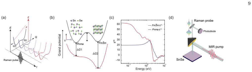
The central object is the optomechanical potential, $G_F(\xi,t)=U(\xi)-\tfrac{1}{2}\vec F^*(\omega_0,t)\cdot\varepsilon^{(1)}(\omega_0;\xi)\cdot\vec F(\omega_0,t)\,V_p$, where $\xi$ is a generalized coordinate along the Pnma-to-Fm3m transition path, $U(\xi)$ is the equilibrium potential, $\varepsilon^{(1)}$ is the real part of the dielectric tensor at the mid-infrared frequency, $\vec F$ is the applied field, and $V_p$ is the unit-cell volume. The mechanism works because the two phases have very different refractive indices, so the field lowers the grand potential of Fm3m more than that of Pnma and reduces the barrier between them; above about 1.1 V/nm in the calculation the barrier vanishes entirely. The same object connects the approach to ordinary impulsive stimulated Raman scattering, since a parabolic $U(\xi)$ reduces the optomechanical force to the standard ISRS expression. The experiments use time-domain Raman scattering (displacive excitation of coherent $A_g$ modes by 800-nm pulses) as the symmetry-sensitive probe of whether the switch has occurred.
What would settle it
Perform femtosecond electron or X-ray diffraction on SnSe under the same 5-µm, ~0.6 V/nm excitation: if the Bragg-peak pattern remains orthorhombic Pnma while the time-domain Raman spectrum loses all four $A_g$ modes, then the Raman suppression is not caused by a structural phase transition; appearance of diffraction peaks consistent with a cubic or higher-symmetry cell would confirm the assignment.
Extended reading notes
Core claim
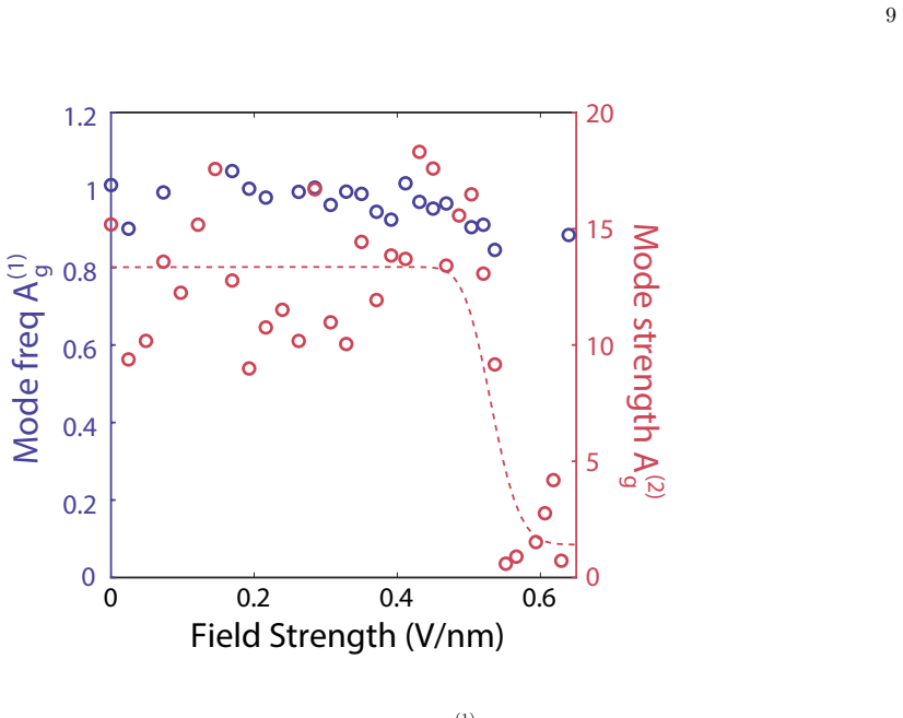
The paper claims that intense, sub-picosecond mid-infrared pulses at 5-µm wavelength act as optical tweezers for the lattice of tin selenide, driving it across a structural phase boundary from the ambient orthorhombic Pnma phase toward the higher-symmetry Fm3m rocksalt structure. The evidence is a sharp threshold in the time-domain Raman response: above an incident field strength of roughly 0.6 V/nm, all four $A_g$ Raman modes are suppressed abruptly and almost completely within a few picoseconds, and this happens without any preceding softening of the mode frequencies. That absence of softening is what separates the observed phase from the thermally driven second-order transition to Cmcm and from the near-infrared-driven distortion toward Immm, both of which show clear softening. The switched state shows a reflectivity enhancement larger than 10 percent at 1520 nm that persists up to about 0.24 ms, and the paper reports that atom-probe measurements under a DC field of about 16 V/nm show a related transformation to a higher-symmetry phase with changed bond-rupture statistics. The authors conclude that a nonresonant optomechanical force, proportional to the derivative of the real dielectric function with respect to a phonon coordinate times the field intensity, can stabilize a metastable phase and perform a diffusionless, low-energy structural switch.
Load-bearing premise
The inference that the sudden loss of $A_g$ Raman oscillations reflects a symmetry-changing structural transition assumes that the 800-nm time-domain Raman probe still reports $A_g$ mode activity faithfully in the mid-infrared-excited state; if the mid-infrared pulse instead creates a strongly absorbing or screened near-surface layer that suppresses the probe, the modes could appear to vanish without any bulk phase change.
Editorial extensions
If this is right
- Above roughly 0.6 V/nm of 5-µm mid-infrared excitation, SnSe switches within a few picoseconds to a metastable phase in which all four $A_g$ Raman modes are silent; the paper identifies this as a transition toward the higher-symmetry Fm3m rocksalt structure.
- The switched state is distinct from thermally induced and carrier-induced states because no phonon softening precedes the mode disappearance, ruling out a second-order transition to Cmcm or a small Immm distortion.
- The phase exhibits a reflectivity enhancement above 10 percent that lives for up to about 0.24 ms, long enough for device readout, and the sample survives 100,000 excitation cycles with little fatigue.
- The estimated switching energy density, about $3\times10^{-3}$ aJ/nm$^3$, is several orders of magnitude below the energy used in thermal phase-change materials such as Ge2Sb2Te5.
- A DC electric field of roughly 16 V/nm produces an analogous transformation to a higher-symmetry phase in atom probe tomography, evidenced by changes in bond-rupture statistics and a sixfold detector histogram.
Reading between the lines
- If the mechanism is general, any material with a large refractive-index contrast between two polymorphs should be switchable at similarly low energy densities; screening the real-part dielectric contrast of candidate phase-change materials could predict new optomechanical switches before any laser work.
- The lifetime of the switched state increases with field strength in the paper's data, so pulse-shaping or two-color excitation might trade off threshold against retention; extending the slow decay from 0.24 ms toward seconds would make the effect useful for non-volatile storage, a direction the paper leaves open.
- A direct structural probe, such as femtosecond electron or X-ray diffraction, would test the phase assignment independently of the Raman probe and could reveal whether the transition passes through the metallic state the authors say the theory predicts en route to Fm3m.
- The same optomechanical term, $\tfrac{1}{2}\partial\varepsilon/\partial\xi\,|F|^2 V_p$, should apply to other collective coordinates such as ferroelectric polarization or magnetic order, suggesting a unified nonresonant route to coupled order parameters that the authors do not develop.
Signed reviews
Editorial analysis
A structured set of objections, weighed in public.
Referee Report
Summary. The paper reports experiments and calculations on SnSe in which intense 5-µm mid-infrared pulses are claimed to drive a nonthermal, nonresonant structural phase transition from the orthorhombic Pnma phase toward a higher-symmetry (Fm-3m or Immm) phase. The main optical evidence is an abrupt suppression of the A_g coherent-phonon oscillations in time-domain Raman scattering above a critical field strength of about 0.6 V/nm, without the softening seen in heating or near-infrared controls, together with a long-lived (>10%, millisecond) reflectivity enhancement. Supporting evidence is provided by atom probe tomography, where a field-dependent probability of multiple events is interpreted as a DC-field-driven transition to a higher-symmetry phase at about 15.8 V/nm. Density functional theory calculations of the field-dependent grand potential are used to argue that the Pnma-to-Fm-3m barrier is lowered by the optical field, enabling a barrierless transition at higher fields.
Significance. If the central inference is correct, this work would demonstrate a genuinely new route to structural phase control: using nonresonant, dispersive optomechanical forces rather than resonant absorption or nonlinear phononics, with potentially very low energy cost and ultrafast response. The paper combines several complementary techniques (transient reflectivity, time-domain Raman, APT, DFT) and includes parameter-free first-principles calculations that are not fit to the observed threshold, which is a strength. The long-lived reflectivity signal and the fatigue resistance are also notable. However, the structural assignment relies on indirect, symmetry-based reasoning from Raman mode suppression, and the APT evidence is statistically fragile; both need to be strengthened before the conclusions can be considered established.
major comments (4)
- [Section IV, Fig. 2c,d] The central claim that the abrupt loss of all A_g Raman oscillations signals a symmetry-changing structural transition assumes that the 800-nm time-domain Raman probe faithfully reports A_g mode activity in the MIR-excited state. This is not demonstrated. Section III describes a 'huge background signal' and Fig. 3 shows a >10% reflectivity enhancement and millisecond-lived optical-constant changes, so the 800-nm field penetration, the displacive excitation efficiency, and the probe collection efficiency can all be strongly modified in the high-field state. A MIR-generated absorbing or screened near-surface layer would suppress coherent-phonon excitation even if the bulk crystal symmetry is unchanged. The paper provides no high-field-state calibration of the probe, no surviving reference Raman mode (e.g., a B_g mode that would remain active in the proposed phase), and no direct structural probe. Without such a control, the mode-suppression data are consistent with a genuine symmetry change but do not uniquely establish one.
- [Section IV and Supplementary Note II] The Raman data cannot distinguish between the Pnma-to-Fm-3m and Pnma-to-Immm transitions, because the authors themselves state (Supplementary Note II) that all four A_g modes become Raman inactive in both higher-symmetry phases. The abstract and introduction specifically invoke the Fm-3m topological crystalline insulator phase, and Fig. 1 and Section VII model the transition to Fm-3m, but the experimental Raman mode suppression is equally consistent with Immm or an average structure of even lower symmetry that breaks the Raman activity. The conclusion should either be restricted to 'a higher-symmetry phase whose identity is not uniquely determined' or supported by a direct structural measurement, such as time-resolved X-ray or electron diffraction.
- [Section VI, Fig. 4] The APT evidence for a DC-field-driven phase transition is a post hoc two-parabola fit to the probability of multiple events (PME) versus field, with voltage-mode data points excluded from the fits because 'the evaporation mechanism is different.' The transition at about 15.8 V/nm is inferred from the crossing of the two fitted parabolas, but no statistical justification is given for preferring two parabolas over a single smooth curve (the quoted R² values of 0.98 versus 0.69 do not account for the additional parameters), and the voltage-mode exclusion is not justified quantitatively. Furthermore, at high field the PME exceeds 90%, which the authors state degrades the detector-histogram resolution; the claimed 'sixfold symmetry' in laser-assisted mode is therefore not robust. These data are suggestive, but they cannot serve as independent confirmation of the optomechanical mechanism without additional structural evidence or a more rigorous analysis.
- [Section VII, Fig. 5] The theoretical modeling is described as predicting that the Pnma-to-Fm-3m barrier vanishes at approximately 1.1 V/nm, while the experimental mode-suppression threshold is approximately 0.6 V/nm and the free-energy crossover occurs at about 0.5 V/nm. The paper only claims 'qualitative agreement' (Supplementary Fig. 21c) but does not discuss the factor-of-two discrepancy between the theoretical barrierless field and the measured threshold. If the theory is being used to support the interpretation that the observed transition is the predicted optomechanical one, the mismatch should be addressed explicitly, for example by considering local-field enhancements, finite-temperature effects, or the influence of the 800-nm Raman probe pulse on the effective potential.
minor comments (6)
- [Abstract] The phrase 'the A_g Raman modes disappear abruptly' is imprecise; the time-domain Raman data in Fig. 2d show suppression of three of the four A_g modes (A_g(1), A_g(2), A_g(4)), and the manuscript should state which modes are observed and which are not resolved.
- [Section IV] The sentence 'and allows us to infer the atomic transformation via the time domain response of associated vibrational modes' is a verbatim repetition from Section II and makes the text read awkwardly; it should be removed or rewritten.
- [Fig. 2b caption] The phrase 'less pronouced' is a typo and should read 'less pronounced'.
- [Section V] The text 'after 105 MIR excitation cycles' is missing a superscript; it should read 'after 10^5 MIR excitation cycles' (as elsewhere in Supplementary Fig. 16).
- [Section VI] The phrase 'as evidence by atom-probe techniques' should be 'as evidenced by atom-probe techniques'.
- [Supplementary Fig. 2 caption] The caption contains a subject-verb agreement error: 'the amplitude of the fast decay signal show' should be 'the amplitude of the fast decay signal shows'.
Circularity Check
No significant circularity: the central claim rests on parameter-free DFT, independent group-theoretical mode analysis, and direct time-domain Raman/APT measurements; self-citations are motivational rather than load-bearing.
full rationale
Walking the paper's derivation chain, the main claim is that MIR fields above a critical strength abruptly suppress the Ag Raman modes in SnSe and that this signals a symmetry-changing structural transition toward a higher-symmetry phase. The support for this is not definitionally tied to the measurements. First, the theoretical basis (Section VII and Supplementary Fig. 21) is a first-principles HSE-DFT calculation of the grand potential G_F(ξ,t) = U(ξ) − (1/2) F*(ω0,t)·ε^(1)(ω0;ξ)·F(ω0,t)V_p along the Pnma→Fm3m path; the parameters come from DFT-computed dielectric functions and potentials, not from fitting the experimental threshold. The paper's theory predicts energetic preference near ~0.5 V/nm and barrierless behavior near ~1.1 V/nm, while the observed Raman suppression occurs near ~0.6 V/nm; the theory is not tuned to reproduce that value. Second, the inference from mode disappearance to a symmetry change is grounded in an external group-theoretical fact (Supplementary Note II): all four Ag modes become inactive for the higher-symmetry phases, whereas only two vanish for the Cmcm phase. This is a mathematical implication about the probe, not an input that guarantees the observed suppression. Third, the prior self-citations (e.g., refs. [4,5]) motivate the optomechanical mechanism, but the present paper re-derives the relevant energetics with its own DFT calculations, so the citation chain is not load-bearing. The APT evidence uses a two-parabola fit to PME data, but this is descriptive data modeling rather than a derivation of the phase change, and the sixfold detector histogram supplies an independent symmetry indicator. Finally, the one place where an experimental value enters theory—Supplementary Note V, where the amplitude of the mode-strength coefficient is taken from measured Ag mode strengths—is used only to illustrate Raman force directions, not to predict the phase transition or the threshold. Possible concerns that the MIR pulse changes the sensitivity of the 800-nm time-domain Raman probe in the high-field state are experimental-control or interpretation risks, not circularity, because the paper does not define the structural phase in terms of that probe response. On the provided text, no claim reduces to its own inputs by construction, and no fitted parameter is renamed as a prediction.
Assumptions & free parameters
free parameters (4)
- Polynomial coefficients a1-a4 in U(xi)
- Polynomial coefficients b1-b5 in eps(xi)
- Phenomenological damping =
5e-13 s
- Raman-force mode weights w_n
assumptions (5)
- domain assumption DFT with the HSE hybrid functional gives quantitatively reliable dielectric functions and energy barriers for SnSe.
- domain assumption The Pnma-to-Fm3m transition can be captured by a single reaction coordinate xi along the calculated path.
- standard math All four Ag Raman modes become inactive in Fm3m and Immm, while two remain active in Cmcm.
- domain assumption The 800-nm displacive probe excites and detects Ag coherent phonons with constant efficiency regardless of MIR pre-excitation.
- domain assumption SnSe is transparent at 5 um, so MIR excitation is nonresonant and heating or carrier generation is negligible.
Cite this review
Pith. "Pith review of Nonresonant optomechanical control of structural phases." pith.science (2026). https://pith.science/paper/JMID26OE
@misc{pith2026260808899,
  author       = {Pith},
  title        = {Pith review of: Nonresonant optomechanical control of structural phases},
  year         = {2026},
  howpublished = {\url{https://pith.science/paper/JMID26OE}},
  note         = {Machine review of arXiv:2608.08899}
}
abstract
Optical tweezers demonstrate how light can exert forces to trap, repel, and manipulate microscopic particles without absorption. Recent theory has suggested that such forces can extend beyond particle manipulation to drive structural phase transitions in solids. Here we apply this optomechanical principle to tin selenide (SnSe), a material where proximity to several different structural phases gives rise to its high thermoelectric figure of merit and makes it a candidate for a switchable topological crystalline insulator. Whereas the force for standard optical tweezers arises from a gradient in the intensity of a light field, the optomechanical force is mediated by a gradient in the dielectric constant as a function of phonon coordinate. Unlike conventional methods that rely on resonant excitation and absorption through the imaginary part of the dielectric function, this approach operates dispersively through the real part and can be directly driven by Raman processes, enabling selective transitions with reduced energy cost and ultrafast response. Using time-domain Raman scattering, we show that above a critical mid-infrared field strength the $A_g$ Raman modes disappear abruptly without softening, signaling the formation of a new structural phase. This phase, distinct from those induced by heating or carrier excitation, exhibits large-amplitude and long-lived modulations in its optical response. Complementing this observation, we show also evidence for an equivalent DC-field-driven structural phase transformation to a higher symmetry phase, as observed by atom probe tomography. Our study demonstrates the concept of nonresonant optomechanical phase control and defines novel opportunities for synthesizing hidden structural phases with unique functional properties.
Figures


Figures from the paper (26 more)




Reference graph
Works this paper leans on
-
[1]
Ashkin, Acceleration and trapping of particles by ra- diation pressure, Phys
A. Ashkin, Acceleration and trapping of particles by ra- diation pressure, Phys. Rev. Lett.24, 156 (1970)
1970
-
[2]
Ashkin, J
A. Ashkin, J. M. Dziedzic, J. E. Bjorkholm, and S. Chu, Observation of a single-beam gradient force optical trap for dielectric particles, Opt. Lett.11, 288 (1986)
1986
-
[3]
S. M. Block, L. S. B. Goldstein, and B. J. Schnapp, Bead movement by single kinesin molecules studied with opti- cal tweezers, Nature348, 348 (1990)
1990
-
[4]
J. Zhou, H. Xu, Y. Li, R. Jaramillo, and J. Li, Opto- mechanics driven fast martensitic transition in two- dimensional materials, Nano Lett.18, 7794 (2018)
2018
-
[5]
J. Zhou, S. Zhang, and J. Li, Normal-to-topological insulator martensitic phase transition in group-IV monochalcogenides driven by light, NPG Asia Mater.12, 2 (2020)
2020
-
[6]
J. Shi, C. Heide, H. Xu, Y. Shen, M. Henstridge, I. Sedwick, A. Mangu, X. Peng, S. Zhang, M. Trigo, T. F. Heinz, J. Li, K. A. Nelson, E. Baldini, J. Zhou, S. Ghimire, D. A. Reis, and A. M. Lindenberg, Non- resonant raman control of ferroelectric polarization, Ad- vanced Materialsn/a, e10524
-
[7]
Fausti, R
D. Fausti, R. I. Tobey, N. Dean, S. Kaiser, A. Dienst, M. C. Hoffmann, S. Pyon, T. Takayama, H. Takagi, and A. Cavalleri, Light-induced superconductivity in a stripe- ordered cuprate, Science331, 189 (2011)
2011
-
[8]
E. J. Sie, C. M. Nyby, C. D. Pemmaraju, S. J. Park, X. Shen, J. Yang, M. C. Hoffmann, B. K. Ofori-Okai, R. Li, A. H. Reid, S. Weathersby, E. Mannebach, N. Finney, D. Rhodes, D. Chenet, A. Antony, L. Bali- cas, J. Hone, T. P. Devereaux, T. F. Heinz, X. Wang, and A. M. Lindenberg, An ultrafast symmetry switch in a Weyl semimetal, Nature565, 61 (2019)
2019
Show all 94 references
-
[9]
T. F. Nova, A. S. Disa, M. Fechner, and A. Cavalleri, Metastable ferroelectricity in optically strained SrTiO3, Science364, 1075 (2019)
2019
-
[10]
X. Li, T. Qiu, J. Zhang, E. Baldini, J. Lu, A. M. Rappe, and K. A. Nelson, Terahertz field–induced ferroelectric- ity in quantum paraelectric SrTiO 3, Science364, 1079 (2019)
2019
-
[11]
M. Liu, H. Y. Hwang, H. Tao, A. C. Strikwerda, K. Fan, G. R. Keiser, A. J. Sternbach, K. G. West, S. Kitti- watanakul, J. Lu, S. A. Wolf, F. G. Omenetto, X. Zhang, K. A. Nelson, and R. D. Averitt, Terahertz-field-induced insulator-to-metal transition in vanadium dioxide meta- ...
2012
-
[12]
Baum, D.-S
P. Baum, D.-S. Yang, and A. H. Zewail, 4D visualiza- tion of transitional structures in phase transformations by electron diffraction, Science318, 788 (2007)
2007
-
[13]
Stojchevska, I
L. Stojchevska, I. Vaskivskyi, T. Mertelj, P. Kusar, D. Svetin, S. Brazovskii, and D. Mihailovic, Ultrafast switching to a stable hidden quantum state in an elec- tronic crystal, Science344, 177 (2014)
2014
-
[14]
Zhang, X
J. Zhang, X. Tan, M. Liu, S. W. Teitelbaum, K. W. Post, F. Jin, K. A. Nelson, D. N. Basov, W. Wu, and R. D. Averitt, Cooperative photoinduced metastable phase control in strained manganite films, Nat. Mater. 15, 956 (2016)
2016
-
[15]
V. A. Stoica, N. Laanait, C. Dai, Z. Hong, Y. Yuan, Z. Zhang, S. Lei, M. R. McCarter, A. Yadav, A. R. Damodaran, S. Das, G. A. Stone, J. Karapetrova, D. A. 9 Raman pulse E t Q Q1 Q2 ε MIR pump SnSe Raman probe Photodiode (d) Sn Se (b) /uni0394G1 /uni0394G2 a cb Pnma Fm3m Grand...
2019
-
[16]
Huang, S
Y. Huang, S. Yang, S. Teitelbaum, G. De la Pe˜ na, T. Sato, M. Chollet, D. Zhu, J. L. Niedziela, D. Bansal, A. F. May, A. M. Lindenberg, O. Delaire, D. A. Reis, and M. Trigo, Observation of a novel lattice instability in ultrafast photoexcited SnSe, Phys. Rev. X12, 011029 (2022)
2022
-
[17]
Schlauderer, C
S. Schlauderer, C. Lange, S. Baierl, T. Ebnet, C. P. Schmid, D. C. Valovcin, A. K. Zvezdin, A. V. Kimel, R. V. Mikhaylovskiy, and R. Huber, Temporal and spec- tral fingerprints of ultrafast all-coherent spin switching, Nature569, 383 (2019)
2019
-
[18]
Y. Yan, J. Gamble, Edward B., and K. A. Nelson, Impul- sive stimulated scattering: General importance in fem- tosecond laser pulse interactions with matter, and spec- troscopic applications, J. Chem. Phys.83, 5391 (1985)
1985
-
[19]
Fahy and R
S. Fahy and R. Merlin, Reversal of ferroelectric domains by ultrashort optical pulses, Phys. Rev. Lett.73, 1122 (1994)
1994
-
[20]
H. Xu, J. Zhou, Y. Li, R. Jaramillo, and J. Li, Op- tomechanical control of stacking patterns of h-bn bilayer, Nano Research12, 2634 (2019)
2019
-
[21]
Zhou and S
J. Zhou and S. Zhang, Terahertz optics-driven phase transition in two-dimensional multiferroics, npj 2D Ma- terials and Applications5, 16 (2021)
2021
-
[22]
J. Zhou, H. Xu, Y. Shi, and J. Li, Terahertz driven re- versible topological phase transition of monolayer transi- tion metal dichalcogenides, Advanced Science8, 2003832 (2021)
2021
-
[23]
J. Zhou, S. Mao, and S. Zhang, Noncontacting optostric- tion driven anisotropic and inhomogeneous strain in two- dimensional materials, Phys. Rev. Research2, 022059 (2020)
2020
-
[24]
Lai and C
A. Lai and C. A. Schuh, Direct electric-field induced phase transformation in paraelectric zirconia via electri- cal susceptibility mismatch, Phys. Rev. Lett.126, 015701 (2021)
2021
-
[25]
F¨ orst, C
M. F¨ orst, C. Manzoni, S. Kaiser, Y. Tomioka, Y. Tokura, R. Merlin, and A. Cavalleri, Nonlinear phononics as an ultrafast route to lattice control, Nature Physics7, 854 (2011)
2011
-
[26]
Wuttig and N
M. Wuttig and N. Yamada, Phase-change materials for rewriteable data storage, Nat. Mater.6, 824 (2007)
2007
-
[27]
Mujica, A
A. Mujica, A. Rubio, A. Mu˜ noz, and R. J. Needs, High-pressure phases of group-IV, III–V, and II–VI com- pounds, Rev. Mod. Phys.75, 863 (2003)
2003
-
[28]
L.-D. Zhao, G. Tan, S. Hao, J. He, Y. Pei, H. Chi, H. Wang, S. Gong, H. Xu, V. P. Dravid, C. Uher, G. J. Snyder, C. Wolverton, and M. G. Kanatzidis, Ultra- high power factor and thermoelectric performance in hole-doped single-crystal snse, Science351, 141 (2016), https://www.s...
2016 doi
-
[29]
B. Qin, M. G. Kanatzidis, and L.-D. Zhao, The development and impact of tin selenide on thermoelectrics, Science386, eadp2444 (2024), https://www.science.org/doi/pdf/10.1126/science.adp2444
2024 doi
-
[30]
Z. Wang, J. Wang, Y. Zang, Q. Zhang, J.-A. Shi, T. Jiang, Y. Gong, C.-L. Song, S.-H. Ji, L.-L. Wang, L. Gu, K. He, W. Duan, X. Ma, X. Chen, and Q.-K. Xue, Molecular beam epitaxy-grown SnSe in the rock- salt structure: An artificial topological crystalline insu- lator material,...
2015
-
[31]
W. Jin, S. Vishwanath, J. Liu, L. Kong, R. Lou, Z. Dai, J. T. Sadowski, X. Liu, H.-H. Lien, A. Chaney, Y. Han, M. Cao, J. Ma, T. Qian, S. Wang, M. Dobrowolska, J. Furdyna, D. A. Muller, K. Pohl, H. Ding, J. I. Dadap, H. G. Xing, and R. M. Osgood, Electronic structure of the me...
2017
-
[32]
Y. Sun, Z. Zhong, T. Shirakawa, C. Franchini, D. Li, Y. Li, S. Yunoki, and X.-Q. Chen, Rocksalt SnS and SnSe: Native topological crystalline insulators, Phys. Rev. B88, 235122 (2013)
2013
-
[33]
D. A. Rehn, Y. Li, E. Pop, and E. J. Reed, Theoretical potential for low energy consumption phase change mem- ory utilizing electrostatically-induced structural phase transitions in 2D materials, Npj Comput. Mater.4, 2 (2018)
2018
-
[34]
J. Yu, A. Yue, and O. Stafsudd, Growth and electronic properties of the SnSe semiconductor, J. Cryst. Growth 54, 248 (1981)
1981
-
[35]
H. R. Chandrasekhar, R. G. Humphreys, U. Zwick, and M. Cardona, Infrared and Raman spectra of the iv-vi compounds SnS and SnSe, Phys. Rev. B15, 2177 (1977). 12
1977
-
[36]
Kuramochi and T
H. Kuramochi and T. Tahara, Tracking ultrafast struc- tural dynamics by time-domain raman spectroscopy, J. Am. Chem. Soc.143, 9699 (2021)
2021
-
[37]
A. M. Weiner, D. E. Leaird, G. P. Wiederrecht, and K. A. Nelson, Femtosecond multiple-pulse impulsive stimulated raman scattering spectroscopy, J. Opt. Soc. Am. B8, 1264 (1991)
1991
-
[38]
H. J. Zeiger, J. Vidal, T. K. Cheng, E. P. Ippen, G. Dres- selhaus, and M. S. Dresselhaus, Theory for displacive excitation of coherent phonons, Phys. Rev. B45, 768 (1992)
1992
-
[39]
C. W. Li, J. Hong, A. F. May, D. Bansal, S. Chi, T. Hong, G. Ehlers, and O. Delaire, Orbitally driven giant phonon anharmonicity in SnSe, Nat. Phys.11, 1063 (2015)
2015
-
[40]
Zhang, H
J. Zhang, H. Zhu, X. Wu, H. Cui, D. Li, J. Jiang, C. Gao, Q. Wang, and Q. Cui, Plasma-assisted synthe- sis and pressure-induced structural transition of single- crystalline SnSe nanosheets, Nanoscale7, 10807 (2015)
2015
-
[41]
F. Liu, P. Parajuli, R. Rao, P. C. Wei, A. Karunarathne, S. Bhattacharya, R. Podila, J. He, B. Maruyama, G. Priyadarshan, J. R. Gladden, Y. Y. Chen, and A. M. Rao, Phonon anharmonicity in single-crystalline SnSe, Phys. Rev. B98, 224309 (2018)
2018
-
[42]
Shirane and Y
G. Shirane and Y. Yamada, Lattice-dynamical study of the 110 K phase transition in SrTiO 3, Phys. Rev.177, 858 (1969)
1969
-
[43]
L. D. Landau, E. M. Lifˇ sic, E. M. Lifshitz, and L. Pitaevskii,Statistical physics: theory of the condensed state, Vol. 9 (Butterworth-Heinemann, 1980)
1980
-
[44]
M. T. Dove, Theory of displacive phase transitions in minerals, Am. Mineral.82, 213 (1997)
1997
-
[45]
I. S. Aranson and L. Kramer, The world of the com- plex Ginzburg-Landau equation, Rev. Mod. Phys.74, 99 (2002)
2002
-
[46]
Y. Han, J. Yu, H. Zhang, F. Xu, K. Peng, X. Zhou, L. Qiao, O. V. Misochko, K. G. Nakamura, G. M. Vana- core, and J. Hu, Photoinduced ultrafast symmetry switch in SnSe, J. Phys. Chem. Lett.13, 442 (2022)
2022
-
[47]
Y. Ye, Y. Xian, J. Cai, K. Lu, Z. Liu, T. Shi, J. Du, Y. Leng, R. Wei, W. Wang, X. Liu, G. Bi, and J. Qiu, Linear and nonlinear optical properties of few-layer ex- foliated SnSe nanosheets, Adv. Opt. Mater.7, 1800579 (2019)
2019
-
[48]
S. Yin, Y. Han, T. Yan, Q. Fu, T. Xu, and W. Wu, Ultrafast carrier dynamics in SnSe thin film studied by femtosecond transient absorption technique, Physica B Condens. Matter622, 413347 (2021)
2021
-
[49]
J. M. Flitcroft, I. Pallikara, and J. M. Skelton, Thermo- electric properties of pnma and rocksalt sns and snse, Solids3, 155 (2022)
2022
-
[50]
Cojocaru-Mir´ edin, Y
O. Cojocaru-Mir´ edin, Y. Yu, J. K¨ ottgen, T. Ghosh, C.-F. Sch¨ on, S. Han, C. Zhou, M. Zhu, and M. Wuttig, Atom probe tomography: a local probe for chemical bonds in solids, Advanced Materials36, 2403046 (2024)
2024
-
[51]
M. A. Khan, S. P. Ringer, and R. Zheng, Atom probe to- mography on semiconductor devices, Advanced Materials Interfaces3, 1500713 (2016)
2016
-
[52]
Jannasch, A
A. Jannasch, A. F. Demir¨ ors, P. D. J. van Oostrum, A. van Blaaderen, and E. Sch¨ affer, Nanonewton opti- cal force trap employing anti-reflection coated, high- refractive-index titania microspheres, Nature Photonics 6, 469 (2012)
2012
-
[53]
Merlin, Generating coherent THz phonons with light pulses, Solid State Commun.102, 207 (1997)
R. Merlin, Generating coherent THz phonons with light pulses, Solid State Commun.102, 207 (1997)
1997
-
[54]
A. L. Lacaita and A. Redaelli, The race of phase change memories to nanoscale storage and applications, Micro- electron. Eng.109, 351–356 (2013)
2013
-
[55]
J. W. Mclver, B. Schulte, F.-U. Stein, T. Matsuyama, G. Jotzu, G. Meier, and A. Cavalleri, Light-induced anomalous hall effect in graphene, Nat. Phys.16, 38 (2020)
2020
-
[56]
B. C. Pein, C. K. Lee, L. Shi, J. Shi, W. Chang, H. Y. Hwang, J. Scherer, I. Coropceanu, X. Zhao, X. Zhang, V. Bulovi´ c, M. G. Bawendi, A. P. Willard, and K. A. Nelson, Terahertz-driven Stark spectroscopy of CdSe and CdSe–CdS core–shell quantum dots, Nano Lett.19, 8125 (2019)
2019
-
[57]
Hohenberg and W
P. Hohenberg and W. Kohn, Inhomogeneous electron gas, Phys. Rev.136, B864 (1964)
1964
-
[58]
Kohn and L
W. Kohn and L. J. Sham, Self-consistent equations in- cluding exchange and correlation effects, Phys. Rev.140, A1133 (1965)
1965
-
[59]
Kresse and J
G. Kresse and J. Furthm¨ uller, Efficient iterative schemes forab initiototal-energy calculations using a plane-wave basis set, Phys. Rev. B54, 11169 (1996)
1996
-
[60]
Kresse and J
G. Kresse and J. Furthm¨ uller, Efficiency ofab-initiototal energy calculations for metals and semiconductors using a plane-wave basis set, Comput. Mater. Sci.6, 15 (1996)
1996
-
[61]
P. E. Bl¨ ochl, Projector augmented-wave method, Phys. Rev. B50, 17953 (1994)
1994
-
[62]
J. Heyd, G. E. Scuseria, and M. Ernzerhof, Hybrid func- tionals based on a screened Coulomb potential, J. Chem. Phys.118, 8207 (2003). Supplementary Information Nonresonant optomechanical control of structural phases∗ (Dated: August 11, 2026) I. DET AILS OF MIR-PUMP TIME-DOMAI...
2003
-
[63]
Zhang, H
J. Zhang, H. Zhu, X. Wu, H. Cui, D. Li, J. Jiang, C. Gao, Q. Wang, Q. Cui,Nanoscale2015,710807
-
[64]
J. Zhou, S. Zhang, J. Li,NPG Asia Mater.2020,12, 1 2
2020
-
[65]
L. Das, A. Guleria, S. Adhikari,RSC Adv.2015,5 61390
2015
-
[66]
Y. Yao, A. J. Hoffman, C. F. Gmachl,Nat. Photon. 2012,6, 7 432
2012
-
[67]
Sch¨ adle, B
T. Sch¨ adle, B. Mizaikoff,Appl. Spectrosc.2016,70, 10 1625
2016
-
[68]
C. M. Watts, X. Liu, W. J. Padilla,Adv. Mater.2012, 24, 23 OP98
2012
-
[69]
F. Liu, P. Parajuli, R. Rao, P. C. Wei, A. Karunarathne, S. Bhattacharya, R. Podila, J. He, B. Maruyama, G. Priyadarshan, J. R. Gladden, Y. Y. Chen, A. M. Rao, Phys. Rev. B2018,98224309
-
[70]
J. Shi, C. Heide, H. Xu, Y. Shen, M. Henstridge, I. Sedwick, A. Mangu, X. Peng, S. Zhang, M. Trigo, T. F. Heinz, J. Li, K. A. Nelson, E. Baldini, J. Zhou, S. Ghimire, D. A. Reis, A. M. Lindenberg,Advanced Materials n/a, n/a e10524
-
[71]
Merlin,Solid State Commun.1997,102, 2 207
R. Merlin,Solid State Commun.1997,102, 2 207
1997
-
[72]
E. P. Silaeva, L. Arnoldi, M. L. Karahka, B. Deconihout, A. Menand, H. J. Kreuzer, A. Vella,Nano Letters2014, 14, 11 6066, pMID: 25271987
-
[73]
L. Tegg, L. T. Stephenson, J. M. Cairney,Microscopy and Microanalysis2024,30, 3 466
-
[74]
D. R. Kingham,Surface Science1982,116, 2 273
-
[75]
Barkhuijsen, R
H. Barkhuijsen, R. de Beer, W. Bov´ ee, D. van Ormondt, J. Magn. Reson.1985,61, 3 465
1985
-
[76]
J. J. Led, H. Gesmar,Chem. Rev.1991,91, 7 1413
1991
-
[77]
Huang, S
Y. Huang, S. Yang, S. Teitelbaum, G. De la Pe˜ na, T. Sato, M. Chollet, D. Zhu, J. L. Niedziela, D. Bansal, A. F. May, A. M. Lindenberg, O. Delaire, D. A. Reis, M. Trigo,Phys. Rev. X2022,12011029
-
[78]
M. I. Aroyo, J. M. Perez-Mato, C. Capillas, E. Kroumova, S. Ivantchev, G. Madariaga, A. Kirov, H. Won- dratschek,Zeitschrift f¨ ur Kristallographie-Crystalline Materials2006,221, 1 15
-
[79]
Henstridge, M
M. Henstridge, M. F¨ orst, E. Rowe, M. Fechner, A. Cav- alleri,Nat. Phys.2022,18, 4 457
2022
-
[80]
Zalden, F
P. Zalden, F. Quirin, M. Schumacher, J. Siegel, S. Wei, A. Koc, M. Nicoul, M. Trigo, P. Andreasson, H. Enquist, M. J. Shu, T. Pardini, M. Chollet, D. Zhu, H. Lemke, I. Ronneberger, J. Larsson, A. M. Lindenberg, H. E. Fis- 4 cher, S. Hau-Riege, D. A. Reis, R. Mazzarello, M. Wut...
-
[81]
E. J. Sie, C. M. Nyby, C. D. Pemmaraju, S. J. Park, X. Shen, J. Yang, M. C. Hoffmann, B. K. Ofori-Okai, R. Li, A. H. Reid, S. Weathersby, E. Mannebach, N. Finney, D. Rhodes, D. Chenet, A. Antony, L. Bali- cas, J. Hone, T. P. Devereaux, T. F. Heinz, X. Wang, A. M. Lindenberg,Na...
-
[82]
X. Li, T. Qiu, J. Zhang, E. Baldini, J. Lu, A. M. Rappe, K. A. Nelson,Science2019,364, 6445 1079
-
[83]
T. F. Nova, A. S. Disa, M. Fechner, A. Cavalleri,Science 2019,364, 6445 1075
2019
-
[84]
M. Liu, H. Y. Hwang, H. Tao, A. C. Strikwerda, K. Fan, G. R. Keiser, A. J. Sternbach, K. G. West, S. Kitti- watanakul, J. Lu, S. A. Wolf, F. G. Omenetto, X. Zhang, K. A. Nelson, R. D. Averitt,Nature2012,487, 7407 345
-
[85]
Zhang, X
J. Zhang, X. Tan, M. Liu, S. W. Teitelbaum, K. W. Post, F. Jin, K. A. Nelson, D. N. Basov, W. Wu, R. D. Averitt, Nat. Mater.2016,15, 9 956
2016
-
[86]
V. A. Stoica, N. Laanait, C. Dai, Z. Hong, Y. Yuan, Z. Zhang, S. Lei, M. R. McCarter, A. Yadav, A. R. Damodaran, S. Das, G. A. Stone, J. Karapetrova, D. A. Walko, X. Zhang, L. W. Martin, R. Ramesh, L.-Q. Chen, H. Wen, V. Gopalan, J. W. Freeland,Nat. Mater.2019, 18, 4 377
2019
-
[87]
Stojchevska, I
L. Stojchevska, I. Vaskivskyi, T. Mertelj, P. Kusar, D. Svetin, S. Brazovskii, D. Mihailovic,Science2014, 344, 6180 177
-
[88]
Kogar, A
A. Kogar, A. Zong, P. E. Dolgirev, X. Shen, J. Straqua- dine, Y.-Q. Bie, X. Wang, T. Rohwer, I.-C. Tung, Y. Yang, R. Li, J. Yang, S. Weathersby, S. Park, M. E. Kozina, E. J. Sie, H. Wen, P. Jarillo-Herrero, I. R. Fisher, X. Wang, N. Gedik,Nat. Phys.2020,16, 2 159
2020
-
[89]
Fausti, R
D. Fausti, R. I. Tobey, N. Dean, S. Kaiser, A. Dienst, M. C. Hoffmann, S. Pyon, T. Takayama, H. Takagi, A. Cavalleri,Science2011,331, 6014 189
-
[90]
Mitrano, A
M. Mitrano, A. Cantaluppi, D. Nicoletti, S. Kaiser, A. Perucchi, S. Lupi, P. Di Pietro, D. Pontiroli, M. Ricc` o, S. R. Clark, D. Jaksch, A. Cavalleri,Nature2016,530, 7591 461
-
[91]
B¨ uttner, B
F. B¨ uttner, B. Pfau, M. B¨ ottcher, M. Schneider, G. Mercurio, C. M. G¨ unther, P. Hessing, C. Klose, A. Wittmann, K. Gerlinger, L.-M. Kern, C. Str¨ uber, C. von Korff Schmising, J. Fuchs, D. Engel, A. Churikova, S. Huang, D. Suzuki, I. Lemesh, M. Huang, L. Caretta, D. Weder...
2021
-
[92]
Stupakiewicz, K
A. Stupakiewicz, K. Szerenos, D. Afanasiev, A. Kirilyuk, A. V. Kimel,Nature2017,542, 7639 71
-
[93]
M. Rini, R. Tobey, N. Dean, J. Itatani, Y. Tomioka, Y. Tokura, R. W. Schoenlein, A. Cavalleri,Nature2007, 449, 7158 72
-
[94]
Schlauderer, C
S. Schlauderer, C. Lange, S. Baierl, T. Ebnet, C. P. Schmid, D. C. Valovcin, A. K. Zvezdin, A. V. Kimel, R. V. Mikhaylovskiy, R. Huber,Nature2019,569, 7756 383. 5 10-2 10-1 100 101 -100 -50 0 50 100 150 200ε Fm3m ε(1) Pnma εyy (1) Pnma εyy (2) Fm3m ε(2) 10-2 10-1 100 101 -100 ...
Reviewed August 14, 2026 · model on record in the stance chip above.
Discussion (0). Continue with ORCID to comment.