REVIEW 2 major objections 6 minor 44 references
UPI-Net: Semantic Contour Detection in Placental Ultrasound
T0 review · 2 major / 6 minor · reviewed 2026-08-14 · deepseek-v4-flash
Pith's one-line read UPI-Net, a deep network with lightweight global-context blocks, detects the utero-placental interface in ultrasound better than existing contour detectors at similar computational cost.
desk verdict Well-executed application paper whose main result is plausible but depends on a single-annotator ground truth; deserves review, with annotation reliability checks before publication. read the letter →
The pith
A machine-rendered reading of the paper's core claim, the machinery that carries it, and where it could break.
The reading
What carries the argument
The argument is carried by two lightweight feature-refinement blocks added to a nested multi-scale edge-detection backbone: a global context (GC) block, which pools features across the whole image with a spatial softmax and recalibrates channels, and a convolutional group-wise enhancement (CGE) block, which splits features into groups, applies group convolution and normalization, and gates the input with sigmoid importance maps. GC blocks refine the first three convolutional stages, CGE blocks refine the last two, all refined features are concatenated into a fused output, and a Coord-Conv layer prepends normalized coordinates so the network can learn placental geometry implicitly. This mechanism gives the detector a global view early, suppresses UPI-like false edges, and promotes high-level semantic entities such as the placenta itself.
What would settle it
Have two or more independent obstetric specialists re-trace the UPI in the 4,871 images, or a sizeable random subset, measure inter-observer agreement, and rerun the nested 10-fold comparison on the new labels; if the median ODS gap between UPI-Net and HED shrinks to overlap or reverses, the claimed ordering is an artifact of the single-annotator ground truth.
Extended reading notes
Core claim
The central claim is that UPI detection improves when the network models global context of placental geometry rather than relying only on local edge cues. UPI-Net achieves this by attaching global context blocks to early convolutional stages, convolutional group-wise enhancement blocks to later stages, and a Coord-Conv layer at the input, then aggregating the refined features into a fused output with an additional side-output supervision. In a subject-level nested 10-fold cross-validation on 4,871 images from 49 scans, UPI-Net reports median ODS F-measure 0.458 (first to third quartile 0.430-0.479) and OIS 0.493, ahead of HED, CASENet, DS-FPN, and DCAN, with 14.7 million parameters and 53.5 G FLOPs.
Load-bearing premise
The load-bearing assumption is that the single annotator's traced UPI curves, drawn by a computer scientist under an obstetric specialist's guidance, are correct enough to serve as ground truth; if those curves are biased or noisy, the reported F-measures and the ranking they produce would not measure true UPI detection quality.
Editorial extensions
If this is right
- UPI-Net reports median ODS F-measure 0.458, exceeding the best baseline HED at 0.427, so on this dataset the global-context design improves semantic contour detection at comparable cost.
- Ablation results show that both the Coord-Conv layer and the conv-5 side-output supervision contribute to the final ODS of 0.458, compared with 0.438 and 0.444 when each is removed.
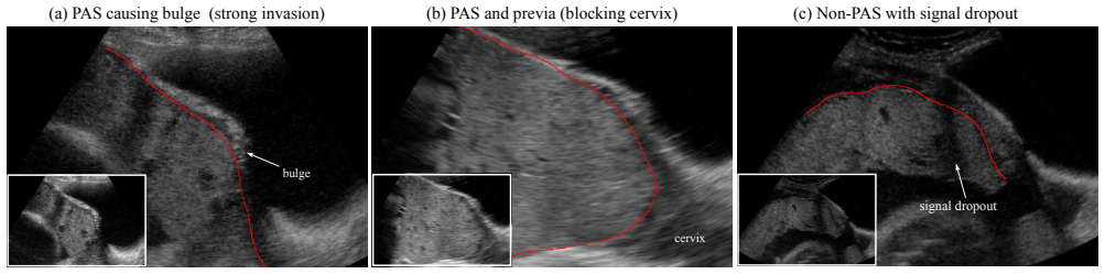
- Activation maps from the CGE blocks reveal that the network learns a semantic representation of the placenta itself without placenta-location supervision, which the paper suggests could help operators interpret the scene.
- The architecture adds only about 1.2 G FLOPs over HED, so it could fit into existing ultrasound analysis pipelines without major hardware demands.
- The paper argues that the same contour-detection approach could be adapted to other clinical scenarios involving edge or contour detection in breast, liver, heart, and brain imaging.
Reading between the lines
- Because all labels come from one annotator guided by one specialist, the reported absolute F-measures and the ranking of methods are conditional on that single ground truth; an independent multi-observer labeling study could change both and would be the natural next test.
- A practical extension would be to quantify uncertainty of the predicted contour, since the paper reports point estimates and quartiles but no per-image confidence; a probabilistic UPI map would be more actionable for clinicians deciding whether a PAS risk flag is reliable.
- The method's gains are measured on F-measure over traced curves; an evaluation against clinically meaningful outcomes, such as whether detected UPI irregularity correlates with confirmed PAS at delivery, would test whether the contour accuracy translates into diagnostic utility.
Editorial analysis
A structured set of objections, weighed in public.
Referee Report
Summary. The paper proposes UPI-Net, a deep convolutional network for detecting the utero-placental interface (UPI) in 2D placental ultrasound images, formulated as a semantic contour detection problem. UPI-Net augments a VGG-16 backbone with lightweight global context (GC) blocks on early convolutional stages, convolutional group-wise enhancement (CGE) blocks on later stages, a CoordConv input layer, and multi-scale feature aggregation with one side-output and a fused output. The authors evaluate on 4,871 images from 49 scans using subject-level nested 10-fold cross-validation, comparing against HED, CASENet, DS-FPN, and DCAN with standard contour metrics (ODS, OIS). They report that UPI-Net achieves the best median ODS of 0.458 [0.430, 0.479], compared to 0.427 [0.409, 0.442] for the best baseline HED, with modest additional computational cost. An ablation study shows that both the CoordConv layer and the side-output supervision improve performance.
Significance. If the empirical claims hold, UPI-Net is a useful step toward automated prenatal risk assessment for placenta accreta spectrum disorders, and the evaluation protocol is methodologically more careful than typical medical imaging studies: subject-level splitting avoids data leakage across scans, and nested cross-validation reduces selection bias in hyperparameter tuning. The architecture combines existing components (GC blocks, SGE-inspired CGE blocks, CoordConv) in a sensible way for the UPI geometry. The main weaknesses are that all metrics are computed against a single-annotator ground truth without any inter-observer agreement, and that the reported performance advantage over baselines is not supported by statistical significance testing. These issues directly affect the strength of the central claim that UPI-Net 'yields the highest performance,' so they need to be addressed before publication.
major comments (2)
- [Sec. 4.1] The UPI ground-truth annotations were produced by a single computer scientist 'under the guidance of' an obstetric specialist, with no inter-observer agreement, verification protocol, or annotation uncertainty reported. Since every performance metric in Table 1 measures agreement with this one annotator's tracing style, the 0.031 median ODS advantage over HED could be explained by annotator-specific bias rather than by genuine improvement in UPI detection. Please provide evidence of annotation reproducibility, for example a second expert annotation on a subset of images with inter-observer F-measure or boundary-distance statistics, or at minimum an explicit discussion of this limitation and its potential impact on the reported results.
- [Sec. 4.6 / Table 1] The claim that UPI-Net 'yields the highest performance' is based on median ODS across 10 folds, but no statistical significance test is reported. The interquartile range of UPI-Net ([0.430, 0.479]) overlaps with that of HED ([0.409, 0.442]), and with only 10 folds the difference could easily arise by chance. Please report paired statistical tests (e.g., Wilcoxon signed-rank) on the fold-wise ODS and OIS values for UPI-Net versus each baseline, and if the differences are not significant, temper the abstract's claim accordingly.
minor comments (6)
- [Fig. 6 / Table 1] The citation for DCAN is inconsistent: Table 1 uses [6], while Figure 6 uses [7]; reference [7] in the bibliography is DeepLab, not DCAN. Please unify the citation.
- [Sec. 4.5] There is a typo: 'hyper-paramater' should be 'hyper-parameter'.
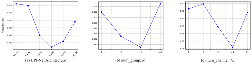
- [Sec. 4.4 / Fig. 5] The hyperparameter search is reported only via validation loss curves; reporting validation ODS (or a table of validation ODS values) would help the reader assess how sensitive the final test performance is to the chosen GC/CGE configuration and NG/NC values.
- [Sec. 4.4] The word 'optimial' should be 'optimal' in the sentence describing the iterative hyperparameter search.
- [Fig. 8 caption] The caption reads '453 rd kernel'; it should be '453rd kernel'.
- [Sec. 4.1] The annotation description uses the placeholder names 'X' and 'Y' ('annotated by X (a computer scientist) under the guidance of Y (an obstetric specialist)'). This is unusual in a formal paper; please use the authors' initials or a more descriptive phrasing.
Circularity Check
No circular derivation: UPI-Net's reported advantage is an empirical comparison against external benchmarks under nested cross-validation; self-cited dataset and baseline are not load-bearing.
full rationale
The central claim is the Table 1 comparison of median ODS/OIS across test folds. UPI-Net's architecture and training are specified independently in Sec. 3, and the evaluation uses a subject-level nested 10-fold cross-validation that tunes hyperparameters on validation splits and reports held-out test performance (Sec. 4.2). No equation defines the reported F-measure in terms of a fitted parameter or a prior result; the loss is a standard class-balanced cross-entropy over the annotated UPI maps. The two self-citations, [9] for the ultrasound dataset and [31] for the DS-FPN baseline, are provenance and baseline references: the dataset provides external images and labels, and DS-FPN is one of four compared methods rather than a premise that forces UPI-Net's score. The CGE block is a modification of [22] justified by validation experiments, not by asserting the conclusion. The single-annotator ground truth with no inter-observer agreement (Sec. 4.1) is a legitimate validity/generalizability concern, but it is a missing-support issue about label reliability, not a circular derivation. No fitted parameter is renamed as a prediction, and no uniqueness theorem is imported from the authors' prior work.
Assumptions & free parameters
free parameters (3)
- GC/CGE block configuration =
3G-2C (GC on conv-1..3, CGE on conv-4..5)
- num_group NG =
16
- num_channel NC =
32
assumptions (4)
- domain assumption ImageNet-pretrained VGG-16 features transfer to placental ultrasound.
- domain assumption Manual UPI annotations are correct ground truth.
- domain assumption Sagittal slices of 3D volumes are treated as independent samples within a subject-level CV.
- standard math Class-balancing weight omega = |Y-|/|Y+| equalizes expected gradient contributions.
Cite this review
Pith. "Pith review of UPI-Net: Semantic Contour Detection in Placental Ultrasound." pith.science (2026). https://pith.science/paper/2GTTGRIA
@misc{pith2026190900229,
  author       = {Pith},
  title        = {Pith review of: UPI-Net: Semantic Contour Detection in Placental Ultrasound},
  year         = {2026},
  howpublished = {\url{https://pith.science/paper/2GTTGRIA}},
  note         = {Machine review of arXiv:1909.00229}
}
read the original abstract
Semantic contour detection is a challenging problem that is often met in medical imaging, of which placental image analysis is a particular example. In this paper, we investigate utero-placental interface (UPI) detection in 2D placental ultrasound images by formulating it as a semantic contour detection problem. As opposed to natural images, placental ultrasound images contain specific anatomical structures thus have unique geometry. We argue it would be beneficial for UPI detectors to incorporate global context modelling in order to reduce unwanted false positive UPI predictions. Our approach, namely UPI-Net, aims to capture long-range dependencies in placenta geometry through lightweight global context modelling and effective multi-scale feature aggregation. We perform a subject-level 10-fold nested cross-validation on a placental ultrasound database (4,871 images with labelled UPI from 49 scans). Experimental results demonstrate that, without introducing considerable computational overhead, UPI-Net yields the highest performance in terms of standard contour detection metrics, compared to other competitive benchmarks.
Figures


Figures from the paper (5 more)



Reference graph
Works this paper leans on
-
[7]
L.-C. Chen et al. Deeplab: Semantic image segmentation with deep convolutional nets, atrous convolution, and fully connected crfs. IEEE TPAMI, 40(4):834–848, 2017. 2
work page 2017
-
[6]
H. Chen et al. Dcan: deep contour-aware networks for accu- rate gland segmentation. In CVPR, pages 2487–2496, 2016. 6
work page 2016
-
[1]
P. Arbelaez et al. Contour detection and hierarchical image segmentation. IEEE T-PAMI, 33(5):898–916, 2011. 2, 5
work page 2011
-
[2]
A. Aslam et al. Improved edge detection algorithm for brain tumor segmentation. Procedia Computer Science, 58:430– 437, 2015. 2
work page 2015
-
[3]
G. Bertasius et al. High-for-low and low-for-high: Efficient boundary detection from deep object features and its appli- cations to high-level vision. In ICCV, pages 504–512, 2015. 2
work page 2015
-
[4]
GCNet: Non-local Networks Meet Squeeze-Excitation Networks and Beyond
Y . Cao et al. Gcnet: Non-local networks meet squeeze-excitation networks and beyond. arXiv preprint arXiv:1904.11492, 2019. 2, 3, 4
work page Pith review arXiv 1904
-
[5]
G. C. Cawley and N. L. Talbot. On over-fitting in model selection and subsequent selection bias in performance eval- uation. JMLR, 11(Jul):2079–2107, 2010. 5
work page 2010
-
[8]
¨O. C ¸ ic ¸ek et al. 3d u-net: learning dense volumetric segmen- tation from sparse annotation. In MICCAI, pages 424–432. Springer, 2016. 5
work page 2016
Show all 44 references
-
[9]
Collins, G
S. Collins, G. Stevenson, J. Noble, L. Impey, and A. Welsh. Influence of power doppler gain setting on virtual organ computer-aided analysis indices in vivo: can use of the in- dividual sub-noise gain level optimize information? Ultra- sound in Obstetrics & Gynecology, 40(1):75...
2012
-
[10]
Dollar et al
P. Dollar et al. Supervised learning of edges and object boundaries. In CVPR, volume 2, pages 1964–1971. IEEE,
1964
-
[11]
Gao et al
S.-H. Gao et al. Res2net: A new multi-scale backbone archi- tecture. arXiv preprint arXiv:1904.01169, 2019. 2
1904 arXiv
-
[12]
Gibson et al
E. Gibson et al. Automatic multi-organ segmentation on ab- dominal ct with dense v-networks. IEEE TMI, 37(8):1822– 1834, 2018. 5
2018
-
[13]
Hariharan et al
B. Hariharan et al. Semantic contours from inverse detectors. In ICCV, pages 991–998, 2011. 2
2011
-
[14]
He et al
K. He et al. Delving deep into rectifiers: Surpassing human- level performance on imagenet classification. InICCV, pages 1026–1034, 2015. 2, 6
2015
-
[15]
He et al
K. He et al. Deep residual learning for image recognition. In CVPR, pages 770–778, 2016. 2
2016
-
[16]
Hu et al
H. Hu et al. Relation networks for object detection. In CVPR, pages 3588–3597, 2018. 2
2018
-
[17]
Hu et al
J. Hu et al. Squeeze-and-excitation networks. In CVPR, pages 7132–7141, 2018. 2, 4
2018
-
[18]
Jauniaux et al
E. Jauniaux et al. Figo consensus guidelines on placenta acc- reta spectrum disorders: Prenatal diagnosis and screening. IJGO, 140(3):274–280, 2018. 1
2018
-
[19]
Jauniaux et al
E. Jauniaux et al. Placenta accreta spectrum: pathophysi- ology and evidence-based anatomy for prenatal ultrasound imaging. AJOG, 218(1):75–87, 2018. 1
2018
-
[20]
Lawrence et al
S. Lawrence et al. Neural network classification and prior class probabilities. In Neural networks: tricks of the trade , pages 299–313. Springer, 1998. 3
1998
-
[21]
Lee et al
C.-Y . Lee et al. Deeply-supervised nets. In Artificial Intelli- gence and Statistics, pages 562–570, 2015. 4
2015
-
[22]
Li et al
X. Li et al. Spatial group-wise enhance: Improving semantic feature learning in convolutional networks. arXiv preprint arXiv:1905.09646, 2019. 2, 4, 5
1905 arXiv
-
[23]
Li et al
Y . Li et al. Attention-guided unified network for panoptic segmentation. In CVPR, pages 7026–7035, 2019. 2
2019
-
[24]
Lin et al
T.-Y . Lin et al. Feature pyramid networks for object detec- tion. In CVPR, pages 2117–2125, 2017. 3, 4
2017
-
[25]
Liu et al
R. Liu et al. An intriguing failing of convolutional neural net- works and the coordconv solution. In NeurIPS, pages 9628– 9639, 2018. 5
2018
-
[26]
Long et al
J. Long et al. Fully convolutional networks for semantic seg- mentation. In CVPR, pages 3431–3440, 2015. 2
2015
-
[27]
Merkow et al
J. Merkow et al. Dense volume-to-volume vascular boundary detection. In MICCAI, pages 371–379. Springer, 2016. 2
2016
-
[28]
A. A. Novikov et al. Fully convolutional architectures for multiclass segmentation in chest radiographs. IEEE TMI , 37(8):1865–1876, 2018. 5
2018
-
[29]
Park et al
J. Park et al. Bam: Bottleneck attention module. arXiv preprint arXiv:1807.06514, 2018. 2
2018 arXiv
-
[30]
Prasad et al
M. Prasad et al. Learning class-specific edges for object de- tection and segmentation. In Computer Vision, Graphics and Image Processing, pages 94–105. Springer, 2006. 2
2006
-
[31]
Qi et al
H. Qi et al. Automatic lacunae localization in placental ul- trasound images via layer aggregation. In MICCAI, pages 921–929. Springer, 2018. 3, 6
2018
-
[32]
Sabour et al
S. Sabour et al. Dynamic routing between capsules. In NeurIPS, pages 3856–3866, 2017. 2
2017
-
[33]
Shahrokni et al
A. Shahrokni et al. Classifier-based contour tracking for rigid and deformable objects. In BMVC, 2005. 2
2005
-
[34]
Vaswani et al
A. Vaswani et al. Attention is all you need. In NeurIPS, pages 5998–6008, 2017. 2
2017
-
[35]
Wang et al
X. Wang et al. Non-local neural networks. In CVPR, pages 7794–7803, 2018. 2, 3, 4
2018
-
[36]
Wang et al
Y . Wang et al. Deep attentive features for prostate segmenta- tion in 3d transrectal ultrasound. IEEE TMI, 2019. 5
2019
-
[37]
Woo et al
S. Woo et al. Cbam: Convolutional block attention module. In ECCV, pages 3–19, 2018. 2
2018
-
[38]
Wu and K
Y . Wu and K. He. Group normalization. In ECCV, pages 3–19, 2018. 4
2018
-
[39]
Xie and Z
S. Xie and Z. Tu. Holistically-nested edge detection. In ICCV, pages 1395–1403, 2015. 2, 3, 5, 6
2015
-
[40]
Yu et al
F. Yu et al. Dilated residual networks. In CVPR, pages 472– 480, 2017. 2
2017
-
[41]
Yu and V
F. Yu and V . Koltun. Multi-scale context aggregation by di- lated convolutions. ICLR, 2016. 2
2016
-
[42]
Yu et al
Z. Yu et al. Casenet: Deep category-aware semantic edge detection. In CVPR, pages 5964–5973, 2017. 2, 3, 5, 6
2017
-
[43]
Zhang et al
H. Zhang et al. Context encoding for semantic segmentation. In CVPR, pages 7151–7160, 2018. 2
2018
-
[44]
Zhang et al
H. Zhang et al. Self-attention generative adversarial net- works. pages 7354–7363, 2019. 2
2019
Reviewed August 14, 2026 · model on record in the stance chip above.
Discussion (0). Continue with ORCID to comment.