REVIEW 5 major objections 4 minor 42 references
From Classification to Localization and Clinical Validation: Large-Scale Development of a Deep Learning System for Thoracic Disease Detection on Chest Radiographs in Thailand
T0 review · 5 major / 4 minor · reviewed 2026-08-02 · deepseek-v4-flash
Pith's one-line read This paper claims that a chest X-ray AI trained on 874,858 Thai images reaches near-ceiling accuracy at its home hospital, keeps most of that performance across 13 other hospitals, and localizes lesions so well that radiologists agree with
desk verdict A substantial applied CXR validation with real multi-site numbers, but the missing patient-level split definition and proprietary labeling pipeline mean the headline AUROC should be treated as unverified until the authors close those gaps. read the letter →
The pith
A machine-rendered reading of the paper's core claim, the machinery that carries it, and where it could break.
The reading
What carries the argument
The load-bearing mechanism is a single multi-task network that outputs, for each of nine conditions, both a classification score and a heatmap. It uses a densely connected convolutional encoder with attention layers that compare different image regions, a decoder that restores spatial detail via skip connections, and a probabilistic class-activation-map pooling layer that produces sharper heatmaps while preserving classification. Because the same representation drives both outputs, localization is learned from image-level labels alone, with no pixel annotations during training; pixel annotations are used only to evaluate the resulting heatmaps. This coupling is what lets the paper report cla
What would settle it
Compare patient identifiers between the training and test sets: if any patient appears in both, recompute the in-domain AUROC on a disjoint subset. Separately, take a random sample of training images, have radiologists re-label them from scratch, and compare with the natural-language-derived labels; a high disagreement rate would show the reported metrics inherit label noise.
Extended reading notes
Core claim
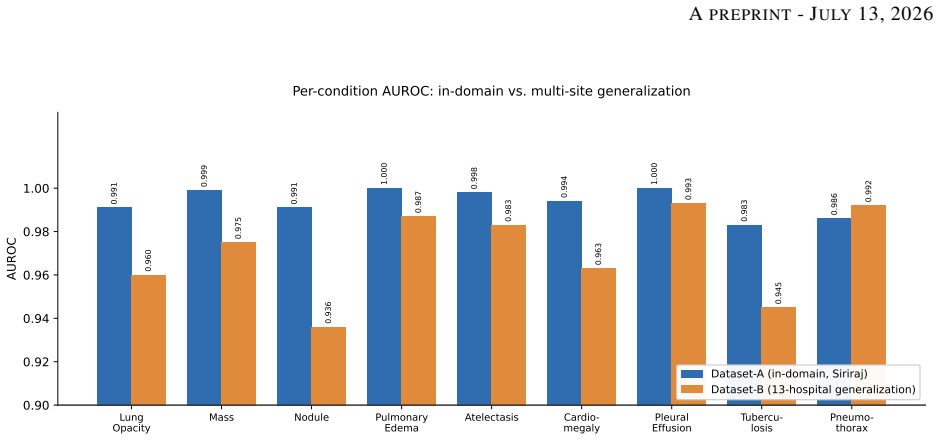
The paper's central claim is that a locally developed chest X-ray model can combine high in-domain discrimination with cross-site generalization and clinically acceptable localization. Trained on 874,858 frontal radiographs with paired reports from one large Bangkok hospital, the system achieved a mean AUROC of 0.994 on 19,871 radiologist-verified held-out cases, and 0.970 on 5,992 independent cases from 13 hospitals. On 4,549 annotated cases, it localized lesions with a mean lesion-localization fraction of 77.9% at 0.59 false localizations per image. In a usability study, five thoracic radiologists showed 93.6% classification concordance and 94.7% localization concordance, with a standard u
Load-bearing premise
The central claims rest on the held-out test sets being free of patients who also appear in training; the paper does not state whether the split was by patient or by image, and if patients overlap, the headline 0.994 AUROC would be inflated.
Editorial extensions
If this is right
- If the model is deployed as a second reader, it can flag abnormal studies and point radiologists to the region of interest, potentially shortening reading time.
- The small drop from 0.994 to 0.970 AUROC across 13 hospitals indicates that a model trained at one site can retain ranking ability elsewhere without retraining.
- Fixed operating points chosen in-domain lose sensitivity on external data, especially for diffuse findings; per-site threshold adjustment may be needed.
- A concordance rate near 94% with radiologists suggests the system's outputs are clinically actionable rather than merely statistically discriminative.
- The combination of classification and localization in a single model avoids the need for separate detection pipelines, simplifying deployment.
Reading between the lines
- Because the paper does not state whether the held-out split was at patient level, a reader should treat the in-domain 0.994 as an upper bound until a patient-disjoint split is confirmed; this is a testable extension, not a claim the paper makes.
- The training labels come from a proprietary natural-language model reviewed by radiologists; the error rate of that labeler is not quantified, so the true ceiling of the system is unknown. An audit that compares fully manual labels to the pipeline's labels on a random sample would bound this.
- The usability numbers rest on five radiologists; a larger multi-reader study across more sites would give a more stable estimate of the 89 score and concordance rates.
- The fixed-threshold sensitivity drop on external data suggests that deployment would benefit from site-specific threshold calibration, which could raise sensitivity without sacrificing AUROC.
Editorial analysis
A structured set of objections, weighed in public.
Referee Report
Summary. The paper presents Inspectra CXR version 5, a deep learning system for multi-label thoracic disease classification and weakly supervised lesion localization on frontal chest radiographs. The model combines a DenseNet-121 backbone with Attend-and-Compare Modules and a PCAM aggregation layer, and was trained on 874,858 frontal CXRs from Siriraj Hospital. The authors report a held-out in-domain mean AUROC of 0.994 on 19,871 radiologist-verified cases, a multi-site generalization mean AUROC of 0.970 on 5,992 cases from 13 Thai hospitals, a mean lesion-localization fraction of 77.9% on 4,549 annotated cases, radiologist concordance of 93.6% (classification) and 94.7% (localization), and a System Usability Scale score of 89.
Significance. If the results hold, this would be a practically valuable validation of a locally developed CXR AI system for a Southeast Asian population, combining in-domain accuracy, cross-site transfer, localization, and usability in a single deployment-oriented study. The scale of the training cohort and the inclusion of an external 13-hospital test set are notable strengths, as is the pixel-annotated localization evaluation. However, the central validity of the headline accuracy numbers is not yet established: the split unit used for the held-out sets is not specified, the training-label generation pipeline is proprietary and unquantified, and the concordance metric counts a substantial number of misses as concordant. These issues are load-bearing for the paper's main claims.
major comments (5)
- [§3.1, §3.3] The held-out test set is not demonstrated to be patient-disjoint from training. Section 3.1 excludes only "duplicate examinations from the same patient on the same date," and Section 3.3 states the 19,871 images were "held out" without specifying the split unit. If the same patient appears in training and test on different dates, patient-specific anatomy or follow-up patterns can inflate the in-domain AUROC. Please report the number of overlapping patients (or perform a patient-disjoint split) for Dataset-A, and similarly for Dataset-B and Dataset-C.
- [§3.2] All training labels originate from a proprietary natural-language labeling model whose accuracy is never reported. The statement that radiologists "reviewed and corrected" these labels does not quantify the fraction audited, the correction rate, or inter-reader agreement. Because the model is trained on these labels, every downstream metric is conditional on an unmeasured label-error process. Please report a random audit of the training labels, the NLP model's performance, or an equivalent assessment.
- [§3.7, Tables 4–5] The concordance metric counts "Add" as concordant, yet for localization "Add" is defined as "The heatmap misses some lesions" and for classification as the system under-calling relative to the radiologist. In Table 9, 753 of 4,549 localization cases (16.6%) and 856 of 19,871 classification cases are "Add." Treating misses as concordant systematically inflates the 93.60%/94.72% headline concordance. Please report the four categories separately and recompute concordance with Add excluded or as a sensitivity analysis.
- [Tables 6–8] All metrics are point estimates without confidence intervals, despite small positive counts (e.g., pneumothorax n=64 in Dataset-A; atelectasis n=118 in Dataset-B) and near-ceiling AUROCs. Report 95% CIs (e.g., bootstrap or DeLong) for AUROC, sensitivity, specificity, and LLF. Without intervals, the 0.994 vs 0.970 gap and per-condition differences cannot be properly assessed.
- [§3.3, §4.4] The relationship among Dataset-A, Dataset-B, and Dataset-C is not stated. If Dataset-C is a subset of Dataset-A, the localization and classification results are not independent; if disjoint, the total case counts need clarification. In addition, the five radiologists in the usability study may include the same readers who verified labels or created annotations; if so, the concordance analysis is partially a consistency check of the labelers. Please clarify reader overlap and case-set overlap.
minor comments (4)
- [§5 (Discussion)] The sentence "Per-condition ROC analysis from case-level prediction scores is planned as future work" conflicts with Tables 6 and 7, which already report per-condition AUROC. Please clarify what additional ROC analysis is intended (e.g., ROC curves with confidence intervals).
- [§3.4] "Cut-Noise" and the "proprietary image-processing quality algorithms" are not described beyond a single sentence. Please provide a specification or a citation so that the method is reproducible.
- [§3.5, §4.2] The operating point selection is described only as balancing sensitivity and specificity at a "clinically appropriate PPV." Please state the actual thresholds applied to Dataset-B and whether these are the same thresholds selected on the validation set.
- [Table 3] The label set is the nine conditions, but Table 3 also lists "Normal" counts. Please state whether Normal is a separate class used in the test evaluation and how normal-only cases are defined relative to the nine-condition labels.
Circularity Check
No significant circularity: the reported AUROC, localization, and usability results are empirical measurements against external radiologist-verified ground truth and reader ratings, not derivations from the model's own outputs.
full rationale
The paper claims no first-principles derivation; its central results are empirical evaluations against external benchmarks. Training labels come from radiologist reports with radiologist review; test labels and localization annotations are explicitly radiologist-verified and are not produced by the evaluated model. AUROC on Datasets A and B is computed against these external labels, LLF is computed against radiologist pixel annotations, and SUS comes from independent radiologist ratings, so the headline numbers are not fitted parameters renamed as predictions. The self-citation [14] motivating local adaptation is background context, not a load-bearing step: the current claims stand on the reported held-out and multi-site test sets. No equation or definition reduces a reported quantity to the training labels or to another claimed result. Remaining concerns such as the unspecified patient-level train/test split, the unquantified proprietary NLP labeler, and the decision to count 'Add' as concordant are external-validity limitations rather than circularity, and do not warrant raising the circularity score.
Assumptions & free parameters
free parameters (5)
- Per-condition operating points =
not reported
- Per-class loss weights =
not reported
- Cut-Noise hyperparameters =
not reported
- IoU threshold for localization metrics =
0.5
- Training schedule =
10 epochs, LR 1e-4, decay 0.1 at epochs 7/9, batch 10
assumptions (5)
- domain assumption ImageNet-pretrained DenseNet-121 features transfer to frontal chest radiographs
- domain assumption Free-text radiologist reports, processed by an unspecified proprietary NLP model, yield sufficiently accurate training labels
- domain assumption The proprietary image-quality filters correctly identify diagnostic-quality images and do not bias disease prevalence
- domain assumption Radiologist-verified labels on the test sets are definitive ground truth
- domain assumption The development and test partitions do not share patients
invented entities (3)
-
Cut-Noise technique
-
Proprietary natural-language labeling model
-
Proprietary image-processing quality algorithms
Cite this review
Pith. "Pith review of From Classification to Localization and Clinical Validation: Large-Scale Development of a Deep Learning System for Thoracic Disease Detection on Chest Radiographs in Thailand." pith.science (2026). https://pith.science/paper/3EWITLMD
@misc{pith2026260709305,
  author       = {Pith},
  title        = {Pith review of: From Classification to Localization and Clinical Validation: Large-Scale Development of a Deep Learning System for Thoracic Disease Detection on Chest Radiographs in Thailand},
  year         = {2026},
  howpublished = {\url{https://pith.science/paper/3EWITLMD}},
  note         = {Machine review of arXiv:2607.09305}
}
read the original abstract
Chest radiography (CXR) remains the most widely used thoracic imaging modality, yet expert interpretation is constrained by a severe shortage of radiologists in Thailand and across Southeast Asia. Local adaptation of deep learning models to Thai data has been shown to substantially improve accuracy on Thai populations. Here we present the development and comprehensive validation of the chest radiograph analysis model in Inspectra CXR version 5, a deep learning system that performs multi-label thoracic disease classification and weakly supervised lesion localization within a single model. The architecture couples a DenseNet-121 backbone with Attend-and-Compare Modules (ACM) and a Probabilistic Class Activation Map (PCAM) aggregation layer, producing a per-condition classification score and heatmap simultaneously. The model was developed on 874,858 frontal chest radiographs with paired radiologist reports from Siriraj Hospital, Bangkok. On a held-out, radiologist-verified in-domain test set of 19,871 cases, it achieved a mean AUROC of 0.994 (mean sensitivity 92.4%, specificity 98.6%) across nine clinically important conditions. On an independent generalization set of 5,992 cases from 13 hospitals across Thailand, the mean AUROC was 0.970, indicating robust transfer across sites. For localization, evaluated on 4,549 radiologist-annotated cases, the model attained a mean lesion-localization fraction (LLF) of 77.9% at 0.59 non-lesion localizations per image. In a usability evaluation with five thoracic radiologists, the system reached a classification concordance of 93.6%, a localization concordance of 94.7%, and a mean System Usability Scale (SUS) score of 89. These results indicate that a locally developed, localization-capable CXR system can deliver high accuracy, generalize across heterogeneous Thai hospitals, and earn the trust of practicing radiologists.
Figures

Reference graph
Works this paper leans on
-
[8]
Deep learning in chest radiography: detection of findings and presence of change.PloS one, 13(10), 2018
Ramandeep Singh, Mannudeep K Kalra, Chayanin Nitiwarangkul, John A Patti, Fatemeh Homayounieh, Atul Padole, Pooja Rao, Preetham Putha, Victorine V Muse, Amita Sharma, et al. Deep learning in chest radiography: detection of findings and presence of change.PloS one, 13(10), 2018
2018
-
[37]
Kalra, Chayanin Nitiwarangkul, John A
Ramandeep Singh, Mannudeep K. Kalra, Chayanin Nitiwarangkul, John A. Patti, Fatemeh Homayounieh, Atul Padole, Pooja Rao, Preetham Putha, Victorine V . Muse, Amita Sharma, and Subba R. Digumarthy. Deep learning in chest radiography: Detection of findings and presence of change.PLOS ONE, 13(10):e0204155, 2018. 11 APREPRINT- JULY23, 2026
2018
-
[1]
Chest radiography in general practice: indications, diagnostic yield and consequences for patient management.Br J Gen Pract, 56(529):574–578, 2006
Anouk M Speets, Yolanda van der Graaf, Arno W Hoes, Sandra Kalmijn, Alfred PE Sachs, Matthieu JCM Rutten, Jan Willem C Gratama, Alexander D Montauban van Swijndregt, and Willem PThM Mali. Chest radiography in general practice: indications, diagnostic yield and consequences for patient management.Br J Gen Pract, 56(529):574–578, 2006
2006
-
[2]
Mortality estimates for south east asia, and indepth mortality surveillance: necessary but not sufficient?International journal of epidemiology, 42(4):1196–1199, 2013
Chalapati Rao. Mortality estimates for south east asia, and indepth mortality surveillance: necessary but not sufficient?International journal of epidemiology, 42(4):1196–1199, 2013
2013
-
[3]
Global tuberculosis report 2019, 2019
World Health Organization. Global tuberculosis report 2019, 2019. Last accessed 13 April 2020
2019
-
[4]
Assessing the accuracy and certainty in interpreting chest x-rays in the medical division.Clinical medicine, 13(4):349, 2013
I Satia, S Bashagha, A Bibi, R Ahmed, S Mellor, and F Zaman. Assessing the accuracy and certainty in interpreting chest x-rays in the medical division.Clinical medicine, 13(4):349, 2013
2013
-
[5]
Thai specialist statistics 2019, 2019
The Medical Council of Thailand. Thai specialist statistics 2019, 2019. Last accessed 13 April 2020
2019
-
[6]
Pranav Rajpurkar, Jeremy Irvin, Kaylie Zhu, Brandon Yang, Hershel Mehta, Tony Duan, Daisy Ding, Aarti Bagul, Curtis Langlotz, Katie Shpanskaya, et al. Chexnet: Radiologist-level pneumonia detection on chest x-rays with deep learning.arXiv preprint arXiv:1711.05225, 2017
arXiv 2017
Show all 42 references
-
[7]
Chexpert: A large chest radiograph dataset with uncer- tainty labels and expert comparison.Proceedings of the AAAI Conference on Artificial Intelligence, 33:590–597, Jul 2019
Jeremy Irvin, Pranav Rajpurkar, Michael Ko, Yifan Yu, Silviana Ciurea-Ilcus, Chris Chute, Henrik Marklund, Behzad Haghgoo, Robyn Ball, Katie Shpanskaya, and et al. Chexpert: A large chest radiograph dataset with uncer- tainty labels and expert comparison.Proceedings of the AAA...
2019
-
[9]
Diagnose like a radiologist: Attention guided convolutional neural network for thorax disease classification.arXiv preprint arXiv:1801.09927, 2018
Qingji Guan, Yaping Huang, Zhun Zhong, Zhedong Zheng, Liang Zheng, and Yi Yang. Diagnose like a radiologist: Attention guided convolutional neural network for thorax disease classification.arXiv preprint arXiv:1801.09927, 2018
2018 arXiv
-
[10]
Xiaosong Wang, Yifan Peng, Le Lu, Zhiyong Lu, Mohammadhadi Bagheri, and Ronald M. Summers. Chestx- ray8: Hospital-scale chest x-ray database and benchmarks on weakly-supervised classification and localization of common thorax diseases.2017 IEEE Conference on Computer Vision an...
2017
-
[11]
Mimic-cxr: A large publicly available database of labeled chest radiographs.arXiv preprint arXiv:1901.07042, 1(2), 2019
Alistair EW Johnson, Tom J Pollard, Seth Berkowitz, Nathaniel R Greenbaum, Matthew P Lungren, Chih-ying Deng, Roger G Mark, and Steven Horng. Mimic-cxr: A large publicly available database of labeled chest radiographs.arXiv preprint arXiv:1901.07042, 1(2), 2019
1901 arXiv
-
[12]
Padchest: A large chest x-ray image dataset with multi-label annotated reports.arXiv preprint arXiv:1901.07441, 2019
Aurelia Bustos, Antonio Pertusa, Jose-Maria Salinas, and Maria de la Iglesia-Vayá. Padchest: A large chest x-ray image dataset with multi-label annotated reports.arXiv preprint arXiv:1901.07441, 2019
1901 arXiv
-
[13]
Seelwan Sathitratanacheewin and Krit Pongpirul. Deep learning for automated classification of tuberculosis-related chest x-ray: Dataset specificity limits diagnostic performance generalizability.arXiv preprint arXiv:1811.07985, 2018
2018
-
[14]
Local adaptation improves accuracy of deep learning model for automated x-ray thoracic disease detection: A thai study.arXiv preprint arXiv:2004.10975, 2020
Isarun Chamveha, Trongtum Tongdee, Pairash Saiviroonporn, and Warasinee Chaisangmongkon. Local adaptation improves accuracy of deep learning model for automated x-ray thoracic disease detection: A thai study.arXiv preprint arXiv:2004.10975, 2020
2004 arXiv
-
[15]
Densely connected convolutional networks
Gao Huang, Zhuang Liu, Laurens Van Der Maaten, and Kilian Q Weinberger. Densely connected convolutional networks. InProceedings of the IEEE conference on computer vision and pattern recognition, pages 4700–4708, 2017
2017
-
[16]
Learning visual context by comparison
Minchul Kim, Jongchan Park, Seil Na, Chang Min Park, and Donggeun Yoo. Learning visual context by comparison. InEuropean Conference on Computer Vision (ECCV), pages 576–592. Springer, 2020
2020
-
[17]
Weakly supervised lesion localization with probabilistic-cam pooling
Wenwu Ye, Jin Yao, Hui Xue, and Yi Li. Weakly supervised lesion localization with probabilistic-cam pooling. arXiv preprint arXiv:2005.14480, 2020
2005 arXiv
-
[18]
Learning to recognize abnormalities in chest x-rays with location-aware dense networks.arXiv preprint arXiv:1803.04565, 2018
Sebastian Guendel, Sasa Grbic, Bogdan Georgescu, Kevin Zhou, Ludwig Ritschl, Andreas Meier, and Dorin Comaniciu. Learning to recognize abnormalities in chest x-rays with location-aware dense networks.arXiv preprint arXiv:1803.04565, 2018
2018 arXiv
-
[19]
Deep residual learning for image recognition
Kaiming He, Xiangyu Zhang, Shaoqing Ren, and Jian Sun. Deep residual learning for image recognition. In Proceedings of the IEEE conference on computer vision and pattern recognition, pages 770–778, 2016
2016
-
[20]
Imagenet: A large-scale hierarchical image database
Jia Deng, Wei Dong, Richard Socher, Li-Jia Li, Kai Li, and Li Fei-Fei. Imagenet: A large-scale hierarchical image database. In2009 IEEE conference on computer vision and pattern recognition, pages 248–255. Ieee, 2009. 10 APREPRINT- JULY23, 2026
2009
-
[21]
Learning deep features for discriminative localization
Bolei Zhou, Aditya Khosla, Agata Lapedriza, Aude Oliva, and Antonio Torralba. Learning deep features for discriminative localization. InProceedings of the IEEE conference on computer vision and pattern recognition, pages 2921–2929, 2016
2016
-
[22]
Jarrel CY Seah, Cyril HM Tang, Quinlan D Buchlak, et al. Effect of a comprehensive deep-learning model on the accuracy of chest x-ray interpretation by radiologists: A retrospective, multireader multicase study.The Lancet Digital Health, 3(8):e496–e506, 2021
2021
-
[23]
Di Capua, Markus Y
Jong Seok Ahn, Shadi Ebrahimian, Shaunagh McDermott, Sanghyup Lee, Laura Naccarato, John F. Di Capua, Markus Y . Wu, Eric W. Zhang, Victorine Muse, Benjamin Miller, Farid Sabzalipour, Bernardo C. Bizzo, Keith J. Dreyer, Parisa Kaviani, Subba R. Digumarthy, and Mannudeep K. Kal...
2022
-
[24]
Al-antari, Pavlína Polášková, and Sergei Strukov
Daniel Kvak, Anna Chromcová, Robert Hrubý, Eva Jan˚ u, Marek Biroš, Marija Pajdakovi´c, Karolína Kvaková, Mugahed A. Al-antari, Pavlína Polášková, and Sergei Strukov. Leveraging deep learning decision-support system in specialized oncology center: A multi-reader retrospective ...
2023
-
[25]
Ai-assisted detection for chest x-rays (aid-cxr): a multi-reader multi-case study protocol.BMJ Open, 14:e080554, 2024
Farhaan Khan, Indrajeet Das, Marusa Kotnik, Louise Wing, Edwin Van Beek, John Murchison, Jong Seok Ahn, Sang Hyup Lee, Ambika Seth, Abdala Trinidad Espinosa Morgado, Howell Fu, Alex Novak, Nabeeha Salik, Alan Campbell, Ruchir Shah, Fergus Gleeson, and Sarim Ather. Ai-assisted ...
2024
-
[26]
Development and validation of deep learning–based automatic detection algorithm for malignant pulmonary nodules on chest radiographs.Radiology, 290(1):218–228, 2019
Ju Gang Nam, Sunggyun Park, Eui Jin Hwang, et al. Development and validation of deep learning–based automatic detection algorithm for malignant pulmonary nodules on chest radiographs.Radiology, 290(1):218–228, 2019
2019
-
[27]
Digumarthy, and Mannudeep K
Hyunsuk Yoo, Sang Hyup Lee, Chiara Daniela Arru, Ruhani Doda Khera, Ramandeep Singh, Sean Siebert, Dohoon Kim, Yuna Lee, Ju Hyun Park, Hye Joung Eom, Subba R. Digumarthy, and Mannudeep K. Kalra. Ai-based improvement in lung cancer detection on chest radiographs: results of a m...
2021
-
[28]
Jong Hyuk Lee, Hye Young Sun, Sunggyun Park, Hyungjin Kim, Eui Jin Hwang, Jin Mo Goo, and Chang Min Park. Performance of a deep learning algorithm compared with radiologic interpretation for lung cancer detection on chest radiographs in a health screening population.Radiology,...
2020
-
[29]
Real-world evaluation of an ai triaging system for chest x-rays: A prospective clinical study.European Journal of Radiology, 181:111783, 2024
Srinath Sridharan, Alicia Seah Xin Hui, Narayan Venkataraman, Prasanna Sivanath Tirukonda, Ram Pratab Jeyaratnam, Sindhu John, Saraswathy Suresh Babu, Perry Liew, Joe Francis, Tsai Koh Tzan, Wong Kang Min, Goh Min Liong, and Charlene Liew Jin Yee. Real-world evaluation of an a...
2024
-
[30]
Kiraly, Sreenivasa Raju Kalidindi, Monde Muyoyeta, Jameson Malemela, Ting Shih, Greg S
Sahar Kazemzadeh, Jin Yu, Shahar Jamshy, Rory Pilgrim, Zaid Nabulsi, Christina Chen, Neeral Beladia, Charles Lau, Scott Mayer McKinney, Thad Hughes, Atilla P. Kiraly, Sreenivasa Raju Kalidindi, Monde Muyoyeta, Jameson Malemela, Ting Shih, Greg S. Corrado, Lily Peng, Katherine ...
2023
-
[31]
Zhi Zhen Qin, Shahriar Ahmed, Mohammad Shahnewaz Sarker, Kishor Paul, Ahammad Shafiq Sikder Adel, Tasneem Naheyan, Rachael Barrett, Sayera Banu, and Jacob Creswell. Tuberculosis detection from chest x-rays for triaging in a high tuberculosis-burden setting: an evaluation of fi...
2021
-
[32]
ClinicalTrials.gov, 2025
Ai assisted detection of chest x-rays (aid-cxr). ClinicalTrials.gov, 2025. NCT06075836
2025
-
[33]
ClinicalTrials.gov, 2025
Radiograph accelerated detection and identification of cancer in the lung (radical). ClinicalTrials.gov, 2025. NCT06044454
2025
-
[34]
ClinicalTrials.gov, 2024
Validity of pleural effusion detection software. ClinicalTrials.gov, 2024. NCT05903287
2024
-
[35]
ClinicalTrials.gov, 2023
Multi-reader retrospective study examining carebot ai cxr 2.0.21-v2.01 implementation in everyday radiology clinical practice. ClinicalTrials.gov, 2023. NCT05963945
2023
-
[36]
Can deep learning reliably recognize abnormality patterns on chest x-rays? a multi-reader study examining one month of ai implementation in everyday radiology clinical practice
Daniel Kvak, Anna Chromcová, Petra Ovesná, Jakub Dandár, Marek Biroš, Robert Hrubý, Daniel Dufek, and Marija Pajdakovi´c. Can deep learning reliably recognize abnormality patterns on chest x-rays? a multi-reader study examining one month of ai implementation in everyday radiol...
2023
-
[38]
Sus: A retrospective.Journal of Usability Studies, 8(2):29–40, 2013
John Brooke. Sus: A retrospective.Journal of Usability Studies, 8(2):29–40, 2013
2013
-
[39]
An empirical evaluation of the system usability scale
Aaron Bangor, Philip T Kortum, and James T Miller. An empirical evaluation of the system usability scale. International Journal of Human–Computer Interaction, 24(6):574–594, 2008
2008
-
[40]
Hyunsuk Yoo, Eun Young Kim, Hyungjin Kim, Ye Ra Choi, Moon Young Kim, Sung Ho Hwang, Young Joong Kim, Young Jun Cho, and Kwang Nam Jin. Artificial intelligence-based identification of normal chest radiographs: A simulation study in a multicenter health screening cohort.Korean ...
2022
-
[41]
Performance of a deep-learning algorithm for referable thoracic abnormalities on chest radiographs: A multicenter study of a health screening cohort.PLOS ONE, 16(2):e0246472, 2021
Eun Young Kim, Young Jae Kim, Won-Jun Choi, Gi Pyo Lee, Ye Ra Choi, Kwang Nam Jin, and Young Jun Cho. Performance of a deep-learning algorithm for referable thoracic abnormalities on chest radiographs: A multicenter study of a health screening cohort.PLOS ONE, 16(2):e0246472, 2021
2021
-
[42]
Deep learning-based detection system for multiclass lesions on chest radiographs: comparison with observer readings.European Radiology, 30:1359–1368, 2020
Sohee Park, Sang Min Lee, Kyung Hee Lee, Kyu-Hwan Jung, Woong Bae, Jooae Choe, and Joon Beom Seo. Deep learning-based detection system for multiclass lesions on chest radiographs: comparison with observer readings.European Radiology, 30:1359–1368, 2020. 12
2020
Reviewed August 2, 2026 · model on record in the stance chip above.
Discussion (0). Continue with ORCID to comment.