REVIEW 4 major objections 6 minor 80 references
AortaDiff: Volume-Guided Conditional Diffusion Models for Multi-Branch Aortic Surface Generation
T0 review · 4 major / 6 minor · reviewed 2026-08-06 · deepseek-v4-flash
Pith's one-line read This paper claims that a volume-conditioned diffusion model can generate aortic centerlines from CT/MRI volumes, and that using those centerlines as automatic prompts for a pretrained segmenter plus NURBS fitting yields CFD-compatible…
desk verdict The pipeline works and the numbers are strong, but the 'minimal dependency on large labeled datasets' claim leans on ScribblePrompt's pretrained priors, not on the diffusion model. read the letter →
The pith
A machine-rendered reading of the paper's core claim, the machinery that carries it, and where it could break.
The reading
What carries the argument
The central object is the centerline-as-a-1D-image: each aortic centerline is represented as 16 ordered points stored in a three-channel 1D image. A vision transformer extracts a feature volume from the input CT/MRI volume, and at each diffusion timestep the model looks up feature vectors at the current centerline point locations, concatenates them with the noisy 1D centerline latent, and uses a 1D U-Net to predict the added noise. This volume-guided conditional denoising lets the model generate patient-specific centerlines without large point-cloud training sets. The second load-bearing mechanism is prompt-based contour extraction: each generated centerline point defines an orthogonal slicing plane through the volume, and the point itself is used as a prompt for ScribblePrompt, a segmentation model pretrained on large-scale medical images, yielding lumen contours that are resampled, aligned with ICP, and fitted with a cubic NURBS surface.
What would settle it
Run the identical AortaDiff pipeline on the AVT volumes but replace ScribblePrompt with a non-trainable intensity-threshold or region-growing contour extractor driven by the same centerline prompts, then recompute final mesh Chamfer distance and Hausdorff distance against the ground-truth meshes; if the errors remain near 0.39 mm CD, the diffusion model is carrying the pipeline, whereas a large jump would show that the claimed end-to-end accuracy is inherited from the external segmenter.
Extended reading notes
Core claim
On the paper's own terms, the discovery is that a compact 1D representation—the centerline encoded as a 16-point 1D image with x, y, and z as channels—is enough of a target for a volume-conditioned diffusion model to capture the aorta's shape and branching topology, and that downstream contour extraction and NURBS fitting convert that sparse signal into a high-fidelity surface. Training on 18 human aorta volumes from the VMR dataset, the model generates centerlines with Chamfer distance 0.33 mm on VMR and 0.50 mm on the held-out AVT dataset, and final NURBS meshes with 0.28 mm and 0.39 mm Chamfer distance respectively, outperforming point-cloud diffusion baselines and supervised segmentation followed by marching cubes or surface nets.
Load-bearing premise
The whole pipeline assumes that ScribblePrompt, a segmentation model pretrained on large amounts of medical image data, will correctly outline the aorta on every cross-sectional slice when given only the diffusion-generated centerline point as a prompt; if that segmentation fails, the final mesh quality collapses regardless of how well the centerline was generated.
Editorial extensions
If this is right
- Researchers can obtain CFD-compatible aortic meshes from CT/MRI volumes without manual contouring, since the pipeline is fully automatic from volume to surface.
- The training requirement drops to tens of labeled volumes, making mesh generation practical for datasets where full mesh labels are scarce.
- The generated NURBS meshes flow directly into CFD solvers, and simulated velocity, pressure, and wall shear stress fields on them match those computed on ground-truth meshes.
- Pathological aortas, including aneurysm and coarctation cases, can be constructed as reliably as healthy ones, supporting in silico experiments for surgical planning.
- The modular design allows centerline generation, segmentation, and surface fitting to be upgraded independently as better components become available.
Reading between the lines
- The reported mesh accuracy is likely inherited in large part from ScribblePrompt's pretrained segmentation ability rather than from the diffusion model alone; a fair decomposition would swap in different segmenters on the same generated centerlines to quantify how much each stage contributes.
- The same centerline-to-contour-to-NURBS recipe could extend to other tubular anatomies, such as coronary or cerebral arteries, where annotated mesh data is equally scarce.
- The fixed 16-point centerline sampling is a strong inductive bias; an adaptive sampling scheme based on local curvature could handle longer or more tortuous vessels without the contour-interference failures observed at 25 points.
- Because the output is a parametric NURBS surface, isogeometric analysis and statistical shape modeling are natural next steps, but the paper only demonstrates steady-state CFD, leaving pulsatile and fluid-structure interaction simulations as open tests.
Editorial analysis
A structured set of objections, weighed in public.
Referee Report
Summary. AortaDiff proposes a three-stage pipeline for constructing 3D aortic surface meshes from CT/MRI volumes: a volume-guided conditional diffusion model (CDM) generates ordered 16-point centerlines for the main aorta and supra-aortic branches; each centerline point is used as a prompt for ScribblePrompt to segment a vessel contour on an orthogonal slice; and the extracted contours are fitted with a NURBS surface to produce a smooth, CFD-compatible mesh. The method is trained on 18 human aorta cases from VMR and tested on four VMR plus eight AVT cases, with quantitative comparisons against point-cloud diffusion baselines for centerlines, segmentation baselines for contours, and marching-cubes/surface-nets pipelines for meshes. The paper also reports qualitative OpenFOAM CFD results comparing AortaDiff meshes with ground-truth meshes. The central claims are full automation, minimal dependency on large labeled datasets, and high geometric fidelity leading to CFD-ready surfaces.
Significance. If the claims hold, the practical contribution is substantial: an automatic volume-to-mesh workflow for the aorta and its branches that avoids manual contouring and uses only tens of aorta-specific training samples for the centerline generator. The modular design is a strength, and the paper ships a large amount of quantitative evaluation across centerline, contour, and mesh stages, including a parameter study and visualization of the diffusion process. The NURBS output is genuinely useful for CFD and for parametric editing. However, the significance is currently undercut by attribution problems: the part that actually produces the vessel surface is ScribblePrompt, an externally pretrained large-scale segmentation model, not the proposed diffusion model. The experimental comparisons therefore do not isolate the contribution of AortaDiff, and the 'minimal dependency on large labeled datasets' claim is overstated as written. The paper is best viewed as presenting a promising workflow whose contribution needs sharper framing and additional ablations.
major comments (4)
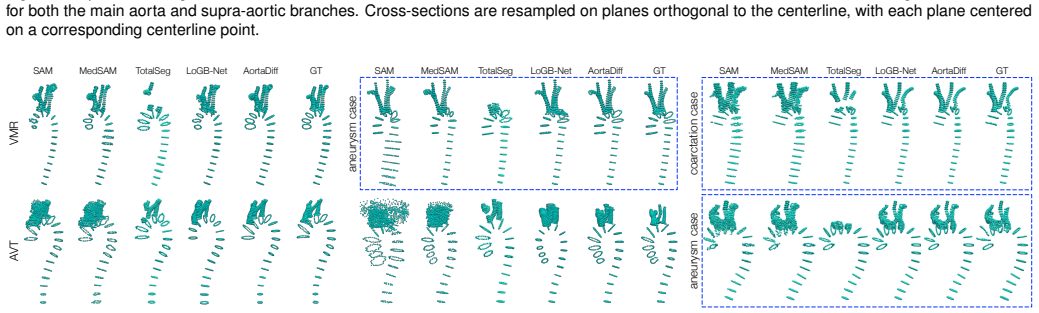
- [3.3, 4.3, 6] The claim of 'minimal dependency on large labeled datasets' is not supported by the experimental design. The proposed CDM generates only 16 coarse centerline points per branch; the actual vessel surface is determined by ScribblePrompt segmentations on orthogonal slices (Section 3.3) and by NURBS fitting. Section 4.3 explicitly credits ScribblePrompt's 'strong prior knowledge gained through large-scale self-supervised pretraining' for the segmentation quality, and Section 6 admits that the CDM-generated centerlines are 'not perfectly centered' and that 'the robustness of the segmentation module mitigates the major impact on performance.' The comparisons against LoGB-Net and TotalSeg in Tables 2-4 are therefore not a clean test of data efficiency: AortaDiff inherits a powerful pretrained segmentation prior while the supervised baselines are trained from comparatively limited aortic data. The authors should either restrict the minimal-dependency claim to the centerline-generation module or add an end-to-end ablation that replaces ScribblePrompt with SAM, MedSAM, or a non-learning contouring method and reports the resulting mesh metrics.
- [4.2, Table 1] The centerline baseline comparison may be unfair. The authors state that DPM, PVD, and DiT-3D were 'adapted' into conditional models 'by following AortaDiff's conditioning strategy,' but no hyperparameter tuning, training duration, or convergence evidence is provided for these baselines. The reported gaps are extremely large (e.g., overall CD 0.33 mm for AortaDiff versus 2.39 mm for DPM on VMR), which raises the concern that the baselines were not trained to their potential, rather than that AortaDiff is intrinsically superior. Please report the baseline hyperparameters, training iterations, learning-rate schedules, and per-seed variance, or use published conditional variants with their own recommended settings.
- [Appendix Section 4, Table A1] The number of centerline points (16) was selected through a parameter study conducted on all test cases from both VMR and AVT. This is test-set-based model selection and makes the reported metrics optimistic: the method and the evaluation set are no longer independent. The centerline point count should be chosen on a held-out validation split or justified through an independent criterion, and the final test metrics should be computed only after fixing that hyperparameter.
- [4.5, Figure 8] The CFD validation is exclusively qualitative. The paper claims 'a high degree of consistency' and 'comparable' velocity, pressure, and WSS fields between AortaDiff and ground-truth meshes, but no quantitative hemodynamic metrics are reported. Since 'CFD-compatible meshes' is a core contribution, the authors should provide numerical comparisons such as pressure-drop error, WSS error, or flow-rate distribution differences across branches. If such metrics are not available, the CFD claim should be softened to 'successful simulation execution' rather than geometric and hemodynamic equivalence.
minor comments (6)
- [Abstract and Section 1] The phrase 'minimal dependency on large labeled datasets' should be qualified to make clear that the dependency applies to aorta-specific annotations; as written, it conflicts with the use of ScribblePrompt, a model trained on large-scale medical imaging data.
- [3.3] ScribblePrompt is described as 'a SAM-based model,' but ScribblePrompt is not a variant of SAM in the original literature; please verify the description and cite the underlying architecture correctly.
- [3.1] The choice of 16 centerline points is introduced in the main text as 'empirical' and only justified later in Appendix Section 4; a forward reference to the parameter study would help the reader.
- [4.2] The definitions of chamfer distance, Hausdorff distance, and Earth mover's distance are given by references only; a one-line formula or a precise reference to the exact definitions used would improve reproducibility.
- [5] The 'Ad-Hoc Expert Feedback' section is anecdotal and not a substitute for the quantitative evaluation presented elsewhere; if retained, it should be clearly labeled as an informal case report rather than a validation result.
- [References] Reference [15] contains a malformed DOI ('arXiv.11474' instead of a full arXiv identifier); please correct it.
Circularity Check
No circularity: the CDM is standard supervised learning and the NURBS/CFD stages are independent; the minimal-data claim is weakened by reliance on ScribblePrompt but no prediction reduces to its own input.
full rationale
Score 1: no circular reduction is exhibited. The centerline model is trained on GT centerlines via the noise-prediction loss (Eq. 1) and evaluated against those GT centerlines (Table 1), which is standard supervised learning and evaluation, not a construction-level tautology. The NURBS surface construction (Eqs. 3-4) and the OpenFOAM CFD stage are independent geometric and numerical transforms that do not feed back into the training objective. The final mesh accuracy does rely heavily on ScribblePrompt, an external SAM-based model pretrained on large-scale medical data (Sec. 3.3, 4.3), and the paper credits 'ScribblePrompt's strong prior knowledge gained through large-scale self-supervised pretraining.' This undercuts the abstract's 'minimal dependency on large labeled datasets' claim, but that is a dependency/borrowed-prior concern, not circularity. The paper's own limitation statement (Sec. 6) acknowledges that CDM-generated centerlines are 'not perfectly centered' and that 'the robustness of the segmentation module mitigates the major impact on performance,' confirming that the external segmenter is load-bearing for mesh quality; still, nothing in the derivation is defined in terms of its own output. Self-citations [4, 14, 15] are related-work and baseline citations, and LoGB-Net is used as an external comparison baseline rather than as justification for AortaDiff's central claim. No specific circular step can be quoted, so no circularity steps are listed.
Assumptions & free parameters
free parameters (3)
- Number of centerline points per branch =
16
- Number of resampled contour points per cross-section =
32
- Diffusion training settings =
100,000 iterations, batch 16, LR 1e-3
assumptions (5)
- standard math Standard DDPM denoising objective (Eq. 1) learns centerline structure from limited samples.
- domain assumption ScribblePrompt pretrained on large medical datasets can segment aortic cross-sections from point prompts.
- ad hoc to paper A 16-point ordered centerline per branch sufficiently captures aortic morphology for contour-based NURBS reconstruction.
- domain assumption NURBS fitting with ICP-aligned contours yields watertight, CFD-compatible surfaces.
- domain assumption Steady incompressible Navier-Stokes with no-slip walls and prescribed inlet/outlet conditions approximates the relevant hemodynamics.
Cite this review
Pith. "Pith review of AortaDiff: Volume-Guided Conditional Diffusion Models for Multi-Branch Aortic Surface Generation." pith.science (2026). https://pith.science/paper/B75AIHYK
@misc{pith2026250713404,
  author       = {Pith},
  title        = {Pith review of: AortaDiff: Volume-Guided Conditional Diffusion Models for Multi-Branch Aortic Surface Generation},
  year         = {2026},
  howpublished = {\url{https://pith.science/paper/B75AIHYK}},
  note         = {Machine review of arXiv:2507.13404}
}
read the original abstract
Accurate 3D aortic construction is crucial for clinical diagnosis, preoperative planning, and computational fluid dynamics (CFD) simulations, as it enables the estimation of critical hemodynamic parameters such as blood flow velocity, pressure distribution, and wall shear stress. Existing construction methods often rely on large annotated training datasets and extensive manual intervention. While the resulting meshes can serve for visualization purposes, they struggle to produce geometrically consistent, well-constructed surfaces suitable for downstream CFD analysis. To address these challenges, we introduce AortaDiff, a diffusion-based framework that generates smooth aortic surfaces directly from CT/MRI volumes. AortaDiff first employs a volume-guided conditional diffusion model (CDM) to iteratively generate aortic centerlines conditioned on volumetric medical images. Each centerline point is then automatically used as a prompt to extract the corresponding vessel contour, ensuring accurate boundary delineation. Finally, the extracted contours are fitted into a smooth 3D surface, yielding a continuous, CFD-compatible mesh representation. AortaDiff offers distinct advantages over existing methods, including an end-to-end workflow, minimal dependency on large labeled datasets, and the ability to generate CFD-compatible aorta meshes with high geometric fidelity. Experimental results demonstrate that AortaDiff performs effectively even with limited training data, successfully constructing both normal and pathologically altered aorta meshes, including cases with aneurysms or coarctation. This capability enables the generation of high-quality visualizations and positions AortaDiff as a practical solution for cardiovascular research.
Figures


Figures from the paper (10 more)




Reference graph
Works this paper leans on
- [1]
- [2]
- [3]
-
[4]
D. An, P. Du, P. Gu, J.-X. Wang, and C. Wang. Hierarchical LoG Bayesian neural network for enhanced aorta segmentation. In Proceedings of IEEE International Symposium on Biomedical Imaging, pp. 1–5, 2025. doi: 10. 1109/ISBI60581.2025.10980947 6, 7
arXiv 2025
-
[5]
L. Antiga, M. Piccinelli, L. Botti, B. Ene-Iordache, A. Remuzzi, and D. A. Steinman. An image-based modeling framework for patient-specific computational hemodynamics. Medical & Biological Engineering & Computing, 46(11):1097–1112, 2008. doi: 10.1007/S11517-008-0420-1 5
-
[7]
H. G. Barrow, J. M. Tenenbaum, R. C. Bolles, and H. C. Wolf. Parametric correspondence and chamfer matching: Two new techniques for image matching. In Proceedings of International Joint Conference on Artificial Intelligence, pp. 659–663, 1977. doi: 10.5555/1622943.1622971 6
arXiv 1977
-
[8]
P. J. Besl and N. D. McKay. A method for registration of 3-D shapes.IEEE Transactions on Pattern Analysis and Machine Intelligence, 14(2):239– 256, 1992. doi: 10.1109/34.121791 4
-
[9]
S. M. Black, C. Maclean, P. H. Barrientos, K. Ritos, and A. Kazakidi. Reconstruction and validation of arterial geometries for computational fluid dynamics using multiple temporal frames of 4D flow-MRI magnitude images. Cardiovascular Engineering and Technology, 14(5):655–676,
Show all 80 references
-
[11]
M. Danu, C. I. Nita, A. Vizitiu, C. Suciu, and L. M. Itu. Deep learning based generation of synthetic blood vessel surfaces. In Proceedings of International Conference on System Theory, Control and Computing, pp. 662–667, 2019. doi: 10.1109/ICSTCC.2019.8885576 1
2019
-
[12]
L. R. Dice. Measures of the amount of ecologic association between species. Ecology, 26(3):297–302, 1945. doi: 10.2307/1932409 6
1945 doi
-
[13]
Dosovitskiy, L
A. Dosovitskiy, L. Beyer, A. Kolesnikov, D. Weissenborn, X. Zhai, T. Un- terthiner, M. Dehghani, M. Minderer, G. Heigold, S. Gelly, et al. An image is worth 16×16 words: Transformers for image recognition at scale. In Proceedings of International Conference on Learning Represe...
- [14]
-
[15]
P. Du, M. Xu, X. Zhu, and J.-X. Wang. HUG-V AS: A hierarchical NURBS- based generative model for aortic geometry synthesis and controllable editing. arXiv preprint arXiv:2507.11474, 2025. doi: 10.48550/arXiv. 11474 2
2025 doi
- [16]
-
[17]
Fantazzini, M
A. Fantazzini, M. Esposito, A. Finotello, F. Auricchio, B. Pane, C. Basso, G. Spinella, and M. Conti. 3D automatic segmentation of aortic computed tomography angiography combining multi-view 2D convolutional neural networks. Cardiovascular Engineering and Technology , 11:576–586,
-
[18]
S. F. F. Gibson. Using distance maps for accurate surface representation in sampled volumes. In Proceedings of IEEE Symposium on Volume Visualization, pp. 23–30, 1998. doi: 10.1145/288126.288142 2, 7
1998
-
[19]
R. L. Graham, T. S. Woodall, and J. M. Squyres. Open MPI: A flexible high performance MPI. In Proceedings of International Conference on Parallel Processing and Applied Mathematics, pp. 228–239, 2006. doi: 10 .1007/11752578_29 5
2006
-
[20]
Habijan, I
M. Habijan, I. Gali´c, H. Leventi´c, K. Romi´c, and D. Babin. Abdominal aortic aneurysm segmentation from CT images using modified 3D U-Net with deep supervision. In Proceedings of International Symposium on Electronics in Marine, pp. 123–128, 2020. doi: 10.1109/ELMAR49956. 20...
2020
-
[21]
H. K. Hahn, B. Preim, D. Selle, and H.-O. Peitgen. Visualization and interaction techniques for the exploration of vascular structures. In Pro- ceedings of IEEE Visualization Conference, pp. 395–402, 2001. doi: 10. 1109/VISUAL.2001.964538 3
2001
-
[22]
Hausdorff
F. Hausdorff. Grundzüge der Mengenlehre. Veit & Comp, Leipzig, 1914. 6
1914
-
[23]
W. He, Y . Cao, Y . Li, Y . Miao, W. Shi, F. He, F. Yan, Z. Jiang, and H. Zhang. A study on CT aorta segmentation using vessel enhancement diffusion DPM PVD Dit-3D AortaDiff GT VMRAVT Fig. 2: Comparison of centerline generation methods on the remaining test cases from the VMR ...
2015 doi
-
[24]
Heimann, B
T. Heimann, B. van Ginneken, M. Styner, Y . Arzhaeva, V . Aurich, C. Bauer, A. Beck, C. Becker, R. Beichel, G. Bekes, et al. Comparison and evaluation of methods for liver segmentation from CT datasets. IEEE Transactions on Medical Imaging, 28(8):1251–1265, 2009. doi: 10.1109/...
2009 doi
-
[25]
J. Ho, A. Jain, and P. Abbeel. Denoising diffusion probabilistic models. In Proceedings of Advances in Neural Information Processing Systems,
- [26]
-
[27]
Kahng, N
M. Kahng, N. Thorat, D. H. P. Chau, F. B. Viégas, and M. Wattenberg. GAN Lab: Understanding complex deep generative models using inter- active visual experimentation. IEEE Transactions on Visualization and Computer Graphics, 25(1):310–320, 2019. doi: 10.1109/TVCG.2018. 2864500 3
2019 doi
-
[29]
Kretschmer, C
J. Kretschmer, C. Godenschwager, B. Preim, and M. Stamminger. In- teractive patient-specific vascular modeling with sweep surfaces. IEEE Transactions on Visualization and Computer Graphics, 19(12):2828–2837,
-
[30]
T. P. Kuipers, P. Konduri, H. A. Marquering, and E. J. Bekkers. Generating cerebral vessel trees of acute ischemic stroke patients using conditional VMR GTAortaDiffLoGB-NetMedSAMSAM T otalSeg AVT Fig. 3: Comparison of contours extracted from different segmentation methods on t...
2024
-
[31]
Lawonn, S
K. Lawonn, S. Glaßer, A. Vilanova, B. Preim, and T. Isenberg. Occlusion- free blood flow animation with wall thickness visualization. IEEE Trans- actions on Visualization and Computer Graphics, 22(1):728–737, 2016. doi: 10.1109/TVCG.2015.2467961 2
2016
-
[32]
R. Li, X. Li, P.-A. Heng, and C.-W. Fu. Point cloud upsampling via disentangled refinement. In Proceedings of IEEE/CVF Conference on Computer Vision and Pattern Recognition, pp. 344–353, 2021. doi: 10. 1109/CVPR46437.2021.00041 3
2021
-
[33]
W. Lin, H. Liu, L. Gu, and Z. Gao. A geometry-constrained deformable attention network for aortic segmentation. In Proceedings of Interna- tional Conference on Medical Image Computing and Computer Assisted Interventions, pp. 287–296, 2022. doi: 10.1007/978-3-031-16443-9_28 2
2022 doi
-
[34]
W. E. Lorensen and H. E. Cline. Marching cubes: A high resolution 3D surface construction algorithm. In Proceedings of ACM SIGGRAPH Conference, pp. 163–169, 1987. doi: 10.1145/37401.37422 2, 5, 7
1987
-
[35]
Luo and W
S. Luo and W. Hu. Diffusion probabilistic models for 3D point cloud generation. In Proceedings of IEEE/CVF Conference on Computer Vision and Pattern Recognition, pp. 2837–2845, 2021. doi: 10.1109/CVPR46437 .2021.00286 1, 2, 3, 5
2021
-
[36]
J. Lyu, Y . Fu, M. Yang, Y . Xiong, Q. Duan, C. Duan, X. Wang, X. Xing, D. Zhang, J. Lin, et al. Generative adversarial network-based noncontrast CT angiography for aorta and carotid arteries. Radiology, 309(2):e230681,
-
[37]
J. Ma, Y . He, F. Li, L. Han, C. You, and B. Wang. Segment anything in medical images. Nature Communications, 15(1):654:1–654:9, 2024. doi: 10.1038/s41467-024-44824-z 6
2024 doi
-
[38]
Meuschke, T
M. Meuschke, T. Günther, P. Berg, R. Wickenhöfer, B. Preim, and K. La- wonn. Visual analysis of aneurysm data using statistical graphics. IEEE AVT GT LoGB-Net + marching cubes AortaDiff EFDs + marching cubes LoGB-Net + surface nets VMR Fig. 4: Comparison of mesh construction m...
-
[39]
Meuschke, U
M. Meuschke, U. Niemann, B. Behrendt, M. Gutberlet, B. Preim, and K. Lawonn. GUCCI - guided cardiac cohort investigation of blood flow data. IEEE Transactions on Visualization and Computer Graph- ics, 29(3):1876–1892, 2023. doi: 10.1109/TVCG.2021.3134083 2
2023
-
[40]
doi: 10.1148/radiol.230681 3
-
[41]
Meuschke, S
M. Meuschke, S. V oß, O. Beuing, B. Preim, and K. Lawonn. Com- bined visualization of vessel deformation and hemodynamics in cerebral aneurysms. IEEE Transactions on Visualization and Computer Graphics, 23(1):761–770, 2017. doi: 10.1109/TVCG.2016.2598795 2
2017
-
[42]
Mistelbauer, C
G. Mistelbauer, C. Rössl, K. Bäumler, B. Preim, and D. Fleischmann. Implicit modeling of patient-specific aortic dissections with elliptic Fourier descriptors. Computer Graphics Forum, 40(3):423–434, 2021. doi: 10. 1111/CGF.14318 1, 7
2021
-
[43]
S. Mo, E. Xie, R. Chu, L. Hong, M. Nießner, and Z. Li. DiT-3D: Exploring plain diffusion transformers for 3D shape generation. In Proceedings of Advances in Neural Information Processing Systems, 2023. 5
2023
-
[44]
Moccia, E
S. Moccia, E. D. Momi, S. E. Hadji, and L. S. Mattos. Blood vessel segmentation algorithms - review of methods, datasets and evaluation metrics. Computer Methods and Programs in Biomedicine, 158:71–91,
-
[45]
Meuschke, S
M. Meuschke, S. Oeltze-Jafra, O. Beuing, B. Preim, and K. Lawonn. Clas- sification of blood flow patterns in cerebral aneurysms. IEEE Transactions on Visualization and Computer Graphics, 25(7):2404–2418, 2019. doi: 10 .1109/TVCG.2018.2834923 2
2019
-
[46]
Myronenko, D
A. Myronenko, D. Yang, Y . He, and D. Xu. Aorta segmentation from 3D CT in MICCAI SEG.A. 2023 challenge. In Proceedings of Interna- tional Conference on Medical Image Computing and Computer Assisted Interventions, pp. 13–18, 2023. doi: 10.1007/978-3-031-53241-2_2 2
2023 doi
-
[47]
A. Q. Nichol and P. Dhariwal. Improved denoising diffusion probabilistic models. In Proceedings of International Conference on Machine Learning, pp. 8162–8171, 2021. 3
2021
-
[48]
Oeltze-Jafra, J
S. Oeltze-Jafra, J. R. Cebral, G. Janiga, and B. Preim. Cluster analysis of vortical flow in simulations of cerebral aneurysm hemodynamics. IEEE Transactions on Visualization and Computer Graphics, 22(1):757–766,
-
[49]
S. V . Patankar and D. B. Spalding. A calculation procedure for heat, mass and momentum transfer in three-dimensional parabolic flows. In Numerical Prediction of Flow, Heat Transfer, Turbulence and Combustion, chap. 5, pp. 54–73. Elsevier, 1983. doi: 10.1016/B978-0-08-030937-8...
1983 doi
-
[50]
Piegl and W
L. Piegl and W. Tiller. The NURBS Book (Monographs in Visual Commu- nication). Springer, second ed., 1996. 4
1996
-
[51]
Montalt-Tordera, E
J. Montalt-Tordera, E. Pajaziti, R. Jones, E. Sauvage, R. Puranik, A. A. V . Singh, C. Capelli, J. Steeden, S. Schievano, and V . Muthurangu. Auto- matic segmentation of the great arteries for computational hemodynamic assessment. Journal of Cardiovascular Magnetic Resonance, ...
-
[52]
Ronneberger, P
O. Ronneberger, P. Fischer, and T. Brox. U-Net: Convolutional networks for biomedical image segmentation. In Proceedings of International Con- ference on Medical Image Computing and Computer-Assisted Intervention, pp. 234–241, 2015. doi: 10.1007/978-3-319-24574-4_28 4
2015 doi
-
[53]
Rubner, C
Y . Rubner, C. Tomasi, and L. J. Guibas. A metric for distributions with ap- plications to image databases. In Proceedings of IEEE/CVF International Conference on Computer Vision, pp. 59–66, 1998. doi: 10.1109/iccv.1998 .710701 6
1998 doi
-
[54]
Selle, B
D. Selle, B. Preim, A. Schenk, and H. Peitgen. Analysis of vascula- ture for liver surgical planning. IEEE Transactions on Medical Imaging, 21(11):1344–1357, 2002. doi: 10.1109/TMI.2002.801166 3
2002
-
[55]
A. M. Shaker, M. Maaz, H. A. Rasheed, S. H. Khan, M. Yang, and F. S. Khan. UNETR++: Delving into efficient and accurate 3D medical image segmentation. IEEE Transactions on Medical Imaging, 43(9):3377–3390,
- [57]
-
[58]
Swillens, L
A. Swillens, L. Lanoye, J. D. Backer, N. Stergiopulos, P. R. Verdonck, F. Vermassen, and P. Segers. Effect of an abdominal aortic aneurysm on wave reflection in the aorta. IEEE Transactions on Biomedical En- gineering, 55(5):1602–1611, 2008. doi: 10.1109/TBME.2007.913994 1
2008
-
[59]
L. Radl, Y . Jin, A. Pepe, J. Li, C. Gsaxner, F.-H. Zhao, and J. Egger. A VT: Multicenter aortic vessel tree CTA dataset collection with ground truth segmentation masks. Data in Brief, 40:107801, 2022. doi: 10.1016/j.dib. 2022.107801 5
2022
-
[60]
Updegrove, N
A. Updegrove, N. M. Wilson, J. Merkow, H. Lan, A. L. Marsden, and S. C. Shadden. SimVascular: An open source pipeline for cardiovascular simulation. Annals of Biomedical Engineering, 45(3):525–541, 2017. 1
2017
-
[61]
T. P. Vagenas, K. Georgas, and G. K. Matsopoulos. Deep learning-based segmentation and mesh reconstruction of the aortic vessel tree from CTA images. In Proceedings of International Conference on Medical Image Computing and Computer Assisted Interventions, pp. 80–94, 2023. doi...
2023 doi
-
[62]
Valette, J
S. Valette, J. Chassery, and R. Prost. Generic remeshing of 3D triangular meshes with metric-dependent discrete V oronoi diagrams.IEEE Transac- tions on Visualization and Computer Graphics, 14(2):369–381, 2008. doi: 10.1109/TVCG.2007.70430 5
2008
-
[63]
Wasserthal, H.-C
J. Wasserthal, H.-C. Breit, M. T. Meyer, M. Pradella, D. Hinck, A. W. Sauter, T. Heye, D. T. Boll, J. Cyriac, S. Yang, et al. TotalSegmentator: Robust segmentation of 104 anatomic structures in CT images. Radiology: Artificial Intelligence, 5(5):e230024, 2023. doi: 10.1148/rya...
2023 doi
-
[64]
N. M. Wilson, A. K. Ortiz, and A. B. Johnson. The vascular model repository: A public resource of medical imaging data and blood flow simulation results. Journal of Medical Devices, 7(4):0409231, 2013. doi: 10.1115/1.4025983 5
2013 doi
-
[65]
J. M. Wolterink, T. Leiner, and I. Isgum. Blood vessel geometry synthesis using generative adversarial networks. arXiv preprint arXiv:1804.04381,
-
[66]
H. E. Wong, M. Rakic, J. V . Guttag, and A. V . Dalca. ScribblePrompt: Fast and flexible interactive segmentation for any biomedical image. In Proceedings of European Conference on Computer Vision, pp. 207–229,
-
[67]
L. Wu, D. Wang, C. Gong, X. Liu, Y . Xiong, R. Ranjan, R. Krishnamoorthi, V . Chandra, and Q. Liu. Fast point cloud generation with straight flows. In Proceedings of IEEE/CVF Conference on Computer Vision and Pattern Recognition, pp. 9445–9454, 2023. doi: 10.1109/CVPR52729.202...
2023
-
[68]
J. Tao, X. Huang, F. Qiu, C. Wang, J. Jiang, C. Shene, Y . Zhao, and D. Yu. VesselMap: A web interface to explore multivariate vascular data. Computers & Graphics, 59:79–92, 2016. doi: 10.1016/J.CAG.2016.05. 024 2
2016 doi
-
[69]
M. Ye, D. Yang, M. Kanski, L. Axel, and D. Metaxas. Neural deformable models for 3D bi-ventricular heart shape reconstruction and modeling from 2D sparse cardiac magnetic resonance imaging. In Proceedings of IEEE/CVF International Conference on Computer Vision, pp. 14247– 1425...
2023
-
[70]
Y . Zeng, V . M. Patel, H. Wang, X. Huang, T.-C. Wang, M.-Y . Liu, and Y . Balaji. JeDi: Joint-image diffusion models for finetuning-free personal- ized text-to-image generation. In Proceedings of IEEE/CVF Conference on Computer Vision and Pattern Recognition, pp. 6786–6795, 2...
2024
-
[71]
Zhang, Z
B. Zhang, Z. Lai, S. Liu, X. Xie, X. Zhou, Z. Hou, X. Ma, B. Liu, K. Li, and M. Song. SDLU-Net: A similarity-based dynamic linking network for the automated segmentation of abdominal aorta aneurysms and branching vessels. Biomedical Signal Processing and Control, 100:106991, 2...
2025
-
[72]
L. Zhou, Y . Du, and J. Wu. 3D shape generation and completion through point-voxel diffusion. In Proceedings of IEEE/CVF International Confer- ence on Computer Vision, pp. 5806–5815, 2021. doi: 10.1109/ICCV48922 .2021.00577 1, 3, 5
2021
-
[73]
Y . Zhou, H. Chang, X. Lu, and Y . Lu. DenseUNet: Improved image classification method using standard convolution and dense transposed convolution. Knowledge-Based Systems, 254:109658, 2022. doi: 10.1016/j .knosys.2022.109658 2
2022
- [75]
-
[77]
doi: 10.1007/978-3-031-73661-2_12 4
-
[79]
Xiong, Y
X. Xiong, Y . Ding, C. Sun, Z. Zhang, X. Guan, T. Zhang, H. Chen, H. Liu, Z. Cheng, L. Zhao, et al. A cascaded multi-task generative frame- work for detecting aortic dissection on 3-D non-contrast-enhanced com- puted tomography. IEEE Journal of Biomedical and Health Informatic...
2022
-
[2013]
doi: 10.1109/TVCG.2013.169 3
2013 doi
-
[2016]
doi: 10.1109/TVCG.2015.2467203 2
2015
-
[2018]
doi: 10.1016/j.cmpb.2018.02.001 1
2018 doi
-
[2019]
doi: 10.1109/TVCG.2018.2864509 2
2018
-
[2020]
doi: 10.1007/s13239-020-00481-z 1
-
[2022]
doi: 10.1186/s12968-022-00891-z 2
-
[2023]
doi: 10.1007/s13239-023-00679-x 3
-
[2024]
doi: 10.1109/TMI.2024.3398728 2
2024
Reviewed August 6, 2026 · model on record in the stance chip above.
Discussion (0). Continue with ORCID to comment.