REVIEW 4 major objections 5 minor 35 references
Volume Rendering of Human Hand Anatomy
T0 review · 4 major / 5 minor · reviewed 2026-08-12 · deepseek-v4-flash
Pith's one-line read Per-tissue transfer functions make moving hand MRI anatomy readable
desk verdict Solid hand-MRI rendering recipe with a weakly supported perceptual claim and a reproducibility gap. read the letter →
The pith
A machine-rendered reading of the paper's core claim, the machinery that carries it, and where it could break.
The reading
What carries the argument
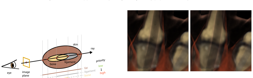
The load-bearing object is the per-tissue transfer function assignment. A transfer function is the mapping that turns each MRI scalar value into an RGB color and an opacity; here, instead of one global mapping, each tissue has its own mapping, and the tissue at a sample point is determined by ray-mesh intersections and the priority rule bone > tendon > muscle > ligament > fat. This assignment is what lets the renderer show several overlapping anatomical structures at once, keep boundaries smooth without voxel-level staircase artifacts, and vary appearance between the two styles. The two concrete transfer-function families in Tables 1 and 2 carry the result: non-fat tissues use a fixed tissue color modulated by MRI value, and fat uses histogram-frequency or intensity-modulated color and opacity, which is what makes the interior-emphasized and fat-emphasized styles behave differently.
What would settle it
Take one MRI frame and have an anatomist label the visible tissues directly on slices; render the same frame with the proposed transfer functions, then compare the rendered tissue boundaries with the labeled anatomy in regions where meshes overlap, for example ligament-bone interfaces. If a substantial fraction of rendered boundaries disagree with the labeled anatomy, the priority rule is not anatomically correct.
Extended reading notes
Core claim
On the paper's own terms, the central discovery is that high-quality volume rendering of the human hand is driven more by how MRI values are mapped per tissue than by the ray-casting algorithm itself. The method starts from known organ segmentations (bones, muscles, tendons, ligaments, skin, with fat defined as everything else inside the skin), intersects each camera ray with the organ meshes, and labels each ray segment by the highest-priority organ in that interval, with priority order bone > tendon > muscle > ligament > fat. Each material then gets its own transfer function; the paper gives two complete families, an interior-emphasized style that makes bones, muscles, tendons, and ligaments opaque with MRI-derived texture, and a fat-emphasized style that makes the subcutaneous fat the focus and reveals superficial veins without contrast medium. Because sampling happens inside ray segments rather than on a voxel grid, staircase artifacts disappear, and stochastic jittering with corrected opacities removes wood-grain banding. The paper's demonstration is that these choices produce clearer, more consistent renders than surface rendering and than an earlier volume-rendering method, across five animated MRI sequences.
Load-bearing premise
The load-bearing premise is that the supplied organ segmentations are accurate and that the fixed priority order bone > tendon > muscle > ligament > fat always puts a sample point in the correct tissue where meshes overlap, and the paper does not validate either against ground truth.
Editorial extensions
If this is right
- Rendering MRI hand volumes in motion with interior-emphasized transfer functions exposes bone marrow, compact bone, spongy bone, and palmar muscle texture that surface rendering cannot show from the same data.
- The fat-emphasized style makes superficial veins such as the dorsal metacarpal and palmar digital veins visible without injected contrast medium, because their MRI intensity differs from surrounding fat.
- The priority rule removes artifacts caused by overlapping segmented meshes, for example where ligaments intersect bones or tendons intersect muscles, so no surface penetration artifacts appear.
- Because the method runs on CPU at several seconds per frame, it can render complete animation sequences offline; the authors use it to visualize five hand motion sequences and note that it makes FEM instabilities in the fat layer easier to spot.
- Compared with voxel-level segmentation tools, ray-segment sampling avoids staircase artifacts at tissue boundaries, giving cleaner renders of segmented anatomy.
Reading between the lines
- The authors do not validate their material assignments against ground truth; a natural next step would be a blinded radiological study measuring whether the rendered boundaries match expert-labeled anatomy in overlap regions.
- The two styles are hand-tuned, so the same pipeline could support automatic or semi-automatic transfer-function optimization per tissue, for example by maximizing boundary contrast or matching a perceptual target.
- If the vein visibility without contrast agent is reproducible across subjects, it could motivate testing this rendering approach as a non-invasive screening tool for superficial hand vasculature, but that clinical claim is beyond the paper.
- Because the method relies only on segmented meshes and MRI values, it should transfer to other articulated joints such as the wrist, foot, or knee where segmented MRI volumes and animated meshes exist, though the paper does not test this.
Signed reviews
Editorial analysis
A structured set of objections, weighed in public.
Referee Report
Summary. The paper presents a volume rendering pipeline for MRI datasets of the human hand. Given an MRI volume and pre-existing segmentations of bones, muscles, tendons, ligaments, and skin, the method casts rays, intersects the organ meshes to decompose each ray into segments, assigns each sample a single material using a fixed priority rule (Equation 7), and applies per-tissue transfer functions before back-to-front compositing. The paper contributes two families of hand-designed transfer functions (an "interior-emphasized" style and a "fat-emphasized" style), and it addresses two rendering artifacts: staircase artifacts are avoided by performing material assignment through ray-mesh intersection rather than voxel-level segmentation, and wood-grain artifacts are reduced by stochastic jittering with the opacity correction in Equation 6. The method is evaluated on five animated MRI sequences with three camera configurations; the authors report CPU rendering times and compare their results qualitatively with surface rendering and with the earlier volume rendering method of Rhee et al. [28].
Significance. The rendering machinery is standard and the mathematical presentation of ray casting is sound; the opacity correction under jittered sampling (Equation 6) is correct, and the use of ray-mesh intersections to assign materials is a reasonable way to avoid voxel-level staircase artifacts. The paper gives useful and concrete transfer function recipes for hand MRI data, and it makes use of a publicly available dataset, which aids reproducibility of the inputs. The reported performance numbers (3.7-4.7 seconds per 1024x1024 frame on a CPU renderer) are also useful. However, the central claim of the paper—that the proposed transfer functions and rendering method improve the clarity and interpretability of hand anatomy visualization—is supported only by the authors' own qualitative descriptions of a small set of rendered images. There is no user study, no perceptual or task-based metric, and no independent anatomical ground truth. Because visualization quality is the paper's main contribution, this missing evidence is the key weakness and must be addressed before the claims can be accepted at face value.
major comments (4)
- [Abstract, Section 5.2, Section 6] The paper's central claim is that the method "improves hand anatomy visualization" and that the choice of transfer function "significantly influences the clarity and interpretability" of the resulting images. The only support for this claim is the authors' qualitative description of Figures 14-18. There is no user study, no perceptual metric, no objective image-quality measure, and no independent anatomical ground truth. Moreover, the comparison with Rhee et al. in Figure 18 uses different MRI data and only "similar" poses, so it is not a controlled comparison. Since the paper's stated contribution is specifically about visualization quality rather than computational performance, this gap is load-bearing. Either soften the claims substantially or add evidence such as a user study with task-based questions, an expert evaluation, or quantitative metrics (e.g., contrast, boundary accuracy, or structure visibility).
- [Section 4.2, Tables 1 and 2] The transfer functions are hand-tuned on the same five MRI sequences that are later used for the evaluation; Tables 1 and 2 state that "All values were carefully chosen for best visualization." This makes the side-by-side comparisons self-confirming: the method is inherently likely to look good on the exact data for which its parameters were optimized. The paper should provide a sensitivity analysis or ablation for the transfer function parameters, and ideally evaluate on a held-out MRI scan or subject to show that the parameters generalize rather than overfit the showcased sequences.
- [Section 4.1, Equation (7)] The material priority rule (bone > tendon > muscle > ligament > fat) is assumed to correctly resolve every case where organ meshes overlap, and the paper provides no validation of the underlying segmentations. The segmentation accuracy is taken from prior work, but the authors do not report how frequently meshes overlap, whether the fixed priority order ever produces anatomically wrong assignments, or how such errors would appear in the final renders. Because a wrong material assignment directly changes the transfer function and hence the displayed anatomy, this assumption needs at least a qualitative failure analysis or a comparison of the material assignment against a voxel-level segmentation or an independent ground truth.
- [Section 4.2, Tables 1 and 2] The power-law transfer function for muscle and fat is given as C = max(min(a*(s/smax)^b, 1.0), 0.0) * C_material, but the numeric values of a and b are never reported. Without these values, other researchers cannot reproduce the exact transfer functions or the rendered images. Please state the values of a and b (or provide an equally specific selection procedure), and likewise clarify how rho_fat(s) is normalized in the interior-emphasized style beyond the general description in the text.
minor comments (5)
- [Section 5.2, Figure 18] The comparison with Rhee et al. should explicitly state that the MRI data, poses, and camera setups are not identical, and that the comparison is therefore illustrative rather than a controlled head-to-head evaluation.
- [Section 4.2.1] The statement that the wrist region has "lower quality" MRI signal would be stronger if supported by a quantitative measure (e.g., signal-to-noise ratio) or at least a labeled example in Figure 6 or Figure 12 showing the affected region.
- [Table 4] The header of Table 4 appears garbled in the rendered text ("nf t(hr)" and the following row of numbers). Please reformat the table so that the column headers are clear and the timing entries are easy to read.
- [Section 5.2] The statement that volume rendering is useful to "debug" FEM simulations is interesting, but no specific example of a detected instability is shown; a small annotated example would make this claim concrete.
- [General] The paper does not state whether the rendering code or transfer function parameter files will be released. Given the importance of reproducibility for hand-crafted transfer functions, please add a statement about code/data availability.
Circularity Check
No significant circularity: transfer functions are explicitly hand-crafted design choices, and the qualitative comparisons do not reduce to the paper's own inputs.
full rationale
The paper's central contribution is a set of hand-crafted per-tissue transfer functions for volume rendering hand MRI data. Tables 1 and 2 state the transfer functions explicitly and note that 'All values were carefully chosen for best visualization,' which is an open design choice rather than a hidden fit to a target outcome. The rendered images are direct outputs of these stated functions, so the observation that the functions produce their intended emphasis is self-consistent but not circular. The material assignment priority rule (Equation 7) is an explicit design decision for resolving mesh overlaps, not a consequence of the rendered results. The stochastic-jittering and opacity-correction discussion (Equation 6) is a standard numerical correction, not a result that presupposes the paper's conclusions. The use of the authors' own MRI dataset and meshes from Wang et al. (2019, 2020) is data and geometry sourcing; the visualization-quality claim is not entailed by the dataset itself, and those prior works are cited as external sources rather than as a justification for the transfer-function design. The absence of a user study or objective perceptual metric is an evaluation limitation, but under the hard rules it is not a form of circularity because the paper does not derive its claim from a metric that it also defines. Overall, no step in the derivation chain reduces to its own inputs or to an unverified self-citation in a load-bearing way.
Assumptions & free parameters
free parameters (3)
- Per-tissue color and opacity constants =
Bone (244,214,145), opacity 1.0; muscle (255,98,56), opacity 1.0; ligament (170,170,170), opacity 1.0; tendon…
- Parameters a and b in the power-law transfer function =
Not specified in the paper
- Fat histogram normalization rho_fat(s)/rho_fat_max =
Computed from the MRI histogram of the fat region
assumptions (5)
- standard math Volume rendering integral and discrete compositing (Eqs. 2-5) are valid for the MRI data
- domain assumption The input MRI scans have been segmented into accurate organ meshes
- ad hoc to paper The material priority rule (bone > tendon > muscle > ligament > fat, Eq. 7) correctly resolves overlapping meshes
- domain assumption MRI signal intensity is consistent enough within each tissue type to use a single transfer function per tissue
- ad hoc to paper The hand-crafted transfer function parameters and styles are suitable for the intended visualization goals
Cite this review
Pith. "Pith review of Volume Rendering of Human Hand Anatomy." pith.science (2026). https://pith.science/paper/L34FYQXO
@misc{pith2026241118630,
  author       = {Pith},
  title        = {Pith review of: Volume Rendering of Human Hand Anatomy},
  year         = {2026},
  howpublished = {\url{https://pith.science/paper/L34FYQXO}},
  note         = {Machine review of arXiv:2411.18630}
}
read the original abstract
We study the design of transfer functions for volumetric rendering of magnetic resonance imaging (MRI) datasets of human hands. Human hands are anatomically complex, containing various organs within a limited space, which presents challenges for volumetric rendering. We focus on hand musculoskeletal organs because they are volumetrically the largest inside the hand, and most important for the hand's main function, namely manipulation of objects. While volumetric rendering is a mature field, the choice of the transfer function for the different organs is arguably just as important as the choice of the specific volume rendering algorithm; we demonstrate that it significantly influences the clarity and interpretability of the resulting images. We assume that the hand MRI scans have already been segmented into the different organs (bones, muscles, tendons, ligaments, subcutaneous fat, etc.). Our method uses the hand MRI volume data, and the geometry of its inner organs and their known segmentation, to produce high-quality volume rendering images of the hand, and permits fine control over the appearance of each tissue. We contribute two families of transfer functions to emphasize different hand tissues of interest, while preserving the visual context of the hand. We also discuss and reduce artifacts present in standard volume ray-casting of human hands. We evaluate our volumetric rendering on five challenging hand motion sequences. Our experimental results demonstrate that our method improves hand anatomy visualization, compared to standard surface and volume rendering techniques.
Figures


Figures from the paper (11 more)




Reference graph
Works this paper leans on
-
[28]
T. Rhee, J. P. Lewis, U. Neumann, and K. Nayak. Scan-based volume animation driven by locally adaptive articulated registrations. IEEE Transactions on Visualization and Computer Graphics, 17(3):368–379, 2010
work page 2010
-
[1]
S. Arens and G. Domik. A survey of transfer functions suitable for volume rendering. In VG@ Eurographics, pp. 77–83, 2010
work page 2010
-
[2]
Hand and Wrist Anatomy website, 2022
Arthritis. Hand and Wrist Anatomy website, 2022. https://www.arthritis.org/health-wellness/about-arthritis/where- it-hurts/hand-and-wrist-anatomy
work page 2022
-
[3]
S. Bruckner, S. Grimm, A. Kanitsar, and M. E. Groller. Illustrative context-preserving exploration of volume data. IEEE Transactions on Visualization and Computer Graphics, 12(6):1559–1569, 2006
work page 2006
-
[4]
J. J. Caban and P. Rheingans. Texture-based transfer functions for direct volume rendering. IEEE Transactions on Visualization and Computer Graphics, 14(6):1364–1371, 2008
work page 2008
- [5]
-
[6]
G. G. Cameron and P. E. Undrill. Rendering volumetric medical image data on a simd-architecture computer. In Proceedings of the Third Eurographics Workshop on Rendering, pp. 135–145, 1992
work page 1992
-
[7]
C. Clinic. Blood Vessels website, 2022. https://my.clevelandclinic.org/health/body/21640-blood-vessels
work page 2022
Show all 35 references
-
[8]
Correa and K.-L
C. Correa and K.-L. Ma. Size-based transfer functions: A new vol- ume exploration technique. IEEE transactions on visualization and computer graphics, 14(6):1380–1387, 2008
2008
-
[9]
Engel, M
K. Engel, M. Hadwiger, J. M. Kniss, A. E. Lefohn, C. R. Salama, and D. Weiskopf. Real-time volume graphics. In ACM Siggraph 2004 Course Notes, pp. 29–es. 2004
2004
-
[10]
F. A. Fellner. Introducing cinematic rendering: a novel technique for post-processing medical imaging data. Journal of Biomedical Science and Engineering, 9(3):170–175, 2016
2016
-
[11]
Hadwiger, C
M. Hadwiger, C. Berger, and H. Hauser. High-quality two-level volume rendering of segmented data sets on consumer graphics hardware. In IEEE Visualization, 2003. VIS 2003., pp. 301–308. IEEE, 2003
2003
-
[12]
Kindlmann and J
G. Kindlmann and J. W. Durkin. Semi-automatic generation of transfer functions for direct volume rendering. In IEEE Symposium on Volume Visualization (Cat. No. 989EX300), pp. 79–86. IEEE, 1998
1998
-
[13]
Kindlmann, R
G. Kindlmann, R. Whitaker, T. Tasdizen, and T. Moller. Curvature- based transfer functions for direct volume rendering: Methods and applications. In IEEE Visualization, 2003. VIS 2003. , pp. 513–520. IEEE, 2003
2003
-
[14]
Kniss, G
J. Kniss, G. Kindlmann, and C. Hansen. Interactive volume rendering using multi-dimensional transfer functions and direct manipulation widgets. In Proceedings Visualization, 2001. VIS’01., pp. 255–562. IEEE, 2001
2001
-
[15]
Kniss, G
J. Kniss, G. Kindlmann, and C. Hansen. Multidimensional transfer functions for interactive volume rendering. IEEE Transactions on visualization and computer graphics, 8(3):270–285, 2002
2002
-
[16]
Kroes, F
T. Kroes, F. H. Post, and C. P. Botha. Exposure render: An interactive photo-realistic volume rendering framework. PloS one, 7(7):e38586, 2012
2012
-
[17]
Kr ¨uger and R
J. Kr ¨uger and R. Westermann. Acceleration techniques for gpu-based volume rendering. In Visualization Conference, IEEE, pp. 38–38. IEEE Computer Society, 2003
2003
-
[18]
Lacroute and M
P. Lacroute and M. Levoy. Fast volume rendering using a shear-warp factorization of the viewing transformation. In Proceedings of the 21st annual conference on Computer graphics and interactive techniques, pp. 451–458, 1994
1994
-
[19]
D. H. Laidlaw, K. W. Fleischer, and A. H. Barr. Partial-volume bayesian classification of material mixtures in mr volume data using voxel his- tograms. IEEE transactions on medical imaging, 17(1):74–86, 1998
1998
-
[20]
M. Levoy. Display of surfaces from volume data. IEEE Computer graphics and Applications, 8(3):29–37, 1988
1988
-
[21]
Ljung, J
P. Ljung, J. Kr ¨uger, E. Groller, M. Hadwiger, C. D. Hansen, and A. Ynnerman. State of the art in transfer functions for direct volume rendering. In Computer Graphics Forum, vol. 35, pp. 669–691. Wiley Online Library, 2016
2016
-
[22]
W. E. Lorensen and H. E. Cline. Marching cubes: A high resolution 3d surface construction algorithm. ACM siggraph computer graphics, 21(4):163–169, 1987
1987
-
[23]
Lundstrom, P
C. Lundstrom, P. Ljung, and A. Ynnerman. Local histograms for design of transfer functions in direct volume rendering. IEEE Transactions on visualization and computer graphics, 12(6):1570–1579, 2006
2006
-
[24]
N. Max. Optical models for direct volume rendering. IEEE Transac- tions on Visualization and Computer Graphics, 1(2):99–108, 1995
1995
-
[25]
J. H. Medicine. Anatomy of the Hands website, 2022. https://www.hopkinsmedicine.org/health/treatment-tests-and- therapies/anatomy-of-the-hand
2022
-
[26]
Meißner, J
M. Meißner, J. Huang, D. Bartz, K. Mueller, and R. Crawfis. A practical evaluation of popular volume rendering algorithms. In Proceedings of the 2000 IEEE symposium on Volume visualization, pp. 81–90, 2000
2000
-
[27]
Rezk-Salama, M
C. Rezk-Salama, M. Hadwiger, T. Ropinski, and P. Ljung. Advanced illumination techniques for gpu-based volume raycasting. ACM SIG- GRAPH Asia Course Notes, 10(1508044.1508045), 2008
2008
-
[29]
C. R. Salama. Gpu-based monte-carlo volume raycasting. In 15th Pacific Conference on Computer Graphics and Applications (PG’07), pp. 411–414. IEEE, 2007
2007
-
[30]
Tappenbeck, B
A. Tappenbeck, B. Preim, and V . Dicken. Distance-based transfer function design: Specification methods and applications. In SimVis, pp. 259–274, 2006
2006
-
[31]
Totsuka and M
T. Totsuka and M. Levoy. Frequency domain volume rendering. In Proceedings of the 20th annual conference on Computer graphics and interactive techniques, pp. 271–278, 1993
1993
-
[32]
Van Gelder and K
A. Van Gelder and K. Kim. Direct volume rendering with shading via three-dimensional textures. In Proceedings of 1996 Symposium on Volume Visualization, pp. 23–30. IEEE, 1996
1996
-
[33]
B. Wang, G. Matcuk, and J. Barbi ˇc. Hand modeling and simulation using stabilized magnetic resonance imaging. ACM Trans. on Graphics (SIGGRAPH 2019), 38(4), 2019
2019
-
[34]
B. Wang, G. Matcuk, and J. Barbi ˇc. Hand MRI dataset, 2020. http://www.jernejbarbic.com/hand-mri-dataset
2020
-
[35]
L. A. Westover. Splatting: a parallel, feed-forward volume rendering algorithm. PhD thesis, The University of North Carolina at Chapel Hill, 1991
1991
Reviewed August 12, 2026 · model on record in the stance chip above.
Discussion (0). Continue with ORCID to comment.