REVIEW 4 major objections 5 minor 1 cited by
Atlas is Your Perfect Context: One-Shot Customization for Generalizable Foundational Medical Image Segmentation
T0 review · 4 major / 5 minor · reviewed 2026-08-03 · deepseek-v4-flash
Pith's one-line read One annotated atlas is enough to customize frozen medical segmentation models to new clinical contexts.
desk verdict AtlasSegFM is a practical, well-evaluated one-shot customization pipeline for segmentation foundation models, but the adaptive fusion module's contribution is under-validated and the SOTA claim needs more statistics. read the letter →
The pith
A machine-rendered reading of the paper's core claim, the machinery that carries it, and where it could break.
The reading
What carries the argument
The load-bearing object is the registered atlas mask Matlas, produced by test-time registration of a single annotated atlas to each query volume. Registration is carried out by a lightweight VoxelMorph-derived network optimized on the fly with a similarity loss, after rigid and affine pre-registration; this yields a global but coarse structural prior. Matlas does double duty: it becomes the prompt that contextualizes the frozen foundation model, and it is the second input to the fusion module. Fusion is implemented as a learnable Kalman filter, Mfinal = Mfm + K·(Matlas − Mfm), where K is a sigmoid-bounded gain predicted by a small 3D convolutional network with multi-scale pooling; this lets
What would settle it
Run AtlasSegFM on a population with large anatomical variation (for example, pediatric, post-surgical, or heavily deformed patients) using one atlas drawn from the far end of the distribution, and measure whether final Dice tracks the quality of the registered atlas mask. If final accuracy collapses to foundation-model-only levels when the atlas prior is poor — or if a strong Matlas fails to produce a good final segmentation — the claim that a single atlas can carry the context is falsified.
Extended reading notes
Core claim
The paper's central claim is that the context needed to customize a segmentation foundation model can be supplied entirely by an atlas at inference time, without any task-specific training. Given a context atlas and a query volume, AtlasSegFM estimates a deformation field by test-time optimization of a VoxelMorph-style registration network, pre-aligned with rigid and affine stages, and warps the atlas mask to produce a coarse structural prior Matlas. That prior is converted into a prompt (center click, bounding box, or the mask itself) matched to the foundation model's preferred input format — the dense mask for nnInteractive, a click for MedSAM2, no prompt for vesselFM — and the foundation
Load-bearing premise
The pipeline assumes that a single atlas and each query image share enough anatomical correspondence for test-time registration to produce a meaningful coarse mask; when a patient's anatomy differs markedly from the atlas, both the prompt and the fusion signal degrade, and the paper's own failure cases show adjacent-organ confusion and missing structures.
Editorial extensions
If this is right
- Clinical sites can customize a single frozen foundation model to a new target structure using one annotated example, removing the need to curate and train on large task-specific datasets.
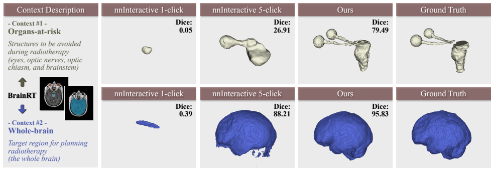
- The hardest cases for foundation models — small, delicate, under-represented structures like optic nerves and veins — receive the largest accuracy gains, because the atlas prior supplies anatomical context the pretrained model lacks.
- The same pipeline accommodates different foundation models and prompt formats, from dense-mask prompting (nnInteractive) to single-click prompting (MedSAM2) to non-promptable domain models (vesselFM), suggesting the atlas-context recipe is model-agnostic.
- On abdominal organs, one-shot AtlasSegFM approaches the accuracy of fully supervised nnU-Net, indicating that atlas-guided customization can be a practical alternative when target-domain annotations are scarce.
- Operating directly on full 3D volumes keeps inference at 1.8 minutes per image with only 1.4 million learnable parameters, well below slice-wise 2D in-context baselines that take 2–18 minutes per image.
Reading between the lines
- The critical path to wider adoption is registration robustness: whenever a patient's anatomy diverges markedly from the chosen atlas, the coarse prior degrades, and the paper's own failure cases (adjacent-organ confusion on Abd-CT, missing aortic-arch segments on AVT) suggest that a multi-atlas selector or atlas ensemble, not a bigger fusion network, is the most direct extension.
- Because the atlas carries the clinician's own delineation conventions, swapping the atlas should let the same frozen foundation model match different physicians' definitions of the same structure — a natural test of whether one-shot customization can encode subjective protocol rather than just anatomy.
- The learned Kalman gain K could be repurposed as a per-voxel confidence map, giving clinicians a principled uncertainty estimate alongside the segmentation; the paper does not report this, but the fusion equation makes it a direct readout.
- A boundary test of the method's scope is lesion segmentation: focal lesions are not consistently present in an atlas, so the structural-correspondence premise breaks down; the paper acknowledges this limitation, implying the method is best suited to anatomically consistent structures.
Editorial analysis
A structured set of objections, weighed in public.
Referee Report
Summary. The paper proposes AtlasSegFM, a one-shot customization framework for medical image segmentation foundation models. Given a single annotated atlas (support) for a new clinical context, AtlasSegFM (1) registers the atlas to each query volume with a test-time optimized VoxelMorph-style network to produce a coarse mask, (2) converts that mask into prompts (click/box/mask) for a frozen interactive or non-interactive foundation model, and (3) fuses the atlas prior and foundation-model prediction with a lightweight convolutional network that predicts a per-voxel gain. The method is evaluated on six datasets (Abd-CT, Abd-MR, AVT, Fe-MRA, OASIS, BrainRT) and claims an average Dice of 80.72%, outperforming uncustomized foundation models and in-context learning baselines, with especially large gains on small structures such as organs-at-risk.
Significance. If the reported results are robust, the contribution is practically valuable: AtlasSegFM leverages off-the-shelf foundation models, requires no target-domain training, uses only one annotated atlas, and processes full 3D volumes at a modest inference cost (1.8 min/image with 1.4M learnable parameters). The paper also includes useful ablations of the registration backend, prompt types, and fusion module, and it makes a credible effort to compare against recent ICL baselines. However, the central claim rests on an adaptive fusion module whose behavior is not sufficiently characterized, and the evaluation lacks uncertainty quantification. The main methodological ideas — atlas-guided prompting and test-time fusion — are worth publishing, but the current evidence does not fully establish the claimed state-of-the-art generality.
major comments (4)
- [§3.5, Eq. (4)–(5); §4.1] The adaptive fusion module g is trained on the single support label with a Dice loss. Since the support is the atlas itself, during fusion training Matlas equals the support label after registration, so the network can minimize loss by predicting K≈1 everywhere. The final Dice exceeding atlas-only Dice suggests that early stopping at 100 iterations or architectural constraints prevent collapse, but this is not analyzed. No ablation is provided against a non-learned fusion rule (e.g., fixed weighted average or uncertainty-weighted fusion), and no sensitivity to fusion training iterations, learning rate, or support choice is reported. The gains attributed to fusion in Table 4 could therefore be partly an artifact of the optimization schedule. Please add these controls and report variance across runs and support selections.
- [Tables 1–3; §4.1] All reported numbers are point estimates without error bars, confidence intervals, or significance tests. The protocol selects one support from the remaining validation set per fold, but no standard deviation across folds or across different atlas choices is given. Two of the six datasets (Fe-MRA and BrainRT) are in-house and not available, so their results cannot be independently checked. This weakens the central claim that the average 80.72% Dice is state-of-the-art: favorable atlas selection or tuning could produce the same aggregated result. Please report per-context means with std/CI, paired significance against the strongest baseline, and per-case results for the in-house datasets (or a release plan).
- [§3.4; Supplementary Table 6] The prompt-type choice is presented as a flexible design, but the supplementary ablation shows that the optimal prompt type is model-specific (Mask for nnInteractive, Click for MedSAM2) and was selected using target-domain labels on Abd-CT. This is effectively a hyperparameter tuned on the evaluation set, which conflicts with the 'no training/fine-tuning' claim and with the claim that the pipeline customizes any foundation model without per-context adjustment. Please state explicitly whether the prompt type was fixed a priori, whether it was selected once on a validation split and then frozen, and whether the same choice transfers across datasets. If prompt selection used target labels, the protocol should be described as including light validation-set tuning.
- [§5, Limitations; Fig. 8] The paper acknowledges that the method fails when the query anatomy differs markedly from the atlas, and Fig. 8 shows cases with Dice 29.39, 37.29, and 40.07. This is a structural precondition of the one-atlas approach, not a minor edge case. To support the claim of 'generalizable foundational medical image segmentation', please quantify the frequency and severity of such failures across all datasets, e.g., by stratifying results according to registration quality (atlas-only Dice) or anatomical similarity. Without this, a reader cannot assess the practical deployability of the method beyond the favorable average.
minor comments (5)
- [§3.5] The module in Eq. (4) is called a 'Kalman filter', but it is a static learned weighted average with no temporal or state recursion. Consider renaming it 'learned adaptive fusion' to avoid conceptual mismatch.
- [§5, abstract] The paper states an average Dice of 80.72% but does not specify over which contexts (six datasets vs. seven context-level tasks, since BrainRT has two contexts). Please state the averaging rule.
- [Table 1, footnote] In the supervised group, Iris is listed with target-dataset training, but in Tables 2 and 3 it appears in the 'In-context Models' section. Clarify the training data for each baseline in each table to avoid seeming inconsistent.
- [Supplementary §6] The sentence 'nnInteractive was designed to refine segmentation iteratively based on user interactions (which are essentially sparse masks)' is confusing because masks are dense, not sparse. Rephrase.
- [§4.1] For Fe-MRA, Table 3 reports nnInteractive and vesselFM, but the implementation details state that vesselFM is used for vascular and nnInteractive for other scenarios. Clarify the exact protocol for this dataset.
Circularity Check
No significant circularity: the central claim is an empirical pipeline result, with no derivation that reduces to its own inputs.
full rationale
AtlasSegFM is an empirical one-shot adaptation pipeline, not a first-principles derivation. The only learned components are optimized at test time from the single support (atlas) example: the registration network (Eq. 2) is optimized with an image-similarity loss against the query image, and the fusion gain K (Eqs. 4-5) is optimized with a Dice loss against the support label (Sec. 4.1). This is standard one-shot learning, and the reported evaluations are on held-out query images, not on the support example itself. The fusion module's training supervision is indeed the atlas mask, which in principle could encourage K to collapse toward 1 and copy M_atlas; however, the paper's own ablations (Table 4) show final Dice above the atlas-only Dice, indicating the learned K is not trivially the identity on the evaluated queries. Whether this holds robustly across support choices and hyperparameters is an empirical robustness concern, not a circularity. The paper's self-citations (Iris [6], UTNet [5]) appear only as baselines and related work and are not load-bearing for the central claim. No uniqueness theorem, ansatz-by-citation, or renaming pattern is present. The acknowledged limitations (anatomical mismatch, failure cases in Fig. 8) are boundary conditions of the method, not circular steps. Therefore no specific reduction from the paper's own equations or citations can be exhibited, and the appropriate finding is no significant circularity.
Assumptions & free parameters
free parameters (4)
- Fusion network g parameters =
≈0.1M weights, optimized on the per-context support for 100 iterations (LR 1e-5)
- Registration network parameters (test-time) =
≈1.3M weights, optimized per atlas-query pair for 1000 iterations (LR 1e-4)
- Prompt-type choice per foundation model =
Mask for nnInteractive; Click for MedSAM2
- Hyperparameters (LR, iterations, kernel sizes) =
LR 1e-4/1e-5, 1000/100 iterations, pooling kernels 3/5/7, U-Net 32 channels
assumptions (4)
- domain assumption A single atlas shares sufficient structural correspondence with query images for test-time registration to yield a meaningful prior mask.
- domain assumption The frozen foundation model's prompt-following behavior generalizes from the atlas-derived prompts to the target structures.
- domain assumption A 100-iteration, single-support training of the fusion module yields a fusion rule that transfers to queries.
- standard math The 5-fold cross-validation protocol from SSL-ALP [27] is a valid few-shot evaluation.
Cite this review
Pith. "Pith review of Atlas is Your Perfect Context: One-Shot Customization for Generalizable Foundational Medical Image Segmentation." pith.science (2026). https://pith.science/paper/WMOG2S5R
@misc{pith2026251218176,
  author       = {Pith},
  title        = {Pith review of: Atlas is Your Perfect Context: One-Shot Customization for Generalizable Foundational Medical Image Segmentation},
  year         = {2026},
  howpublished = {\url{https://pith.science/paper/WMOG2S5R}},
  note         = {Machine review of arXiv:2512.18176}
}
read the original abstract
Accurate segmentation of anatomical structures in medical images is essential for diagnosis and treatment planning. While recent interactive segmentation foundation models enhance generalization through large-scale multimodal pretraining, they still depend on precise prompts and can fail in underrepresented clinical contexts (e.g., small organs-at-risk). We present AtlasSegFM, an atlas-guided framework that customizes off-the-shelf foundation models to new clinical contexts with a single annotated example. AtlasSegFM 1) performs atlas-query registration to generate context-aware prompts, 2) refines the segmentation with a frozen foundation model, and 3) applies a lightweight adaptive fusion module to combine atlas priors with foundation-model inputs and predictions. Extensive experiments on six public and in-house datasets across radiotherapy and vascular scenarios show consistent gains, with the largest improvements on small and delicate structures. AtlasSegFM provides a lightweight, deployable solution for one-shot customization of segmentation foundation models in real-world clinical workflows.
Figures


Figures from the paper (6 more)


Forward citations
Cited by 1 Pith paper
-
Automatic LV Localization and Short-Axis Plane Estimation from Arbitrary CMR Slice
A single CMR slice can localize the left ventricle and regress its continuous 3D short-axis orientation, with a proposed PCC embedding achieving 3.39° average angle error across four datasets.
Reference graph
Works this paper leans on
-
[1]
V oxelmorph: a learning framework for deformable medical image registration.IEEE Transactions on Medical Imaging, 38(8):1788–1800, 2019
Guha Balakrishnan, Amy Zhao, Mert R Sabuncu, John Guttag, and Adrian V Dalca. V oxelmorph: a learning framework for deformable medical image registration.IEEE Transactions on Medical Imaging, 38(8):1788–1800, 2019. 3, 4, 5, 7
2019
-
[2]
Fam- net: Frequency-aware matching network for cross-domain few-shot medical image segmentation
Yuntian Bo, Yazhou Zhu, Lunbo Li, and Haofeng Zhang. Fam- net: Frequency-aware matching network for cross-domain few-shot medical image segmentation. InAAAI, pages 1889– 1897, 2025. 7
2025
-
[3]
Uni- verseg: Universal medical image segmentation
Victor Ion Butoi, Jose Javier Gonzalez Ortiz, Tianyu Ma, Mert R Sabuncu, John Guttag, and Adrian V Dalca. Uni- verseg: Universal medical image segmentation. InICCV, pages 21438–21451, 2023. 2, 3, 5, 6, 7
2023
-
[4]
Few-shot medical image segmenta- tion via generating multiple representative descriptors.IEEE Transactions on Medical Imaging, 43(6):2202–2214, 2024
Ziming Cheng, Shidong Wang, Tong Xin, Tao Zhou, Haofeng Zhang, and Ling Shao. Few-shot medical image segmenta- tion via generating multiple representative descriptors.IEEE Transactions on Medical Imaging, 43(6):2202–2214, 2024. 3, 5, 7
2024
-
[5]
Utnet: a hy- brid transformer architecture for medical image segmentation
Yunhe Gao, Mu Zhou, and Dimitris N Metaxas. Utnet: a hy- brid transformer architecture for medical image segmentation. InMICCAI, pages 61–71, 2021. 2
2021
-
[6]
Show and segment: Universal medical image segmentation via in-context learning
Yunhe Gao, Di Liu, Zhuowei Li, Yunsheng Li, Dongdong Chen, Mu Zhou, and Dimitris N Metaxas. Show and segment: Universal medical image segmentation via in-context learning. InCVPR, pages 20830–20840, 2025. 2, 3, 5, 6, 7
2025
-
[7]
3dsam-adapter: Holistic adaptation of sam from 2d to 3d for promptable tumor segmentation.Medical Image Analysis, 98:103324, 2024
Shizhan Gong, Yuan Zhong, Wenao Ma, Jinpeng Li, Zhao Wang, Jingyang Zhang, Pheng-Ann Heng, and Qi Dou. 3dsam-adapter: Holistic adaptation of sam from 2d to 3d for promptable tumor segmentation.Medical Image Analysis, 98:103324, 2024. 3
2024
-
[8]
Synth- morph: learning contrast-invariant registration without ac- quired images.IEEE Transactions on Medical Imaging, 41 (3):543–558, 2021
Malte Hoffmann, Benjamin Billot, Douglas N Greve, Juan Eu- genio Iglesias, Bruce Fischl, and Adrian V Dalca. Synth- morph: learning contrast-invariant registration without ac- quired images.IEEE Transactions on Medical Imaging, 41 (3):543–558, 2021. 3
2021
Show all 54 references
-
[9]
Learning the ef- fect of registration hyperparameters with hypermorph.The Journal of Machine Learning for Biomedical Imaging, 1:003,
Andrew Hoopes, Malte Hoffmann, Douglas N Greve, Bruce Fischl, John Guttag, and Adrian V Dalca. Learning the ef- fect of registration hyperparameters with hypermorph.The Journal of Machine Learning for Biomedical Imaging, 1:003,
-
[10]
Icl-sam: Synergizing in-context learning model and sam in medical image segmentation
Jiesi Hu, Yang Shang, Yanwu Yang, Xutao Guo, Hanyang Peng, and Ting Ma. Icl-sam: Synergizing in-context learning model and sam in medical image segmentation. InMIDL, pages 641–656, 2024. 3
2024
-
[11]
Multi-atlas seg- mentation of biomedical images: a survey.Medical Image Analysis, 24(1):205–219, 2015
Juan Eugenio Iglesias and Mert R Sabuncu. Multi-atlas seg- mentation of biomedical images: a survey.Medical Image Analysis, 24(1):205–219, 2015. 3
2015
-
[12]
nnu-net: a self-configuring method for deep learning-based biomedical image segmentation.Na- ture Methods, 18(2):203–211, 2021
Fabian Isensee, Paul F Jaeger, Simon AA Kohl, Jens Petersen, and Klaus H Maier-Hein. nnu-net: a self-configuring method for deep learning-based biomedical image segmentation.Na- ture Methods, 18(2):203–211, 2021. 3, 5, 6
2021
-
[13]
nninteractive: Redefining 3d promptable segmentation
Fabian Isensee, Maximilian Rokuss, Lars Krämer, Stefan Dinkelacker, Ashis Ravindran, Florian Stritzke, Benjamin Hamm, Tassilo Wald, Moritz Langenberg, Constantin Ulrich, et al. nninteractive: Redefining 3d promptable segmentation. arXiv preprint arXiv:2503.08373, 2025. 1, 2, 3, 6
2025 arXiv
-
[14]
Chaos challenge-combined (ct-mr) healthy abdominal organ segmen- tation.Medical Image Analysis, 69:101950, 2021
A Emre Kavur, N Sinem Gezer, Mustafa Barı¸ s, Sinem Aslan, Pierre-Henri Conze, Vladimir Groza, Duc Duy Pham, Soumick Chatterjee, Philipp Ernst, Sava¸ s Özkan, et al. Chaos challenge-combined (ct-mr) healthy abdominal organ segmen- tation.Medical Image Analysis, 69:101950, 2021. 5
2021
-
[15]
Segment any- thing
Alexander Kirillov, Eric Mintun, Nikhila Ravi, Hanzi Mao, Chloe Rolland, Laura Gustafson, Tete Xiao, Spencer White- head, Alexander C Berg, Wan-Yen Lo, et al. Segment any- thing. InICCV, pages 4015–4026, 2023. 1, 3
2023
-
[16]
Miccai multi-atlas la- beling beyond the cranial vault–workshop and challenge
Bennett Landman, Zhoubing Xu, Juan Igelsias, Martin Styner, Thomas Langerak, and Arno Klein. Miccai multi-atlas la- beling beyond the cranial vault–workshop and challenge. In MICCAI workshop, page 12, 2015. 5, 3
2015
-
[17]
Few shot medical image segmentation with cross attention transformer
Yi Lin, Yufan Chen, Kwang-Ting Cheng, and Hao Chen. Few shot medical image segmentation with cross attention transformer. InMICCAI, pages 233–243, 2023. 3, 5, 7
2023
-
[18]
Swin-umamba†: Adapting mamba-based vision foundation models for medical image segmentation.IEEE Transactions on Medical Imaging, 2024
Jiarun Liu, Hao Yang, Hong-Yu Zhou, Lequan Yu, Yong Liang, Yizhou Yu, Shaoting Zhang, Hairong Zheng, and Shan- shan Wang. Swin-umamba†: Adapting mamba-based vision foundation models for medical image segmentation.IEEE Transactions on Medical Imaging, 2024. 2
2024
-
[19]
Segment anything in context with vision foundation models.International Journal of Computer Vision, 133(10): 7460–7485, 2025
Yang Liu, Muzhi Zhu, Hao Chen, Xinlong Wang, Bo Feng, Hao Wang, Shiyu Li, Raviteja Vemulapalli, and Chunhua Shen. Segment anything in context with vision foundation models.International Journal of Computer Vision, 133(10): 7460–7485, 2025. 3
2025
-
[20]
Robust one-shot segmentation of brain tissues via image-aligned style transformation
Jinxin Lv, Xiaoyu Zeng, Sheng Wang, Ran Duan, Zhiwei Wang, and Qiang Li. Robust one-shot segmentation of brain tissues via image-aligned style transformation. InAAAI, pages 1861–1869, 2023. 3
2023
-
[21]
Segment anything in medical images.Nature Communications, 15(1):654, 2024
Jun Ma, Yuting He, Feifei Li, Lin Han, Chenyu You, and Bo Wang. Segment anything in medical images.Nature Communications, 15(1):654, 2024. 1, 2, 3
2024
-
[22]
Medsam2: Segment anything in 3d medical images and videos.arXiv preprint arXiv:2504.03600,
Jun Ma, Zongxin Yang, Sumin Kim, Bihui Chen, Mohammed Baharoon, Adibvafa Fallahpour, Reza Asakereh, Hongwei Lyu, and Bo Wang. Medsam2: Segment anything in 3d medical images and videos.arXiv preprint arXiv:2504.03600,
-
[23]
One polyp identifies all: One-shot polyp segmentation with sam via cascaded priors and iterative prompt evolution
Xinyu Mao, Xiaohan Xing, Fei Meng, Jianbang Liu, Fan Bai, Qiang Nie, and Max Meng. One polyp identifies all: One-shot polyp segmentation with sam via cascaded priors and iterative prompt evolution. InICCV, pages 24182–24191, 2025. 3
2025
-
[24]
Daniel S Marcus, Tracy H Wang, Jamie Parker, John G Cser- nansky, John C Morris, and Randy L Buckner. Open access series of imaging studies (oasis): cross-sectional mri data in young, middle aged, nondemented, and demented older adults.Journal of Cognitive Neuroscience, 19(9):...
-
[25]
Cross-domain few- shot segmentation via iterative support-query correspondence mining
Jiahao Nie, Yun Xing, Gongjie Zhang, Pei Yan, Aoran Xiao, Yap-Peng Tan, Alex C Kot, and Shijian Lu. Cross-domain few- shot segmentation via iterative support-query correspondence mining. InCVPR, pages 3380–3390, 2024. 5, 7
2024
-
[26]
Dinov2: Learning robust visual features without supervision.arXiv preprint arXiv:2304.07193, 2023
Maxime Oquab, Timothée Darcet, Théo Moutakanni, Huy V o, Marc Szafraniec, Vasil Khalidov, Pierre Fernandez, Daniel Haziza, Francisco Massa, Alaaeldin El-Nouby, et al. Dinov2: Learning robust visual features without supervision.arXiv preprint arXiv:2304.07193, 2023. 4 9
2023 arXiv
-
[27]
Self-supervised learning for few- shot medical image segmentation.IEEE Transactions on Medical Imaging, 41(7):1837–1848, 2022
Cheng Ouyang, Carlo Biffi, Chen Chen, Turkay Kart, Huaqi Qiu, and Daniel Rueckert. Self-supervised learning for few- shot medical image segmentation.IEEE Transactions on Medical Imaging, 41(7):1837–1848, 2022. 3, 5, 7
2022
-
[28]
Avt: Multicenter aortic vessel tree cta dataset collection with ground truth segmentation masks.Data in Brief, 40:107801, 2022
Lukas Radl, Yuan Jin, Antonio Pepe, Jianning Li, Christina Gsaxner, Fen-hua Zhao, and Jan Egger. Avt: Multicenter aortic vessel tree cta dataset collection with ground truth segmentation masks.Data in Brief, 40:107801, 2022. 5, 3
2022
-
[29]
Tyche: Stochastic in-context learning for medical image segmenta- tion
Marianne Rakic, Hallee E Wong, Jose Javier Gonzalez Ortiz, Beth A Cimini, John V Guttag, and Adrian V Dalca. Tyche: Stochastic in-context learning for medical image segmenta- tion. InCVPR, pages 11159–11173, 2024. 2, 5, 6, 7, 3
2024
-
[30]
Sam 2: Segment any- thing in images and videos.arXiv preprint arXiv:2408.00714,
Nikhila Ravi, Valentin Gabeur, Yuan-Ting Hu, Ronghang Hu, Chaitanya Ryali, Tengyu Ma, Haitham Khedr, Roman Rädle, Chloe Rolland, Laura Gustafson, et al. Sam 2: Segment any- thing in images and videos.arXiv preprint arXiv:2408.00714,
-
[31]
U-net: Convolutional networks for biomedical image segmentation
Olaf Ronneberger, Philipp Fischer, and Thomas Brox. U-net: Convolutional networks for biomedical image segmentation. InMICCAI, pages 234–241, 2015. 2, 7
2015
-
[32]
Vm-unet: Vision mamba unet for medical image segmentation.ACM Transactions on Multimedia Computing, Communications and Applications, 2024
Jiacheng Ruan, Jincheng Li, and Suncheng Xiang. Vm-unet: Vision mamba unet for medical image segmentation.ACM Transactions on Multimedia Computing, Communications and Applications, 2024. 2
2024
-
[33]
Few-shot medical image segmentation with high-fidelity prototypes.Medical Image Analysis, 100:103412, 2025
Song Tang, Shaxu Yan, Xiaozhi Qi, Jianxin Gao, Mao Ye, Jianwei Zhang, and Xiatian Zhu. Few-shot medical image segmentation with high-fidelity prototypes.Medical Image Analysis, 100:103412, 2025. 3, 5, 7
2025
-
[34]
Multi-atlas segmen- tation with joint label fusion.IEEE Transactions on Pattern Analysis and Machine Intelligence, 35(3):611–623, 2012
Hongzhi Wang, Jung W Suh, Sandhitsu R Das, John B Pluta, Caryne Craige, and Paul A Yushkevich. Multi-atlas segmen- tation with joint label fusion.IEEE Transactions on Pattern Analysis and Machine Intelligence, 35(3):611–623, 2012. 3
2012
-
[35]
Mixed trans- former u-net for medical image segmentation
Hongyi Wang, Shiao Xie, Lanfen Lin, Yutaro Iwamoto, Xian- Hua Han, Yen-Wei Chen, and Ruofeng Tong. Mixed trans- former u-net for medical image segmentation. InICASSP, pages 2390–2394, 2022. 2
2022
-
[36]
Sam-med3d: A vision foundation model for general-purpose segmentation on volumetric medical im- ages.IEEE Transactions on Neural Networks and Learning Systems, 2025
Haoyu Wang, Sizheng Guo, Jin Ye, Zhongying Deng, Junlong Cheng, Tianbin Li, Jianpin Chen, Yanzhou Su, Ziyan Huang, Yiqing Shen, et al. Sam-med3d: A vision foundation model for general-purpose segmentation on volumetric medical im- ages.IEEE Transactions on Neural Networks and ...
2025
-
[37]
Seggpt: Towards segmenting everything in context
Xinlong Wang, Xiaosong Zhang, Yue Cao, Wen Wang, Chun- hua Shen, and Tiejun Huang. Seggpt: Towards segmenting everything in context. InCVPR, pages 1130–1140, 2023. 2, 5, 7
2023
-
[38]
vesselfm: A foundation model for universal 3d blood vessel segmentation
Bastian Wittmann, Yannick Wattenberg, Tamaz Ami- ranashvili, Suprosanna Shit, and Bjoern Menze. vesselfm: A foundation model for universal 3d blood vessel segmentation. InCVPR, pages 20874–20884, 2025. 3, 6
2025
-
[39]
Scribbleprompt: fast and flexible interactive segmen- tation for any biomedical image
Hallee E Wong, Marianne Rakic, John Guttag, and Adrian V Dalca. Scribbleprompt: fast and flexible interactive segmen- tation for any biomedical image. InECCV, pages 207–229,
-
[40]
Multiverseg: Scalable interactive segmenta- tion of biomedical imaging datasets with in-context guidance
Hallee E Wong, Jose Javier Gonzalez Ortiz, John Guttag, and Adrian V Dalca. Multiverseg: Scalable interactive segmenta- tion of biomedical imaging datasets with in-context guidance. InICCV, pages 20966–20980, 2025. 1, 3
2025
-
[41]
One-prompt to segment all medical images
Junde Wu and Min Xu. One-prompt to segment all medical images. InCVPR, pages 11302–11312, 2024
2024
-
[42]
Eicseg: Universal medical image segmentation via explicit in-context learning.IEEE Transactions on Medical Imaging, 2025
Shiao Xie, Liangjun Zhang, Ziwei Niu, Fanfan Ye, Qiaoyong Zhong, Di Xie, Yen-Wei Chen, and Lanfen Lin. Eicseg: Universal medical image segmentation via explicit in-context learning.IEEE Transactions on Medical Imaging, 2025. 3
2025
-
[43]
Segmamba: Long-range sequential modeling mamba for 3d medical image segmentation
Zhaohu Xing, Tian Ye, Yijun Yang, Guang Liu, and Lei Zhu. Segmamba: Long-range sequential modeling mamba for 3d medical image segmentation. InMICCAI, pages 578–588,
-
[44]
Unlocking the power of sam 2 for few-shot segmentation
Qianxiong Xu, Lanyun Zhu, Xuanyi Liu, Guosheng Lin, Cheng Long, Ziyue Li, and Rui Zhao. Unlocking the power of sam 2 for few-shot segmentation. InICML, 2025. 3
2025
-
[45]
Mapseg: Unified unsupervised domain adaptation for heterogeneous medical image segmentation based on 3d masked autoencoding and pseudo-labeling
Xuzhe Zhang, Yuhao Wu, Elsa Angelini, Ang Li, Jia Guo, Jerod M Rasmussen, Thomas G O’Connor, Pathik D Wadhwa, Andrea Parolin Jackowski, Hai Li, et al. Mapseg: Unified unsupervised domain adaptation for heterogeneous medical image segmentation based on 3d masked autoencoding an...
2024
-
[46]
Segmic: A universal model for medical image segmentation through in-context learning.Pattern Recognition, page 112179, 2025
Jianwei Zhao, Fan Yang, Xin Li, Zicheng Jiao, Qiang Zhai, Xiaomeng Li, De Wu, Huazhu Fu, and Hong Cheng. Segmic: A universal model for medical image segmentation through in-context learning.Pattern Recognition, page 112179, 2025. 3
2025
-
[47]
nn- former: V olumetric medical image segmentation via a 3d transformer.IEEE Transactions on Image Processing, 32: 4036–4045, 2023
Hong-Yu Zhou, Jiansen Guo, Yinghao Zhang, Xiaoguang Han, Lequan Yu, Liansheng Wang, and Yizhou Yu. nn- former: V olumetric medical image segmentation via a 3d transformer.IEEE Transactions on Image Processing, 32: 4036–4045, 2023. 2
2023
-
[48]
Unet++: Redesigning skip connections to exploit multiscale features in image segmen- tation.IEEE Transactions on Medical Imaging, 39(6):1856– 1867, 2019
Zongwei Zhou, Md Mahfuzur Rahman Siddiquee, Nima Tajbakhsh, and Jianming Liang. Unet++: Redesigning skip connections to exploit multiscale features in image segmen- tation.IEEE Transactions on Medical Imaging, 39(6):1856– 1867, 2019. 2
2019
-
[49]
Maup: Training-free multi-center adaptive uncertainty-aware prompting for cross- domain few-shot medical image segmentation
Yazhou Zhu and Haofeng Zhang. Maup: Training-free multi-center adaptive uncertainty-aware prompting for cross- domain few-shot medical image segmentation. InMICCAI, pages 326–336, 2025. 3, 5, 6, 7
2025
-
[50]
Few-shot medical image segmentation via a region-enhanced prototypical transformer
Yazhou Zhu, Shidong Wang, Tong Xin, and Haofeng Zhang. Few-shot medical image segmentation via a region-enhanced prototypical transformer. InMICCAI, pages 271–280, 2023. 5, 7 10 Atlas is Your Perfect Context: One-Shot Customization for Generalizable Foundational Medical Image ...
2023
-
[51]
Different FMs exhibit varying sensitivities to prompt types (e.g., points, boxes, or dense masks) based on their pretraining objectives and architecture
Prompt Engineering and Selection Strategy A critical component of our proposed AtlasSegFM frame- work is the translation of the coarse structural prior provided by the registered atlas into an effective prompt for the Foun- dation Models. Different FMs exhibit varying sensitiv...
-
[52]
and the MedSAM2 [22]. Prompt Generation.Based on the coarse mask Matlas obtained from the registration step, we generate three types of prompts: • Click: The center of the largest connected component in Matlas. • Box: The minimum bounding box enclosingM atlas. • Mask: The dens...
-
[53]
However, their 2D architectures struggle to effectively cap- ture the full spatial context required for accurate segmen- tation in 3D medical data
Additional Visualizations Advancing beyond 2D ICL methods.Current in-context learning methods for medical image segmentation, such as UniverSeg [3] and Tyche [29], perform well on 2D datasets. However, their 2D architectures struggle to effectively cap- ture the full spatial c...
-
[54]
In our method, we employ a deep-learning- based test-time optimization strategy derived from V ox- elMorph [ 1]
Analysis of Registration Modules The quality of the context prompt depends heavily on the registration step. In our method, we employ a deep-learning- based test-time optimization strategy derived from V ox- elMorph [ 1]. Here, we justify this choice by compar- ing it against ...
Reviewed August 3, 2026 · model on record in the stance chip above.
Discussion (0). Sign in to comment.