Non-invasive load measurement in the human tibia via spectral analysis of flexural waves
Pith reviewed 2026-05-17 23:40 UTC · model grok-4.3
The pith
Peak locations in tibial flexural wave spectra vary linearly with compressive force and serve as non-invasive proxies for bone load.
A machine-rendered reading of the paper's core claim, the machinery that carries it, and where it could break.
Core claim
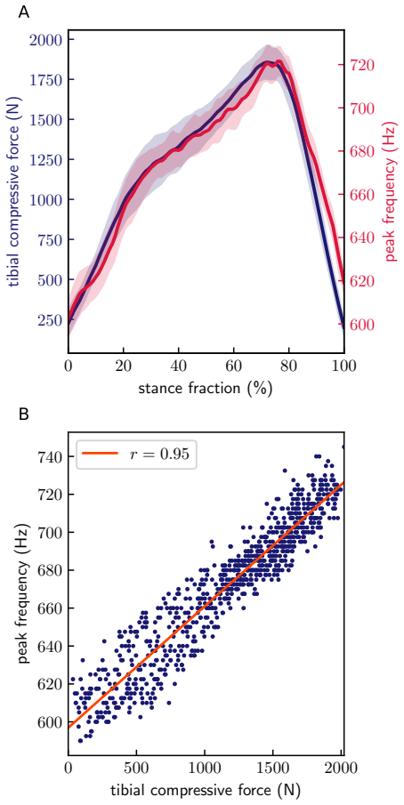
Modelling the tibia as an axially compressed Euler-Bernoulli beam, the authors show that tibial flexural waves have load-dependent frequency spectra. Specifically, under physiological conditions, peak locations in the wave acceleration spectra vary linearly with the compressive force on the tibia and may be used as proxies for the compressive force. Validation with a proof-of-concept wearable system that generates waves via a skin-mounted transducer and records spectra via a skin-mounted accelerometer produces linear relationships in data from nine participants, with Pearson coefficients ranging from 0.81 to 0.99 across swaying and walking trials.
What carries the argument
The linear shift of spectral peak locations with compressive force in flexural waves, as predicted by applying Euler-Bernoulli beam theory to an axially loaded tibia.
If this is right
- The approach enables non-invasive bone force measurement during everyday movements such as walking.
- It supports development of wearable sensors for physiological load monitoring outside laboratory settings.
- Data from the technique can advance research on human locomotion and sports medicine.
- A new class of devices becomes feasible for continuous tracking of forces transmitted by bones.
Where Pith is reading between the lines
- The same spectral approach might extend to monitoring loads in other long bones during dynamic activities.
- Combining wave measurements with standard motion capture could estimate internal force distributions across multiple body segments in real time.
- Long-term recordings could reveal how habitual loading patterns influence bone adaptation or injury risk in athletes.
Load-bearing premise
Soft tissue and skin interfaces do not substantially distort the flexural wave spectra from the underlying bone, allowing skin-mounted sensors to capture the load-dependent behavior predicted by the beam model.
What would settle it
Direct comparison of spectral peak shifts against simultaneously recorded internal tibial forces, for example in cadaver preparations or with implanted sensors, would show whether the predicted linear relationship persists without tissue-induced distortion.
Figures

read the original abstract
Forces transmitted by bones are routinely studied in human biomechanics, but it is challenging to measure them non-invasively, especially outside of laboratory settings. We introduce a technique for non-invasive, in vivo measurement of tibial compressive force using flexural waves propagating in the tibia. Modelling the tibia as an axially compressed Euler-Bernoulli beam, we show that tibial flexural waves have load-dependent frequency spectra. Specifically, under physiological conditions, peak locations in the wave acceleration spectra vary linearly with the compressive force on the tibia and may be used as proxies for the compressive force. We test the validity of this technique using a proof-of-concept wearable system that generates flexural waves via a skin-mounted mechanical transducer and measures the spectra of these waves using a skin-mounted accelerometer. In agreement with beam theory, data from 9 participants demonstrate linear relationships between tibial compressive force and spectral peak location, with Pearson correlation coefficients $r=0.82 - 0.99$ (mean $r=0.93$) for medial-lateral swaying and $r=0.81 - 0.98$ (mean $r=0.93$) for walking trials. This flexural wave-based technique could give rise to a new class of wearable sensors for non-invasive physiological bone load monitoring and measurement, impacting research in human locomotion and sports medicine.
Editorial analysis
A structured set of objections, weighed in public.
Referee Report
Summary. The paper claims that flexural waves in the tibia, modeled as an axially compressed Euler-Bernoulli beam, exhibit load-dependent frequency spectra where peak locations vary linearly with compressive force under physiological conditions. A proof-of-concept wearable system with skin-mounted transducer and accelerometer is used to generate and measure these waves, with experiments on nine participants during medial-lateral swaying and walking showing strong linear relationships (Pearson r = 0.81–0.99, mean r = 0.93) between force and spectral peak location, supporting use as a non-invasive proxy for tibial load.
Significance. If the central claim holds and skin-mounted measurements faithfully capture bone-wave peaks without dominant soft-tissue distortion, the work could enable a new class of wearable sensors for continuous, non-invasive bone-load monitoring in locomotion and sports medicine. The alignment of human data with the beam-theory prediction and the reproducible experimental protocol on multiple participants and tasks are notable strengths.
major comments (2)
- [Methods] The central claim depends on the assumption that acceleration spectra from skin-mounted sensors reproduce the load-dependent peaks of flexural waves inside the tibia as predicted by the axially compressed Euler-Bernoulli equation (EI y'''' + P y'' + μ y_tt = 0) and its dispersion relation ω² = (EI/μ)k⁴ – (P/μ)k². No explicit viscoelastic soft-tissue layer model or control experiment isolating the frequency-dependent transfer function (which can itself change with load via contact pressure or muscle tone) is described; this leaves open the possibility that the reported Pearson correlations arise partly from load-dependent coupling or filtering rather than bone-wave behavior.
- [Results] Details on error propagation, exact peak-detection algorithms, and spectral processing steps from the skin-mounted accelerometer are absent. Without these, it is difficult to evaluate the robustness of the linear fits or to rule out that the high r values (0.81–0.99) partly reflect consistent but non-bone-related load effects on the measurement chain.
minor comments (2)
- [Abstract] The abstract states that peaks 'vary linearly with the compressive force' but does not report the range of forces tested or how they map onto typical in vivo tibial loads during walking.
- [Discussion] Consider adding a brief limitations paragraph discussing possible confounds from changes in muscle tone or skin contact pressure across tasks.
Simulated Author's Rebuttal
We thank the referee for their constructive and detailed comments, which have prompted us to clarify key aspects of our methods and results. We address each major comment below and indicate the revisions made to the manuscript.
read point-by-point responses
-
Referee: [Methods] The central claim depends on the assumption that acceleration spectra from skin-mounted sensors reproduce the load-dependent peaks of flexural waves inside the tibia as predicted by the axially compressed Euler-Bernoulli equation (EI y'''' + P y'' + μ y_tt = 0) and its dispersion relation ω² = (EI/μ)k⁴ – (P/μ)k². No explicit viscoelastic soft-tissue layer model or control experiment isolating the frequency-dependent transfer function (which can itself change with load via contact pressure or muscle tone) is described; this leaves open the possibility that the reported Pearson correlations arise partly from load-dependent coupling or filtering rather than bone-wave behavior.
Authors: We agree that an explicit model of the soft-tissue layer would further strengthen the link between skin-mounted measurements and intraosseous wave behavior. In the revised manuscript we have added a dedicated paragraph in the Methods section that discusses the expected influence of soft tissue, citing literature on wave propagation through layered bone-soft tissue systems and noting that the frequencies employed (well below typical soft-tissue resonance ranges) favor bone-guided flexural modes. We also report that transducer contact force was standardized across trials to limit variability in coupling. While a dedicated control experiment isolating the transfer function would require invasive instrumentation beyond the scope of this proof-of-concept study, the close quantitative agreement between observed peak shifts and the Euler-Bernoulli dispersion relation across nine participants and two distinct tasks provides empirical support that bone-wave physics dominate the recorded spectra. revision: partial
-
Referee: [Results] Details on error propagation, exact peak-detection algorithms, and spectral processing steps from the skin-mounted accelerometer are absent. Without these, it is difficult to evaluate the robustness of the linear fits or to rule out that the high r values (0.81–0.99) partly reflect consistent but non-bone-related load effects on the measurement chain.
Authors: We appreciate this observation and have expanded both the Methods and Results sections in the revised manuscript. We now specify the full spectral processing pipeline (Hann window, 50 % overlap, 4096-point FFT, frequency resolution of 1.2 Hz), the peak-detection algorithm (prominence threshold of 3 dB above local noise floor with parabolic interpolation for sub-bin accuracy), and the error-propagation procedure used for the linear regression slopes and Pearson coefficients (including bootstrap resampling to obtain 95 % confidence intervals). These additions allow readers to assess measurement robustness directly and to evaluate whether non-bone contributions could systematically produce the observed linearity. revision: yes
Circularity Check
No significant circularity; model prediction validated on independent data
full rationale
The paper begins with the standard Euler-Bernoulli beam equation for an axially compressed tibia and derives the dispersion relation whose resonance peaks shift with load P. Under physiological conditions this shift is shown to be linear, providing a qualitative prediction that is then tested experimentally on 9 independent participants using skin-mounted sensors. The reported Pearson r values (0.81–0.99) are obtained from participant data rather than from any fitted constants or self-referential definitions. No self-citation chains, ansatz smuggling, or renaming of known results appear in the derivation; the central claim remains externally falsifiable and is not equivalent to its inputs by construction.
Axiom & Free-Parameter Ledger
axioms (1)
- domain assumption The tibia behaves as an axially compressed Euler-Bernoulli beam under physiological loading conditions
Lean theorems connected to this paper
-
IndisputableMonolith/Foundation/RealityFromDistinction.leanreality_from_one_distinction unclear?
unclearRelation between the paper passage and the cited Recognition theorem.
Modeling the tibia as an axially compressed Euler-Bernoulli beam... EI ∂⁴y/∂x⁴ + P ∂²y/∂x² + ρA ∂²y/∂t² = 0
-
IndisputableMonolith/Cost/FunctionalEquation.leanwashburn_uniqueness_aczel unclear?
unclearRelation between the paper passage and the cited Recognition theorem.
dispersion relation... peak locations... vary linearly with the compressive force
What do these tags mean?
- matches
- The paper's claim is directly supported by a theorem in the formal canon.
- supports
- The theorem supports part of the paper's argument, but the paper may add assumptions or extra steps.
- extends
- The paper goes beyond the formal theorem; the theorem is a base layer rather than the whole result.
- uses
- The paper appears to rely on the theorem as machinery.
- contradicts
- The paper's claim conflicts with a theorem or certificate in the canon.
- unclear
- Pith found a possible connection, but the passage is too broad, indirect, or ambiguous to say the theorem truly supports the claim.
Reference graph
Works this paper leans on
-
[1]
D. Chadefaux, N. Gueguen, A. Thouze, and G. Rao. 3d propagation of the shock-induced vibrations through the whole lower-limb during running. Journal of Biomechanics, 96: 0 109343, 2019
work page 2019
-
[2]
J. F. Doyle. Wave Propagation in Structures. Springer Cham, 2020
work page 2020
- [3]
-
[4]
D. F \"a h and E. St \"u ssi. Phase velocity measurement of flexural waves in human tibia. Journal of biomechanics, 21 0 (11): 0 975--983, 1988
work page 1988
-
[5]
K. F. Graff. Wave motion in elastic solids. Dover Publications, 1991
work page 1991
-
[6]
N. Horvais, P. Samozino, X. Chiementin, J.-B. Morin, and M. Giandolini. Cushioning perception is associated with both tibia acceleration peak and vibration magnitude in heel-toe running. Footwear Science, 11 0 (1): 0 35--44, 2019
work page 2019
-
[7]
W. M. Kohrt, S. A. Bloomfield, K. D. Little, M. E. Nelson, and V. R. Yingling. Physical activity and bone health. Medicine & Science in Sports & Exercise, 36 0 (11): 0 1985--1996, 2004
work page 1985
-
[8]
J. A. Martin, S. C. Brandon, E. M. Keuler, J. R. Hermus, A. C. Ehlers, D. J. Segalman, M. S. Allen, and D. G. Thelen. Gauging force by tapping tendons. Nature communications, 9 0 (1): 0 1592, 2018
work page 2018
-
[9]
E. S. Matijevich, L. M. Branscombe, L. R. Scott, and K. E. Zelik. Ground reaction force metrics are not strongly correlated with tibial bone load when running across speeds and slopes: Implications for science, sport and wearable tech. PloS one, 14 0 (1): 0 e0210000, 2019
work page 2019
-
[10]
E. S. Matijevich, L. R. Scott, P. Volgyesi, K. H. Derry, and K. E. Zelik. Combining wearable sensor signals, machine learning and biomechanics to estimate tibial bone force and damage during running. Human movement science, 74: 0 102690, 2020
work page 2020
-
[11]
C. Milgrom, A. Finestone, A. Hamel, V. Mandes, D. Burr, and N. Sharkey. A comparison of bone strain measurements at anatomically relevant sites using surface gauges versus strain gauged bone staples. Journal of biomechanics, 37 0 (6): 0 947--952, 2004
work page 2004
- [12]
-
[13]
N. Mitsuhashi, K. Fujieda, T. Tamura, S. Kawamoto, T. Takagi, and K. Okubo. Bodyparts3d: 3d structure database for anatomical concepts. Nucleic acids research, 37 0 (suppl\_1): 0 D782--D785, 2009
work page 2009
-
[14]
M. H. Moen, J. L. Tol, A. Weir, M. Steunebrink, and T. C. De Winter. Medial tibial stress syndrome: a critical review. Sports medicine, 39: 0 523--546, 2009
work page 2009
- [15]
-
[16]
A. J. Reiter, J. A. Martin, K. A. Knurr, P. G. Adamczyk, and D. G. Thelen. Achilles tendon loading during running estimated via shear wave tensiometry: a step towards wearable kinetic analysis. Medicine and science in sports and exercise, 56 0 (6): 0 1077, 2024
work page 2024
-
[17]
J.-Y. Rho. An ultrasonic method for measuring the elastic properties of human tibial cortical and cancellous bone. Ultrasonics, 34 0 (8): 0 777--783, 1996
work page 1996
-
[18]
A. G. Robling and C. H. Turner. Mechanical signaling for bone modeling and remodeling. Critical Reviews™ in Eukaryotic Gene Expression, 19 0 (4), 2009
work page 2009
-
[19]
C. T. Rubin. In vivo measurement of bone deformations using strain gauges. Bone mechanics handbook, 2001
work page 2001
-
[20]
S. H. Scott and D. A. Winter. Internal forces at chronic running injury sites. Medicine & Science in Sports & Exercise, 22 0 (3): 0 357--369, 1990
work page 1990
-
[21]
E. St \"u ssi and D. F \"a h. Assessment of bone mineral content by in vivo measurement of flexural wave velocities. Medical and Biological Engineering and Computing, 26 0 (4): 0 349--354, 1988
work page 1988
-
[22]
I. J. Wallace, B. Demes, C. Mongle, O. M. Pearson, J. D. Polk, and D. E. Lieberman. Exercise-induced bone formation is poorly linked to local strain magnitude in the sheep tibia. PloS one, 9 0 (6): 0 e99108, 2014
work page 2014
-
[23]
S. J. Warden, I. S. Davis, and M. Fredericson. Management and prevention of bone stress injuries in long-distance runners. Journal of orthopaedic & sports physical therapy, 44 0 (10): 0 749--765, 2014
work page 2014
-
[24]
R. W. Willy, K. Christensen, B. Hanser, M. Plemmons, and B. C. Ruby. Average vertical loading rate and tibial accelerometry are not valid assessments of internal tibial loads when walking or running with or without load carriage: A cross-sectional laboratory study. Journal of Sports Sciences, 0 0 (0): 0 1--11, 2025
work page 2025
-
[25]
D. A. Winter. Biomechanics and motor control of human movement. John wiley & sons, 2009
work page 2009
- [26]
-
[27]
P. Yang, G.-P. Br \"u ggemann, and J. Rittweger. What do we currently know from in vivo bone strain measurements in humans? Journal of Musculoskeletal and Neuronal Interactions, 11 0 (1): 0 8--20, 2011
work page 2011
-
[28]
P.-F. Yang, M. Sanno, B. Ganse, T. Koy, G.-P. Br \"u ggemann, L. P. M \"u ller, and J. Rittweger. Torsion and antero-posterior bending in the in vivo human tibia loading regimes during walking and running. PloS one, 9 0 (4): 0 e94525, 2014
work page 2014
discussion (0)
Sign in with ORCID, Apple, or X to comment. Anyone can read and Pith papers without signing in.