Portable Medical Imaging in Modern Healthcare: Fundamentals, AI-Based Taxonomy, Image Quality, and Open Challenges
Pith reviewed 2026-05-10 11:27 UTC · model grok-4.3
The pith
Portable medical imaging reaches reliable clinical use when AI methods target quality degradation from motion and hardware limits.
A machine-rendered reading of the paper's core claim, the machinery that carries it, and where it could break.
Core claim
The central claim is that a systematic, quality-centered review of portable medical imaging reveals the direct relationship between image-quality degradation caused by motion artifacts, environmental interference, and hardware limitations and the resulting AI robustness and clinical usability, while providing a taxonomy of AI methods, analyzing devices and datasets, and outlining gaps toward reliable and interpretable systems.
What carries the argument
The AI-based taxonomy of PMI methods, which classifies machine learning, deep learning, transfer learning, and Transformer approaches by their roles in countering modality-specific distortions to support enhancement, reconstruction, quality assessment, and diagnostic tasks.
If this is right
- AI models for PMI must explicitly preserve image quality under unstable conditions to maintain performance in detection and classification.
- Evaluation metrics and public datasets should incorporate portable-specific distortions rather than relying on standard fixed-scanner benchmarks.
- Future PMI systems require greater interpretability to support clinical deployment alongside robustness gains.
- Research should prioritize handling of motion artifacts and hardware limits to close identified gaps in real-world usability.
Where Pith is reading between the lines
- This taxonomy could guide development of cross-modality AI tools that adapt to varying portable hardware without retraining from scratch.
- Field trials measuring diagnostic accuracy before and after quality-aware AI interventions would test the review's practical value.
- Linking PMI quality metrics to telemedicine platforms might extend reliable remote diagnosis in underserved regions.
Load-bearing premise
The literature chosen for the taxonomy and gap analysis is comprehensive and representative of all relevant PMI research.
What would settle it
A new survey of PMI literature that finds no measurable link between explicit quality-focused AI design and improved clinical usability in portable conditions would falsify the central emphasis.
Figures


read the original abstract
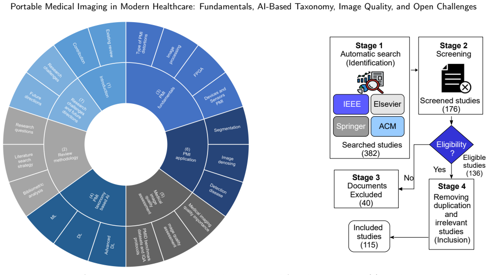
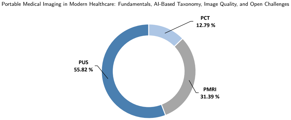
Portable medical imaging (PMI) has emerged as an important solution for point-of-care diagnosis in emergency, rural, and resource-limited settings where conventional imaging infrastructure is not readily available. Modalities such as portable computed tomography, portable magnetic resonance imaging, portable ultrasound, and wireless capsule endoscopy improve access to timely diagnosis, but they remain highly vulnerable to image-quality degradation caused by motion artifacts, environmental interference, hardware limitations, and unstable acquisition conditions. This review provides a systematic and quality-centered synthesis of recent advances in PMI. It introduces a taxonomy of AI-based PMI methods spanning machine learning, deep learning, transfer learning, and Transformer-based approaches, and examines their roles in image enhancement, reconstruction, quality assessment, detection, and classification. The review also analyzes PMI devices, sensing pipelines, modality-specific distortions, evaluation metrics, and publicly available datasets. In contrast to existing surveys that are mainly modality-driven or application-focused, this work emphasizes the relationship between image quality, AI robustness, and clinical usability in portable settings. Finally, it identifies current research gaps and outlines future directions toward reliable, interpretable, and clinically deployable PMI systems.
Editorial analysis
A structured set of objections, weighed in public.
Referee Report
Summary. The manuscript offers a systematic review of portable medical imaging (PMI) technologies, including modalities like portable CT, MRI, ultrasound, and capsule endoscopy. It proposes an AI-based taxonomy covering machine learning, deep learning, transfer learning, and Transformer approaches for tasks such as image enhancement, reconstruction, quality assessment, detection, and classification. The paper analyzes device fundamentals, sensing pipelines, modality-specific distortions, evaluation metrics, and public datasets, while highlighting the interplay between image quality, AI robustness, and clinical usability, and outlining research gaps and future directions.
Significance. If the synthesis is representative, this work could significantly contribute to the field by offering a quality-centered perspective that bridges technical AI advancements with clinical deployment challenges in resource-limited settings. It highlights actionable gaps that could guide the development of more robust and interpretable portable imaging solutions.
major comments (1)
- [Methods or Literature Search Section] The abstract describes the work as a 'systematic' synthesis, but the manuscript lacks a detailed description of the literature selection process, including databases searched, search terms, time period covered, and inclusion/exclusion criteria. Without this, the taxonomy and gap analysis cannot be verified as comprehensive or free from selection bias, which is central to supporting the claims about unique emphasis and open challenges.
minor comments (2)
- [Taxonomy Section] The taxonomy of AI-based PMI methods is presented, but it is unclear how the categorization avoids overlap or ensures coverage of all relevant approaches; for example, the distinction between deep learning and Transformer-based methods may require more explicit justification given that Transformers are a subset of deep learning.
- [Figures] Ensure that all figures, such as the taxonomy diagram, have high resolution and clear legends for readability in the final publication.
Simulated Author's Rebuttal
We thank the referee for the detailed and constructive review. The feedback highlights an important aspect of systematic reviews, and we have revised the manuscript accordingly to enhance transparency.
read point-by-point responses
-
Referee: The abstract describes the work as a 'systematic' synthesis, but the manuscript lacks a detailed description of the literature selection process, including databases searched, search terms, time period covered, and inclusion/exclusion criteria. Without this, the taxonomy and gap analysis cannot be verified as comprehensive or free from selection bias, which is central to supporting the claims about unique emphasis and open challenges.
Authors: We agree that a systematic review requires explicit documentation of the literature search methodology to support claims of comprehensiveness and to allow assessment of potential bias. In the revised manuscript, we have added a dedicated 'Literature Search Strategy' subsection (now Section 2) that details: (1) the databases queried (PubMed, IEEE Xplore, Scopus, Web of Science, and arXiv), (2) the Boolean search strings and keywords employed, (3) the covered time period (2015–2024), and (4) the inclusion/exclusion criteria (peer-reviewed English-language studies on AI methods for portable imaging modalities, excluding non-empirical works and duplicate records). This addition directly addresses the concern and enables verification of the taxonomy and gap analysis. revision: yes
Circularity Check
No circularity: standard literature synthesis without self-referential derivations
full rationale
This is a survey paper whose core output is a taxonomy and gap analysis drawn from reviewed external literature. No equations, fitted parameters, or predictions are generated from the paper's own inputs; the taxonomy classifies existing AI methods rather than deriving them from self-defined quantities. Claims about image-quality/AI-robustness links are interpretive summaries, not reductions by construction. Literature selection methodology, while open to critique on completeness, does not create circularity because the paper does not treat its own selection criteria as a derived result. No self-citation load-bearing, ansatz smuggling, or renaming of known results occurs in the derivation chain.
Axiom & Free-Parameter Ledger
Reference graph
Works this paper leans on
-
[1]
B.E.Kavanagh,K.B.Corney,H.Beks,L.J.Williams,S.E.Quirk,V.L.Versace,Ascopingreviewofthebarriersandfacilitatorstoaccessingandutilising mental health services across regional, rural, and remote australia, BMC health services research 23 (1) (2023) 1060
work page 2023
-
[2]
A. C. Mazari, H. Kheddar, Deep learning-and transfer learning-based models for covid-19 detection using radiography images, in: 2023 International Conference on Advances in Electronics, Control and Communication Systems (ICAECCS), IEEE, 2023, pp. 1–4
work page 2023
-
[3]
O. Ali, A. AlAhmad, H. Kahtan, A review of advanced technologies available to improve the healthcare performance during covid-19 pandemic, Procedia Computer Science 217 (2023) 205–216
work page 2023
-
[4]
W. Bian, P. Li, M. Zheng, C. Wang, A. Li, Y. Li, H. Ni, Z. Zeng, A review of electromagnetic elimination methods for low-field portable mri scanner, in: 2024 5th International Conference on Machine Learning and Computer Application (ICMLCA), IEEE, 2024, pp. 614–618
work page 2024
-
[5]
A.Altaf,M.W.S.Baqai,F.Urooj,M.S.Alam,H.F.Aziz,F.Mubarak,E.A.Knopp,K.M.Siddiqui,S.A.Enam,Utilizationofanultra-low-field,portable magnetic resonance imaging for brain tumor assessment in lower middle-income countries, Surgical Neurology International 14 (2023) 260
work page 2023
- [6]
-
[7]
C.-H. Wang, T. Hwang, Y.-S. Huang, J. Tay, C.-Y. Wu, M.-C. Wu, H. R. Roth, D. Yang, C. Zhao, W. Wang, et al., Deep learning–based localization and detection of malpositioned nasogastric tubes on portable supine chest x-rays in intensive care and emergency medicine: A multi-center retrospective study, Journal of Imaging Informatics in Medicine (2024) 1–11
work page 2024
-
[8]
A. Perez-Sanchez, G. Johnson, N. Pucks, R. N. Soni, T. J. Lund, A. J. Andrade, M.-P. T. Le, J. Solis-McCarthy, T. Wong, A. Ashraf, et al., Comparison of 6 handheld ultrasound devices by point-of-care ultrasound experts: A cross-sectional study, The Ultrasound Journal 16 (1) (2024) 45
work page 2024
-
[9]
A. Groteklaes, T. Dresbach, M. Born, A. Mueller, H. Sabir, Case report: Ultralow-field portable mri improves the diagnosis of congenital hydrocephalus, Frontiers in Pediatrics 13 (2025) 1463314
work page 2025
-
[10]
Q.Cao,R.Deng,Y.Pan,R.Liu,Y.Chen,G.Gong,J.Zou,H.Yang,D.Han,Roboticwirelesscapsuleendoscopy:recentadvancesandupcomingtechnologies, Nature Communications 15 (1) (2024) 4597
work page 2024
-
[11]
H.Edwards,H.Jones,P.Garner,M.Hardy,S.-P.Wilshaw,K.Bielby-Clarke,M.Farrow,Assessingstudentperceptionoftheintegrationofportablewireless ultrasound imaging in undergraduate anatomy education, Clinical Anatomy 36 (5) (2023) 742–753
work page 2023
- [12]
-
[13]
R.Wang,Z.Fang,J.Gu,Y.Guo,S.Zhou,Y.Wang,C.Chang,J.Yu,High-resolutionimagereconstructionforportableultrasoundimagingdevices,EURASIP Journal on Advances in Signal Processing 2019 (2019) 1–12
work page 2019
- [14]
-
[15]
I. Boucherit, H. Kheddar, Reinforced residual encoder–decoder network for image denoising via deeper encoding and balanced skip connections, Big Data and Cognitive Computing 9 (4) (2025) 82
work page 2025
-
[16]
I.Allegretta,B.Marangoni,P.Manzari,C.Porfido,R.Terzano,O.DePascale,G.S.Senesi,Macro-classificationofmeteoritesbyportableenergydispersive x-ray fluorescence spectroscopy (ped-xrf), principal component analysis (pca) and machine learning algorithms, Talanta 212 (2020) 120785
work page 2020
-
[17]
J. E. Iglesias, R. Schleicher, S. Laguna, B. Billot, P. Schaefer, B. McKaig, J. N. Goldstein, K. N. Sheth, M. S. Rosen, W. T. Kimberly, Quantitative brain morphometry of portable low-field-strength mri using super-resolution machine learning, Radiology 306 (3) (2022) e220522
work page 2022
-
[18]
Y. Habchi, Y. Himeur, H. Kheddar, A. Boukabou, S. Atalla, A. Chouchane, A. Ouamane, W. Mansoor, Ai in thyroid cancer diagnosis: Techniques, trends, and future directions, Systems 11 (10) (2023) 519. Y. Habchi et al.:Preprint submitted to ElsevierPage 23 of 27 Portable Medical Imaging in Modern Healthcare: Fundamentals, AI-Based Taxonomy, Image Quality, an...
work page 2023
-
[19]
G. Dong, Y. Ma, A. Basu, Feature-guided cnn for denoising images from portable ultrasound devices, IEEE Access 9 (2021) 28272–28281
work page 2021
-
[20]
D.-K. Hwang, W.-K. Yu, T.-C. Lin, S.-J. Chou, A. Yarmishyn, Z.-K. Kao, C.-L. Kao, Y.-P. Yang, S.-J. Chen, C.-C. Hsu, et al., Smartphone-based diabetic macula edema screening with an offline artificial intelligence, Journal of the Chinese Medical Association 83 (12) (2020) 1102–1106
work page 2020
- [21]
- [22]
-
[23]
Y.-C. Chen, Y.-C. Chu, C.-Y. Huang, Y.-T. Lee, W.-Y. Lee, C.-Y. Hsu, A. C. Yang, W.-H. Liao, Y.-F. Cheng, Smartphone-based artificial intelligence using a transfer learning algorithm for the detection and diagnosis of middle ear diseases: A retrospective deep learning study, EClinicalMedicine 51 (2022)
work page 2022
-
[24]
J. Zhu, B. Shen, A. Abbasi, M. Hoshmand-Kochi, H. Li, T. Q. Duong, Deep transfer learning artificial intelligence accurately stages covid-19 lung disease severity on portable chest radiographs, PloS one 15 (7) (2020) e0236621
work page 2020
-
[25]
P. L. Vidal, J. de Moura, J. Novo, M. Ortega, Multi-stage transfer learning for lung segmentation using portable x-ray devices for patients with covid-19, Expert Systems with Applications 173 (2021) 114677
work page 2021
- [26]
- [27]
-
[28]
M. Cè, G. Oliva, F. L. Rabaiotti, L. Macrì, S. Zollo, A. Aquila, M. Cellina, Portable dynamic chest radiography: literature review and potential bedside applications, Medical Sciences 12 (1) (2024) 10
work page 2024
-
[29]
L. L. Wald, P. C. McDaniel, T. Witzel, J. P. Stockmann, C. Z. Cooley, Low-cost and portable mri, Journal of Magnetic Resonance Imaging 52 (3) (2020) 686–696
work page 2020
-
[30]
A. J. Eggleston, E. Farrington, S. McDonald, S. Aziz, Portable ultrasound technologies for estimating gestational age in pregnant women: a scoping review and analysis of commercially available models, BMJ open 12 (11) (2022) e065181
work page 2022
-
[31]
L. Shaddock, T. Smith, Potential for use of portable ultrasound devices in rural and remote settings in australia and other developed countries: a systematic review, Journal of Multidisciplinary Healthcare (2022) 605–625
work page 2022
- [32]
-
[33]
M. Haji-Hassan, L. M. Lenghel, S. D. Bolboacă, Hand-held ultrasound of the lung: a systematic review, Diagnostics 11 (8) (2021) 1381
work page 2021
-
[34]
S.A.Ibraheem,R.Mahmud,S.MohamadSaini,H.AbuHassan,A.S.Keiteb,A.M.Dirie,Evaluationofdiagnosticperformanceofautomaticbreastvolume scanner compared to handheld ultrasound on different breast lesions: a systematic review, Diagnostics 12 (2) (2022) 541
work page 2022
-
[35]
S. A. Wajid, S. Ahmed, V. Devera, D. Dickerson, A. Thompson, J. Villela, Comparison of the effectiveness of portable ultrasound vs portable x-ray as diagnostic imaging of knee structures in clinical medicine, J Adv Med Pharm Sci 5 (2020) 41–50
work page 2020
-
[36]
D. Kravchenko, M. T. Hagar, M. Vecsey-Nagy, I. Kabat, A. Groteklaes, J. A. Luetkens, D. Kuetting, A. Isaak, T. Emrich, A. Varga-Szemes, et al., Low-field and portable mri technology: advancements and innovations, European Radiology Experimental 9 (1) (2025) 103
work page 2025
-
[37]
A.Jacobi,M.Chung,A.Bernheim,C.Eber,Portablechestx-rayincoronavirusdisease-19(covid-19):Apictorialreview,Clinicalimaging64(2020)35–42
work page 2020
-
[38]
T. Kandemir, Place of portable computed tomography in neurosurgery practice, Journal of Medical Innovation and Technology 1 (2) (2019) 67–69
work page 2019
- [39]
-
[40]
C. N. DesRoche, A. P. Johnson, E. B. Hore, E. Innes, I. Silver, D. Tampieri, B. Y. Kwan, J. O. Jimenez, J. G. Boyd, O. Islam, Feasibility and cost analysis of portable mri implementation in a remote setting in canada, Canadian Journal of Neurological Sciences 51 (3) (2024) 387–396
work page 2024
-
[41]
C. Z. Cooley, M. W. Haskell, S. F. Cauley, C. Sappo, C. D. Lapierre, C. G. Ha, J. P. Stockmann, L. L. Wald, Design of sparse halbach magnet arrays for portable mri using a genetic algorithm, IEEE transactions on magnetics 54 (1) (2017) 1–12
work page 2017
-
[42]
L.Yang,W.He,Y.He,J.Wu,S.Shen,Z.Xu,Activeemisuppressionsystemfora50mtunshieldedportablemriscanner,IEEETransactionsonBiomedical Engineering 69 (11) (2022) 3415–3426
work page 2022
- [43]
-
[44]
W. Qiu, J. Zhou, Y. Chen, M. Su, G. Li, H. Zhao, X. Gu, D. Meng, C. Wang, Y. Xiao, et al., A portable ultrasound system for non-invasive ultrasonic neuro-stimulation, IEEE Transactions on Neural Systems and Rehabilitation Engineering 25 (12) (2017) 2509–2515
work page 2017
-
[45]
M. R. Sobhani, H. Ozum, G. Yaralioglu, A. Ergun, A. Bozkurt, Portable low cost ultrasound imaging system, in: 2016 IEEE International Ultrasonics Symposium (IUS), IEEE, 2016, pp. 1–4
work page 2016
-
[46]
S. Valente, A. Real, J. Gomes-Fonseca, H. R. Torres, E. Lima, P. Morais, J. L. Vilaça, A new handheld ultrasound probe simulator for medical training, in: 2021 IEEE 9th International Conference on Serious Games and Applications for Health (SeGAH), IEEE, 2021, pp. 1–7
work page 2021
-
[47]
O. Paraska, A. Gorban, O. Dynnyk, An innovative solution to optimize ultrasound diagnostics by introducing a portable ultrasound device to the european medical market, in: 62nd International Conference of Machine Design Departments (ICMD 2022), Atlantis Press, 2024, pp. 267–275
work page 2022
-
[48]
G.Ning,J.Wang,H.Liao,Cable-drivenlight-weightingandportablesystemforroboticmedicalultrasoundimaging,IEEETransactionsonMedicalRobotics and Bionics 6 (3) (2024) 1220–1231
work page 2024
-
[49]
S. A. El-Ghany, M. A. Mahmood, A. Abd El-Aziz, An accurate deep learning-based computer-aided diagnosis system for gastrointestinal disease detection using wireless capsule endoscopy image analysis, Applied Sciences 14 (22) (2024) 10243
work page 2024
-
[50]
V. Mitrakos, G. Cummins, F. J. Tauber, B. F. Cox, S. K. Pavuluri, G. S. Wood, M. A. Potter, E. Clutton, S. Cochran, T. Speck, et al., Pressurecap: An endoscopic sensor capsule for real-time gastrointestinal pressure monitoring, Device 2 (5) (2024)
work page 2024
-
[51]
A. Sanaullah, C. Yang, Y. Alexeev, K. Yoshii, M. C. Herbordt, Real-time data analysis for medical diagnosis using fpga-accelerated neural networks, BMC bioinformatics 19 (2018) 19–31
work page 2018
-
[52]
Boyle, Battery-efficient fpga design for portable ai applications (2024)
D. Boyle, Battery-efficient fpga design for portable ai applications (2024)
work page 2024
-
[53]
A. Ibrahim, W. Simon, A. C. Yüzügüler, M. Arditi, J.-P. Thiran, G. De Micheli, 1024-channel single 5w fpga towards high-quality portable 3d ultrasound platform, in: 2017 Design, Automation & Test in Europe Conference & Exhibition (DATE), 2017
work page 2017
-
[54]
J. Xu, Y. Zheng, M. Chirala, M. Almekkawy, An fpga implementation of resource-optimized dynamic digital beamformer for a portable ultrasound imaging sys-tem, Advances in Science, Technology and Engineering Systems 3 (4) (2018) 59–71
work page 2018
-
[55]
W. Wang, Z. Feng, Portable and cost-effective handheld ultrasound system utilizing fpga-based synthetic aperture imaging, IEEE Open Journal of Nanotechnology (2024)
work page 2024
- [56]
- [57]
-
[58]
S. H. Choi, H. W. Jo, K. R. Dong, Y. H. Ryu, Usefulness estimate of noise reduction for mobile abdomen x-ray, Journal of Radiation Industry 15 (1) (2021) 31–37. Y. Habchi et al.:Preprint submitted to ElsevierPage 24 of 27 Portable Medical Imaging in Modern Healthcare: Fundamentals, AI-Based Taxonomy, Image Quality, and Open Challenges
work page 2021
-
[59]
D. C. Ruiz, M. L. Oliveira, H. Gaeta-Araujo, D. Q. Freitas, F. Haiter-Neto, Influence of enhancement filters on the diagnosis of proximal caries lesions in radiographs obtained with a handheld x-ray unit, Oral Radiology (2025) 1–7
work page 2025
-
[60]
H.Chai,X.Bo,L.Guo,C.Peng,Experienceandenlightenmentofhandheldultrasoundapplicationsinmultiplescenariosbasedon5gtechnology.,Advanced Ultrasound in Diagnosis & Therapy (AUDT) 7 (4) (2023)
work page 2023
-
[61]
J.Dai,X.Lei,F.Yu,Highspeedwirelessvideotransmissionforhandheldultrasonicsystem,in:TheFifthInternationalConferenceonBiologicalInformation and Biomedical Engineering, 2021, pp. 1–4
work page 2021
-
[62]
K. D. Krishna, V. Akkala, R. Bharath, P. Rajalakshmi, A. Mohammed, S. Merchant, U. Desai, Computer aided abnormality detection for kidney on fpga based iot enabled portable ultrasound imaging system, Irbm 37 (4) (2016) 189–197
work page 2016
-
[63]
S. Rajendran, A. Porwal, K. Anjali, Anvaya, R. Anuradha, Portable iot devices in healthcare for health monitoring and diagnostics, Internet of Things in bioelectronics: emerging technologies and applications (2024) 263–296
work page 2024
-
[64]
H. H. Gillani, M. A. Qureshi, A. Beghdadi, F. Cheikh, M. Ullah, Distortion classification in computer vision applications: Current progress, challenges, and perspectives, ACM Computing Surveys 58 (6) (2025) 1–36
work page 2025
-
[65]
A.Beghdadi,M.A.Qureshi,S.A.Amirshahi,A.Chetouani,M.Pedersen,Acriticalanalysisonperceptualcontrastanditsuseinvisualinformationanalysis and processing, IEEE Access 8 (2020) 156929–156953
work page 2020
-
[66]
F.J.Mateen,C.Z.Cooley,J.P.Stockmann,D.R.Rice,A.C.Vogel,L.L.Wald,Low-fieldportablebrainmriincnsdemyelinatingdisease,MultipleSclerosis and Related Disorders 51 (2021) 102903
work page 2021
- [67]
- [68]
-
[69]
E.Ljungberg,F.Padormo,M.Poorman,P.Clemensson,N.Bourke,J.C.Evans,J.Gholam,I.Vavasour,S.H.Kollind,S.L.Lafayette,etal.,Characterization of portable ultra-low field mri scanners for multi-center structural neuroimaging, Human Brain Mapping 46 (8) (2025) e70217
work page 2025
-
[70]
M.I.Fuller, K.Ranganathan,S.Zhou, T.N.Blalock,J.A. Hossack,W.F.Walker, Experimentalsystemprototypeofa portable,low-cost,c-scanultrasound imaging device, IEEE Transactions on Biomedical Engineering 55 (2) (2008) 519–530
work page 2008
-
[71]
B. Shin, S. Jeon, J. Ryu, H. J. Kwon, Compressed sensing for elastography in portable ultrasound, Ultrasonic Imaging 39 (6) (2017) 393–413
work page 2017
-
[72]
F.Kording,B.P.Schoennagel,M.T.deSousa,K.Fehrs,G.Adam,J.Yamamura,C.Ruprecht,Evaluationofaportabledopplerultrasoundgatingdevicefor fetal cardiac mr imaging: initial results at 1.5 t and 3t, Magnetic Resonance in Medical Sciences 17 (4) (2018) 308–317
work page 2018
-
[73]
J. Orchard, H. Y. Kim, J. T. Yeow, Plausibility of image reconstruction using a proposed flexible and portable ct scanner, Open Medical Imaging Journal 6 (2012) 1–11
work page 2012
-
[74]
S. Wirth, E. Euler, U. Linsenmaier, S.-M. Heining, D. Kotsianos, K.-J. Pfeifer, W. Mutschler, M. Reiser, C-arm-based mobile computed tomography: a comparison with established imaging on the basis of simulated treatments of talus neck fractures in a cadaveric study, Computer Aided Surgery 9 (1-2) (2004) 27–38
work page 2004
-
[75]
I. M. Mehedi, K. P. Rao, F. M. Alotaibi, H. M. Alkanfery, Intelligent wireless capsule endoscopy for the diagnosis of gastrointestinal diseases, Diagnostics 13 (8) (2023) 1445
work page 2023
-
[76]
D. Xu, H. Zhang, Y. Huang, M. Chen, R. Zheng, Development of machine learning based classification method for carotid plaques using portable 3d ultrasound, in: 2024 IEEE Ultrasonics, Ferroelectrics, and Frequency Control Joint Symposium (UFFC-JS), IEEE, 2024, pp. 1–4
work page 2024
-
[77]
M. Hashir, N. Khalid, N. Mahmood, M. A. Rehman, M. Asad, M. Q. Mehmood, M. Zubair, Y. Massoud, A tinyml based portable, low-cost microwave head imaging system for brain stroke detection, in: 2023 IEEE international symposium on circuits and systems (ISCAS), IEEE, 2023, pp. 1–4
work page 2023
-
[78]
W. T. Kimberly, A. J. Sorby-Adams, A. G. Webb, E. X. Wu, R. Beekman, R. Bowry, S. J. Schiff, A. de Havenon, F. X. Shen, G. Sze, et al., Brain imaging with portable low-field mri, Nature reviews bioengineering 1 (9) (2023) 617–630
work page 2023
-
[79]
W. MU, L. Zheng, D. C. Alexander, J. Gong, W. Yu, S. Y. Huang, A new deep learning structure for improving image quality of a low-field portable mri system
-
[80]
J. Shao, G. Wang, L. Yi, C. Wang, T. Lan, X. Xu, J. Guo, T. Deng, D. Liu, B. Chen, et al., Deep learning empowers lung cancer screening based on mobile low-dose computed tomography in resource-constrained sites, Frontiers in Bioscience-Landmark 27 (7) (2022) 212
work page 2022
discussion (0)
Sign in with ORCID, Apple, or X to comment. Anyone can read and Pith papers without signing in.