Bayesian In Vivo Tracking of Synapses using Joint Poisson Deconvolution and Diffeomorphic Registration
Pith reviewed 2026-05-14 20:37 UTC · model grok-4.3
The pith
A single Bayesian posterior simultaneously builds a probabilistic synapse template, denoises and deconvolves images, infers intensities, registers tissue motion diffeomorphically, and reports uncertainty for in vivo tracking.
A machine-rendered reading of the paper's core claim, the machinery that carries it, and where it could break.
Core claim
By deriving a posterior that incorporates a diffeomorphic mapping for domain warping, a Gaussian point spread function for the imaging process, and a Poisson observation model for raw photon counts, the Bayesian solution simultaneously constructs a probabilistic template of synapse locations, denoises and deconvolves the image data, infers fluorescence intensities, performs diffeomorphic image registration to correct for tissue motion, and provides confidence regions for these parameter estimates.
What carries the argument
The joint posterior over synapse template locations, intensities, and diffeomorphic deformation parameters, under a Poisson likelihood with Gaussian point-spread function.
If this is right
- The method produces uncertainty maps around each synapse location and intensity without separate post-processing.
- It enables direct comparison of the same synapses across multiple imaging sessions in living animals.
- Joint optimization reduces error propagation that occurs when registration and detection are performed sequentially.
- The probabilistic template can be updated incrementally as new time points arrive.
- The framework applies to both simulated 2D+t data and real 3D+t in vivo mouse datasets spanning two weeks.
Where Pith is reading between the lines
- The same joint model could be tested on other sparse fluorescent structures such as dendritic spines or axonal boutons.
- If the diffeomorphic assumption holds across longer time scales, the approach might support studies linking synaptic turnover to behavior.
- Replacing the Gaussian PSF with a measured or learned kernel would be a direct extension that preserves the rest of the pipeline.
- The confidence regions could serve as input for downstream statistical tests on synaptic plasticity across animals.
Load-bearing premise
Synapses can be treated as point sources whose motion is fully captured by one diffeomorphic tissue deformation, with the microscope blur well approximated by a Gaussian and photon counts following Poisson statistics.
What would settle it
A simulation or real dataset in which known synapse positions deviate systematically from any single diffeomorphic warp, or in which the recovered template fails to match ground-truth locations once non-Poisson noise or non-Gaussian blur is introduced.
Figures


read the original abstract
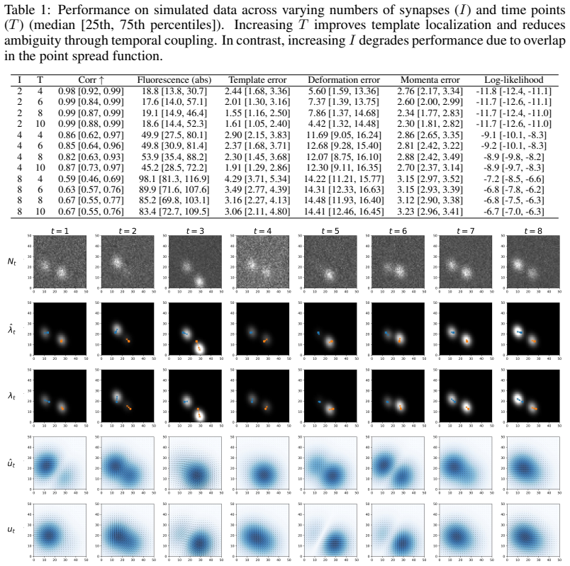
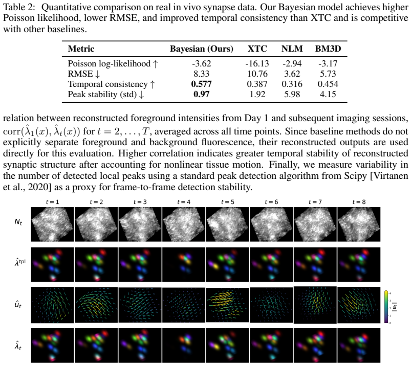
Synapses are densely packed submicron structures that dynamically reorganize during learning and memory formation. Longitudinal \textit{in vivo} imaging of fluorescently tagged synaptic receptors offers a promising opportunity to study large-scale synaptic dynamics and how these processes are disrupted in neurological disease. However, in vivo imaging with 2-photon microscopy uses low laser power and therefore suffers from low signal-to-noise ratio (SNR) and high shot noise, nonlinear tissue motion between days, nonstationary fluctuations in synaptic fluorescence, and significant blur induced by the microscope point spread function (PSF). Together, these factors make it challenging to detect and track synapses, especially in regions with high synaptic density. This paper presents a novel template-based framework for modeling synapses as varying luminance point sources that move under a nonlinear tissue deformation. Taking a unified Bayesian approach, we apply this model to microscopy data by deriving a posterior that incorporates a diffeomorphic mapping for domain warping, a Gaussian point spread function for the imaging process, and a Poisson observation model for raw photon counts. The Bayesian solution simultaneously: (1) Constructs a probabilistic template of synapse locations, (2) denoises and deconvolves the image data, (3) infers fluorescence intensities, (4) performs diffeomorphic image registration to correct for tissue motion, and (5) provides confidence regions for these parameter estimates. We demonstrate the framework on both a 2D+t simulated dataset and a 3D+t longitudinal \textit{in vivo} microscopy dataset of fluorescent synapses imaged in a mouse over two weeks.
Editorial analysis
A structured set of objections, weighed in public.
Referee Report
Summary. The manuscript presents a Bayesian framework for longitudinal in vivo synapse tracking in 2-photon microscopy. Synapses are modeled as varying-luminance point sources whose positions are governed by a probabilistic template; tissue motion is captured by a diffeomorphic deformation field; imaging uses a Gaussian PSF and Poisson photon-count likelihood. The joint posterior is derived to simultaneously construct the template, perform deconvolution and denoising, infer intensities, register images, and supply credible regions. The approach is demonstrated on a simulated 2D+t dataset and a real 3D+t longitudinal dataset acquired over two weeks in mouse cortex.
Significance. If the numerical results hold, the work would provide a coherent probabilistic treatment of several coupled tasks that are currently handled separately in synaptic imaging pipelines. The use of standard components (Poisson likelihood, Gaussian PSF, diffeomorphic group action) together with a point-source template prior yields a well-defined posterior without hidden circularity or identifiability collapse. The reported existence of feasible inference on both simulated and real 3D+t data is a useful existence proof; the availability of confidence regions would be directly useful for downstream biological analysis of synaptic turnover.
minor comments (3)
- [Abstract] Abstract: the claim that the method 'simultaneously' achieves all five listed tasks is strong; the abstract should briefly indicate which quantitative metrics (e.g., localization error, registration Dice, intensity correlation) support each claim on the real data.
- [Methods] Methods section (inferred from derivation): the specific form of the diffeomorphic velocity field parameterization and the numerical scheme used to sample or optimize the joint posterior should be stated explicitly, including any hyper-parameter settings that were held fixed across experiments.
- [Results] Results: while demonstrations on simulated and real data are reported, the manuscript should include at least one ablation (e.g., with vs. without the diffeomorphic term) and direct numerical comparison to a standard sequential pipeline (template estimation followed by separate registration) to quantify the benefit of the joint formulation.
Simulated Author's Rebuttal
We thank the referee for their positive summary and recommendation of minor revision. We are pleased that the unified Bayesian treatment of joint deconvolution, registration, and template inference is viewed as providing a coherent probabilistic framework without identifiability issues, and that the availability of credible regions is recognized as useful for downstream biological analysis.
Circularity Check
No significant circularity; derivation self-contained from stated model
full rationale
The paper constructs a joint posterior over a probabilistic synapse template, per-synapse intensities, and diffeomorphic deformation field by direct application of the imaging model (Gaussian PSF plus Poisson likelihood) and the deformation group action to the point-source template prior. All components are defined independently of the target dataset; no parameter is fitted to a subset of the data and then renamed as a prediction, no self-citation supplies a load-bearing uniqueness theorem or ansatz, and the central claims follow from the standard Bayesian construction without reduction to input quantities by definition. Demonstrations on simulated and real data serve only as existence proofs of numerical feasibility, not as circular validation.
Axiom & Free-Parameter Ledger
axioms (3)
- domain assumption Synapses modeled as varying luminance point sources
- domain assumption Tissue motion captured by diffeomorphic deformation
- domain assumption Imaging process uses Gaussian PSF and Poisson observation model
discussion (0)
Sign in with ORCID, Apple, or X to comment. Anyone can read and Pith papers without signing in.