Attenuation-Resilient Alternating Optimization for Laparoscopic Liver Landmark Detection
Pith reviewed 2026-06-29 18:13 UTC · model grok-4.3
The pith
A2ONet compensates for illumination loss and alternates segmentation with curve modeling to detect liver landmarks more reliably in laparoscopic surgery.
A machine-rendered reading of the paper's core claim, the machinery that carries it, and where it could break.
Core claim
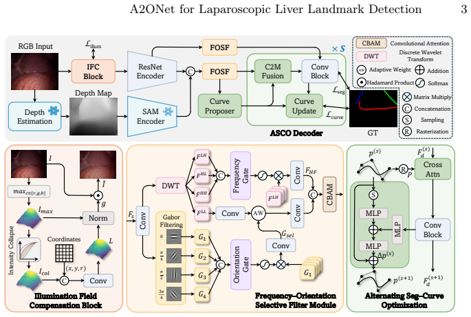
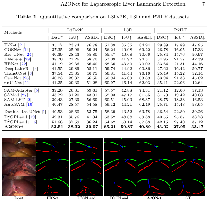
A2ONet mitigates illumination attenuation with an IFC block that adaptively enhances dark regions while preserving structure, uses a lightweight FOSF to suppress texture interference and retain curvilinear cues, and employs an ASCO decoder that iteratively couples dense segmentation with explicit curve modeling to optimize continuity and endpoint accuracy, yielding consistent improvements on the three evaluated datasets.
What carries the argument
The alternating seg-curve optimization (ASCO) decoder that iteratively couples dense segmentation with explicit curve modeling for mutual guidance.
If this is right
- More reliable intraoperative anatomy guidance during laparoscopic liver surgery.
- Improved structural continuity and endpoint localization for detected landmarks.
- Consistent performance gains across the L3D-2K, L3D, and P2ILF datasets.
- Better resilience to underexposed regions and repetitive texture interference.
Where Pith is reading between the lines
- The design may apply to landmark detection in other minimally invasive procedures that share lighting and geometry challenges.
- Deployment would require checking inference speed on standard surgical hardware.
- Testing on data from additional camera systems or patient cohorts could reveal limits to the claimed generalizability.
Load-bearing premise
The assumption that illumination attenuation and pixel-to-curve mismatch are the dominant failure modes and that the IFC block, FOSF, and ASCO decoder mitigate them in a generalizable way.
What would settle it
A new surgical video dataset with similar attenuation and curve challenges where A2ONet shows no accuracy gain over baselines or where ablating any of the three added components produces equivalent results.
Figures

read the original abstract
Liver surface landmark detection is a fundamental prerequisite for anatomical guidance in laparoscopic liver surgery. However, it remains unreliable in practice due to two pervasive challenges: illumination attenuation in underexposed regions and the structural mismatch between pixel-wise localization and continuous curvilinear geometry. To address these limitations, we propose A2ONet, an attenuation-resilient alternating optimization network for robust liver landmark detection. To mitigate illumination attenuation, A2ONet embraces an illumination field compensation (IFC) block that adaptively enhances dark regions while preserving structural consistency. Meanwhile, we introduce a lightweight frequency-orientation selective filter (FOSF) to suppress repetitive texture interference and preserve salient curvilinear cues. Building upon these resilient representations, we design an alternating seg-curve optimization (ASCO) decoder that iteratively couples dense segmentation with explicit curve modeling, enabling mutual guidance to optimize both structural continuity and endpoint localization. Extensive evaluations on L3D-2K, L3D, and P2ILF demonstrate consistent improvements over competitive methods, establishing a more reliable foundation for intraoperative anatomy guidance. Our code will be available at https://github.com/hyperiondk115/A2ONet.
Editorial analysis
A structured set of objections, weighed in public.
Referee Report
Summary. The paper proposes A2ONet for robust liver surface landmark detection in laparoscopic surgery. It introduces an Illumination Field Compensation (IFC) block to handle illumination attenuation, a Frequency-Orientation Selective Filter (FOSF) to reduce texture interference while preserving curvilinear features, and an Alternating Seg-Curve Optimization (ASCO) decoder that iteratively couples segmentation and explicit curve modeling. The central claim is that these components yield consistent improvements over competitive methods on the L3D-2K, L3D, and P2ILF datasets.
Significance. If the empirical gains hold under detailed scrutiny, the work addresses two practically relevant failure modes in surgical vision and could support more reliable intraoperative anatomy guidance. The planned public release of code is a clear strength for reproducibility.
minor comments (3)
- Abstract: the claim of 'consistent improvements' would be strengthened by including at least one or two key quantitative metrics (e.g., mean error reduction or Dice/F1 gains) rather than a purely qualitative statement.
- Ensure that the definitions and integration details of the IFC block, FOSF, and ASCO decoder are presented with sufficient architectural diagrams or pseudocode so that the alternating optimization loop in the decoder can be reproduced from the text alone.
- The evaluation section should explicitly state whether error bars, statistical significance tests, or cross-validation details accompany the reported improvements on the three datasets.
Simulated Author's Rebuttal
We thank the referee for their constructive review and for recommending minor revision. We appreciate the positive assessment of the practical relevance of our work on illumination attenuation and curvilinear geometry challenges in laparoscopic liver landmark detection, as well as the recognition of our planned code release for reproducibility.
Circularity Check
No significant circularity; empirical architecture with independent evaluation
full rationale
The paper introduces three architectural modules (IFC, FOSF, ASCO) to address illumination and curve-modeling issues in laparoscopic landmark detection, then reports performance gains on L3D-2K, L3D, and P2ILF. No equations, parameter-fitting steps, or derivation chains appear in the abstract or described content. The central claim is carried by external dataset evaluations rather than any self-referential definition, fitted-input prediction, or self-citation loop. The work is therefore self-contained against its own benchmarks.
Axiom & Free-Parameter Ledger
free parameters (1)
- Network hyperparameters and training settings
axioms (1)
- domain assumption The L3D-2K, L3D, and P2ILF datasets adequately represent real intraoperative illumination and geometry variations
invented entities (3)
-
Illumination Field Compensation (IFC) block
no independent evidence
-
Frequency-Orientation Selective Filter (FOSF)
no independent evidence
-
Alternating Seg-Curve Optimization (ASCO) decoder
no independent evidence
Reference graph
Works this paper leans on
-
[1]
Medical image analysis99, 103371 (2025)
Ali, S., Espinel, Y., Jin, Y., Liu, P., Güttner, B., Zhang, X., Zhang, L., Dowrick, T., Clarkson, M.J., Xiao, S., et al.: An objective comparison of methods for augmented reality in laparoscopic liver resection by preoperative-to-intraoperative image fu- sion from the miccai2022 challenge. Medical image analysis99, 103371 (2025)
2025
-
[2]
Procedia Computer Science246, 4951–4958 (2024)
Chai, S., Jain, R.K., Teng, S., Liu, J., Li, Y., Tateyama, T., Chen, Y.w.: Lad- der fine-tuning approach for sam integrating complementary network. Procedia Computer Science246, 4951–4958 (2024)
2024
-
[3]
TransUNet: Transformers Make Strong Encoders for Medical Image Segmentation
Chen, J., Lu, Y., Yu, Q., Luo, X., Adeli, E., Wang, Y., Lu, L., Yuille, A.L., Zhou, Y.:Transunet:Transformersmakestrongencodersformedicalimagesegmentation. arXiv preprint arXiv:2102.04306 (2021)
work page internal anchor Pith review Pith/arXiv arXiv 2021
-
[4]
In: Proceedings of the European conference on computer vision (ECCV)
Chen, L.C., Zhu, Y., Papandreou, G., Schroff, F., Adam, H.: Encoder-decoder with atrous separable convolution for semantic image segmentation. In: Proceedings of the European conference on computer vision (ECCV). pp. 801–818 (2018)
2018
-
[5]
In: Proceedings of the IEEE/CVF International Conference on Computer Vision
Chen, T., Zhu, L., Deng, C., Cao, R., Wang, Y., Zhang, S., Li, Z., Sun, L., Zang, Y., Mao, P.: Sam-adapter: Adapting segment anything in underperformed scenes. In: Proceedings of the IEEE/CVF International Conference on Computer Vision. pp. 3367–3375 (2023)
2023
-
[6]
Medical Image Analysis p
Cui, R., Si, W., Li, Z., Wang, K., Pei, J., Heng, P.A., Qin, J.: Depth-induced prompt learning for laparoscopic liver landmark detection. Medical Image Analysis p. 103940 (2026)
2026
-
[7]
In: International Con- ference on Medical Image Computing and Computer-Assisted Intervention
Cui, R., Zhang, J., Pei, J., Wang, K., Heng, P.A., Qin, J.: Topology-constrained learning for efficient laparoscopic liver landmark detection. In: International Con- ference on Medical Image Computing and Computer-Assisted Intervention. pp. 585–594. Springer (2025) 10 L. Liu et al
2025
-
[8]
In: Proceedings of the AAAI Conference on Artificial Intelligence
Guo, D., Si, W., Li, Z., Pei, J., Heng, P.A.: Surgical workflow recognition and blocking effectiveness detection in laparoscopic liver resection with pringle maneu- ver. In: Proceedings of the AAAI Conference on Artificial Intelligence. vol. 39, pp. 3220–3228 (2025)
2025
-
[9]
He,K.,Zhang,X.,Ren,S.,Sun,J.:Deepresiduallearningforimagerecognition.In: Proceedings of the IEEE conference on computer vision and pattern recognition. pp. 770–778 (2016)
2016
-
[10]
arXiv preprint arXiv:2306.13731 , year=
Hu, X., Xu, X., Shi, Y.: How to efficiently adapt large segmentation model (sam) to medical images. arXiv preprint arXiv:2306.13731 (2023)
-
[11]
In: International Conference on Medical Image Computing and Computer- Assisted Intervention
Isensee, F., Wald, T., Ulrich, C., Baumgartner, M., Roy, S., Maier-Hein, K., Jaeger, P.F.: nnu-net revisited: A call for rigorous validation in 3d medical image segmen- tation. In: International Conference on Medical Image Computing and Computer- Assisted Intervention. pp. 488–498. Springer (2024)
2024
-
[12]
In: Proceedings of the IEEE/CVF international conference on computer vision
Kirillov, A., Mintun, E., Ravi, N., Mao, H., Rolland, C., Gustafson, L., Xiao, T., Whitehead, S., Berg, A.C., Lo, W.Y., et al.: Segment anything. In: Proceedings of the IEEE/CVF international conference on computer vision. pp. 4015–4026 (2023)
2023
-
[13]
Koo, B., Robu, M.R., Allam, M., Pfeiffer, M., Thompson, S., Gurusamy, K., David- son, B., Speidel, S., Hawkes, D., Stoyanov, D., et al.: Automatic, global registration inlaparoscopicliversurgery.InternationalJournalofComputerAssistedRadiology and Surgery17(1), 167–176 (2022)
2022
-
[14]
In: Medical imaging with deep learning (2023)
Labrunie, M., Pizarro, D., Tilmant, C., Bartoli, A.: Automatic 3d/2d deformable registration in minimally invasive liver resection using a mesh recovery network. In: Medical imaging with deep learning (2023)
2023
-
[15]
In: International Confer- enceonMedicalImageComputingandComputer-AssistedIntervention
Li, Q., Liu, F., Yang, S., Shen, D., Jin, Y.: Bcrnet: Enhancing landmark detection in laparoscopic liver surgery via bezier curve refinement. In: International Confer- enceonMedicalImageComputingandComputer-AssistedIntervention. pp.77–87. Springer (2025)
2025
-
[16]
IEEE transactions on pattern analysis and machine intelligence11(7), 674–693 (2002)
Mallat, S.G.: A theory for multiresolution signal decomposition: the wavelet repre- sentation. IEEE transactions on pattern analysis and machine intelligence11(7), 674–693 (2002)
2002
-
[17]
Pattern recognition25(12), 1479–1494 (1992)
Mehrotra, R., Namuduri, K.R., Ranganathan, N.: Gabor filter-based edge detec- tion. Pattern recognition25(12), 1479–1494 (1992)
1992
-
[18]
Archives of surgery146(3), 348–356 (2011)
Nguyen, K.T., Marsh, J.W., Tsung, A., Steel, J.J.L., Gamblin, T.C., Geller, D.A.: Comparative benefits of laparoscopic vs open hepatic resection: a critical appraisal. Archives of surgery146(3), 348–356 (2011)
2011
-
[19]
In: International Conference on Medical Image Computing and Computer-Assisted Intervention
Pei, J., Cui, R., Li, Y., Si, W., Qin, J., Heng, P.A.: Depth-driven geometric prompt learning for laparoscopic liver landmark detection. In: International Conference on Medical Image Computing and Computer-Assisted Intervention. pp. 154–164. Springer (2024)
2024
-
[20]
In: International Symposium on Biomedical Simulation
Plantefeve, R., Haouchine, N., Radoux, J.P., Cotin, S.: Automatic alignment of pre and intraoperative data using anatomical landmarks for augmented laparoscopic liver surgery. In: International Symposium on Biomedical Simulation. pp. 58–66. Springer (2014)
2014
-
[21]
In: International Conference on Medical image computing and computer-assisted intervention
Ronneberger, O., Fischer, P., Brox, T.: U-net: Convolutional networks for biomedi- cal image segmentation. In: International Conference on Medical image computing and computer-assisted intervention. pp. 234–241. Springer (2015)
2015
-
[22]
IEEE transactions on pattern analysis and machine intelligence43(10), 3349–3364 (2020) A2ONet for Laparoscopic Liver Landmark Detection 11
Wang, J., Sun, K., Cheng, T., Jiang, B., Deng, C., Zhao, Y., Liu, D., Mu, Y., Tan, M., Wang, X., et al.: Deep high-resolution representation learning for visual recog- nition. IEEE transactions on pattern analysis and machine intelligence43(10), 3349–3364 (2020) A2ONet for Laparoscopic Liver Landmark Detection 11
2020
-
[23]
In: Proceedings of the European conference on computer vision (ECCV)
Woo, S., Park, J., Lee, J.Y., Kweon, I.S.: Cbam: Convolutional block attention module. In: Proceedings of the European conference on computer vision (ECCV). pp. 3–19 (2018)
2018
-
[24]
In: 2018 9th international conference on information technology in medicine and education (ITME)
Xiao, X., Lian, S., Luo, Z., Li, S.: Weighted res-unet for high-quality retina vessel segmentation. In: 2018 9th international conference on information technology in medicine and education (ITME). pp. 327–331. IEEE (2018)
2018
-
[25]
Advances in Neural Information Processing Systems37, 21875–21911 (2024)
Yang, L., Kang, B., Huang, Z., Zhao, Z., Xu, X., Feng, J., Zhao, H.: Depth anything v2. Advances in Neural Information Processing Systems37, 21875–21911 (2024)
2024
-
[26]
In: Proceedings of the IEEE conference on computer vision and pattern recognition
Yu, Z., Feng, C., Liu, M.Y., Ramalingam, S.: Casenet: Deep category-aware se- mantic edge detection. In: Proceedings of the IEEE conference on computer vision and pattern recognition. pp. 5964–5973 (2017)
2017
-
[27]
Zhang, K., Liu, D.: Customized segment anything model for medical image seg- mentation. arXiv preprint arXiv:2304.13785 (2023)
-
[28]
IEEE Transactions on Medical Imaging (2025)
Zhou, J., Gao, B., Wang, K., Pei, J., Heng, P.A., Qin, J.: Landmark-free preoperative-to-intraoperative registration in laparoscopic liver resection. IEEE Transactions on Medical Imaging (2025)
2025
-
[29]
IEEE transac- tions on medical imaging39(6), 1856–1867 (2019)
Zhou, Z., Siddiquee, M.M.R., Tajbakhsh, N., Liang, J.: Unet++: Redesigning skip connections to exploit multiscale features in image segmentation. IEEE transac- tions on medical imaging39(6), 1856–1867 (2019)
2019
discussion (0)
Sign in with ORCID, Apple, or X to comment. Anyone can read and Pith papers without signing in.