Personalized 3D Myocardial Infarct Geometry Reconstruction from Cine MRI for Cardiac Digital Twins
Pith reviewed 2026-06-28 15:27 UTC · model grok-4.3
The pith
A model reconstructs personalized 3D myocardial infarct geometries directly from contrast-free cine MRI for cardiac digital twins.
A machine-rendered reading of the paper's core claim, the machinery that carries it, and where it could break.
Core claim
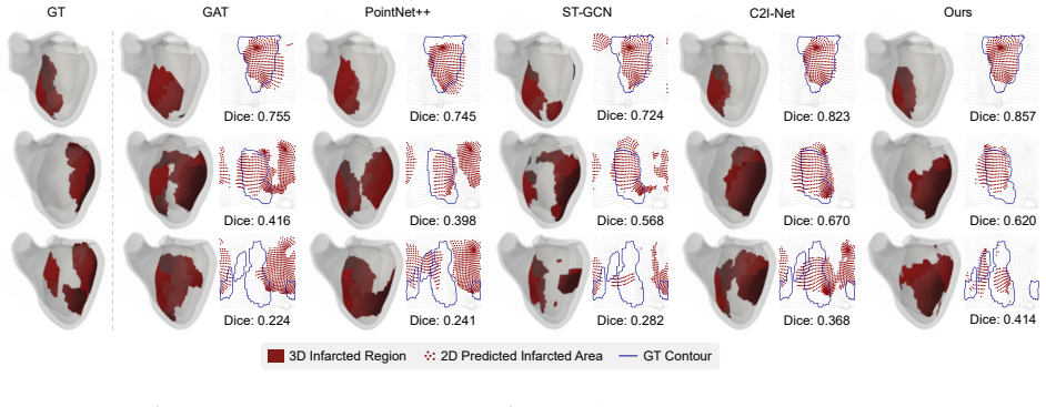
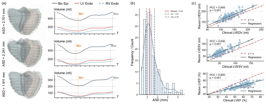
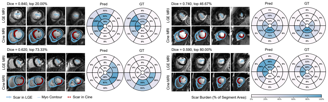
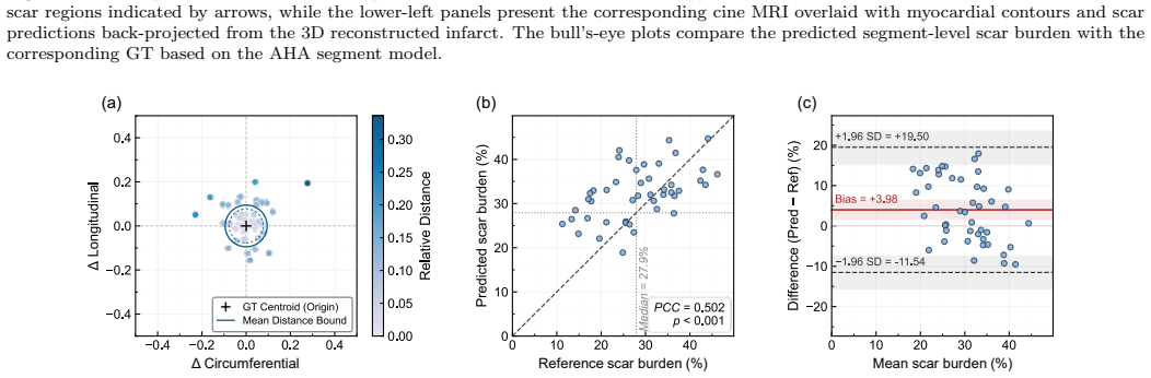
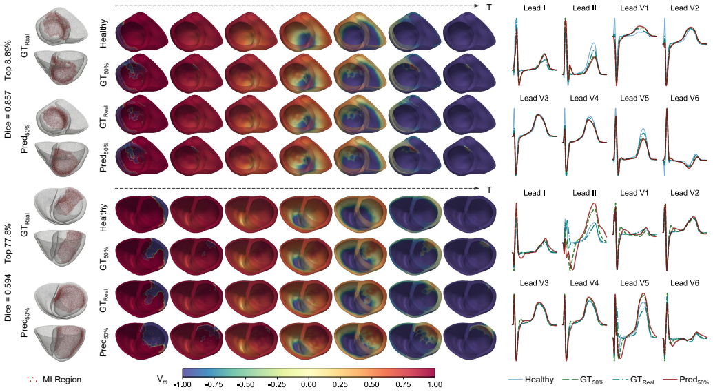
The explicit geometry-motion embedded model with dual-branch adaptive fusion and AHA-17 segment-guided cross-attention reconstructs simulation-ready 3D MI geometries from multi-view cine MRIs, achieving an average Dice score of 0.678 and highly consistent in-silico electrophysiological results with LGE-derived ground truth.
What carries the argument
A 4D biventricular mesh that decouples geometry-aware and motion-aware features, fused via a dual-branch module with multi-scale AHA-17 guided cross-attention to map the infarcted region.
If this is right
- Contrast-free 3D infarct characterization becomes available for renally impaired patients and longitudinal follow-up.
- Cardiac digital twins can incorporate personalized infarct geometries without requiring contrast-enhanced scans.
- In-silico electrophysiological simulations using the reconstructed geometries match LGE-based results.
- The pipeline enables fully automatic extraction of simulation-ready 3D MI shapes from routine multi-view cine MRI.
Where Pith is reading between the lines
- The same motion-geometry decoupling might extend to modeling other regional wall-motion abnormalities such as those in non-ischemic cardiomyopathies.
- If the motion surrogate proves less specific in some populations, fusion with additional non-contrast signals like strain maps could be tested as an incremental improvement.
- Public code release would permit external validation on datasets with varying scanner vendors and patient demographics.
- Embedding the output meshes into real-time CDT platforms could support immediate simulation-based treatment planning.
Load-bearing premise
Abnormal ventricular wall motion visible on cine MRI is sufficiently specific and localized to serve as a reliable surrogate for the true infarct geometry seen on LGE MRI.
What would settle it
Direct comparison in patients imaged with both cine and LGE MRI showing that the cine-derived 3D infarct geometries produce electrophysiological simulation outcomes that differ substantially from LGE-derived ones.
Figures

read the original abstract
Accurate 3D geometric characterization of myocardial infarction (MI) is essential for building cardiac digital twins (CDTs) to precisely simulate infarct-related electrophysiology. Late gadolinium enhancement magnetic resonance imaging (LGE MRI) is the clinical reference for locating MI, yet its reliance on contrast agents restricts use in renally impaired patients and limits longitudinal follow-ups. As an alternative, contrast-free cine MRI visualizes abnormal ventricular wall motion, which is highly indicative of the infarcted area. In this study, we propose a novel explicit geometry-motion embedded model to fully automatically reconstruct personalized, simulation-ready 3D MI geometries directly from multi-view cine MRIs. Specifically, we construct a 4D (3D + t) biventricular mesh to explicitly extract and decouple geometry-aware and motion-aware features. We further design a dual-branch module for adaptive geometry-motion fusion to capture spatiotemporal dependencies for mapping infarcted region. Furthermore, we introduce multi-scale supervision utilizing an AHA-17 segment-guided cross-attention mechanism to steer the prediction, ensuring biophysically consistent reconstruction. Experimental results on 225 cine MRIs demonstrated that the proposed 3D MI reconstruction achieved high performance with an average Dice score of 0.678 $\pm$ 0.011. In the downstream in-silico electrophysiological simulation evaluations, the results were highly consistent with the LGE-derived ground truth, highlighting the great potential of the proposed model for contrast-free scar characterization and seamless integration into CDT modeling. The code will be released publicly upon acceptance of the manuscript for publication.
Editorial analysis
A structured set of objections, weighed in public.
Referee Report
Summary. The paper proposes an explicit geometry-motion embedded model that constructs a 4D biventricular mesh from multi-view cine MRIs, extracts decoupled geometry-aware and motion-aware features, applies a dual-branch adaptive fusion module, and uses AHA-17 segment-guided cross-attention for multi-scale supervision to reconstruct personalized 3D myocardial infarct geometries. On 225 cine MRIs the method reports an average Dice score of 0.678 ± 0.011 against LGE-derived ground truth and states that downstream in-silico electrophysiological simulations are highly consistent with LGE-based results, positioning the approach as a contrast-free alternative for cardiac digital twins.
Significance. If the central motion-to-infarct surrogate assumption holds and the reported performance is shown to be robust, the work would enable contrast-free, simulation-ready 3D scar models for patients ineligible for LGE MRI, directly supporting personalized CDT pipelines. The explicit 4D mesh and AHA-guided supervision are technically interesting design choices that could generalize to other motion-based cardiac tasks. However, the moderate Dice value and absence of comparative or ablation evidence currently limit the assessed impact.
major comments (4)
- [Abstract] Abstract: the headline claim of 'high performance' with Dice 0.678 ± 0.011 is presented without any baseline method, state-of-the-art comparator, or ablation of the dual-branch fusion and AHA cross-attention modules; without these controls it is impossible to determine whether the proposed components drive the result or whether simpler motion-feature baselines would suffice.
- [Abstract] Abstract and experimental section: no information is supplied on patient-level train/validation/test splits, cross-validation strategy, or how the ±0.011 error bar was computed (e.g., standard deviation across folds or bootstrap); these omissions make the single scalar Dice value difficult to interpret as a reliable performance estimate.
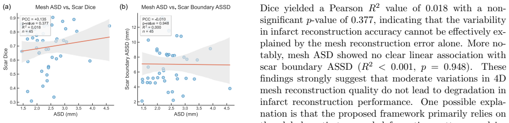
- [Abstract] Abstract: the assertion that cine-MRI wall-motion abnormalities are 'highly indicative of the infarcted area' is treated as given, yet no quantitative analysis of false-positive motion defects (non-infarct ischemia, bundle-branch block, remodeling) or boundary mismatch with LGE is provided; a Dice of ~0.68 implies substantial spatial error that could systematically affect conduction-pathway placement in the downstream EP simulations.
- [Experimental results / downstream evaluation] Downstream evaluation paragraph: the statement that 'results were highly consistent with the LGE-derived ground truth' is qualitative only; no quantitative metrics (e.g., activation-time error, re-entry vulnerability indices, or scar-volume overlap in the EP domain) or details on how the reconstructed 3D mesh is meshed and parameterized for the simulator are supplied.
minor comments (2)
- [Abstract] The manuscript states that 'the code will be released publicly upon acceptance' but provides no link or repository placeholder; adding a footnote with a GitHub URL or Zenodo DOI would improve reproducibility.
- [Methods] Notation for the 4D mesh and the dual-branch module is introduced without an accompanying diagram or equation block; a single figure showing the overall architecture with labeled tensors would clarify the geometry-motion decoupling step.
Simulated Author's Rebuttal
We thank the referee for the constructive and detailed comments. We address each major point below and indicate where revisions will be made to strengthen the manuscript.
read point-by-point responses
-
Referee: [Abstract] Abstract: the headline claim of 'high performance' with Dice 0.678 ± 0.011 is presented without any baseline method, state-of-the-art comparator, or ablation of the dual-branch fusion and AHA cross-attention modules; without these controls it is impossible to determine whether the proposed components drive the result or whether simpler motion-feature baselines would suffice.
Authors: We agree that the abstract would benefit from explicit context on the contribution of the proposed components. The experimental section of the manuscript contains comparisons against baseline methods and ablations of the dual-branch adaptive fusion and AHA-17 cross-attention modules. We will revise the abstract to include a concise statement of these comparative results and the performance gains attributable to the proposed modules. revision: yes
-
Referee: [Abstract] Abstract and experimental section: no information is supplied on patient-level train/validation/test splits, cross-validation strategy, or how the ±0.011 error bar was computed (e.g., standard deviation across folds or bootstrap); these omissions make the single scalar Dice value difficult to interpret as a reliable performance estimate.
Authors: We acknowledge the omission of these experimental details from the abstract. We will add a clear description of the patient-level data partitioning, cross-validation procedure, and the method used to compute the reported error bar to both the abstract and the experimental section in the revised manuscript. revision: yes
-
Referee: [Abstract] Abstract: the assertion that cine-MRI wall-motion abnormalities are 'highly indicative of the infarcted area' is treated as given, yet no quantitative analysis of false-positive motion defects (non-infarct ischemia, bundle-branch block, remodeling) or boundary mismatch with LGE is provided; a Dice of ~0.68 implies substantial spatial error that could systematically affect conduction-pathway placement in the downstream EP simulations.
Authors: The Dice score of 0.678 reflects the expected spatial discrepancy when inferring scar solely from motion. We will expand the discussion to include quantitative analysis of boundary mismatches with LGE and potential sources of false-positive motion defects, together with their possible impact on downstream electrophysiological simulations. revision: yes
-
Referee: [Experimental results / downstream evaluation] Downstream evaluation paragraph: the statement that 'results were highly consistent with the LGE-derived ground truth' is qualitative only; no quantitative metrics (e.g., activation-time error, re-entry vulnerability indices, or scar-volume overlap in the EP domain) or details on how the reconstructed 3D mesh is meshed and parameterized for the simulator are supplied.
Authors: We agree that quantitative metrics and implementation details would strengthen the downstream evaluation. We will add specific quantitative measures (activation-time error, scar-volume overlap, and re-entry indices) and a description of the meshing and parameterization steps used for the electrophysiological simulator in the revised manuscript. revision: yes
Circularity Check
No circularity: empirical supervised model with external LGE ground truth
full rationale
The manuscript presents a supervised deep-learning pipeline that learns a mapping from multi-view cine MRI to 3D infarct geometry, with performance quantified by Dice overlap against independently acquired LGE segmentations on a held-out test set of 225 cases. No equations, ansatzes, or derivations are invoked; the model is trained end-to-end on paired data and evaluated on downstream EP simulations that also use the same external LGE labels. The assumption that wall-motion abnormalities correlate with infarct location is an empirical hypothesis tested by the reported Dice of 0.678, not a definitional or self-referential step. No self-citations appear in the provided text, and the central result does not reduce to any fitted parameter renamed as a prediction. The derivation chain is therefore self-contained against external benchmarks.
Axiom & Free-Parameter Ledger
axioms (1)
- domain assumption Abnormal wall motion on cine MRI is a reliable surrogate for infarct location
Reference graph
Works this paper leans on
-
[1]
Functional Imaging and Modeling of the Heart , year=
An Open-Source End-to-End Pipeline for Generating 3D+t Biventricular Meshes from Cardiac Magnetic Resonance Imaging , author=. Functional Imaging and Modeling of the Heart , year=
-
[2]
Medical image analysis , volume=
Universal ventricular coordinates: A generic framework for describing position within the heart and transferring data , author=. Medical image analysis , volume=. 2018 , publisher=
2018
-
[3]
The Lancet , volume=
Acute myocardial infarction , author=. The Lancet , volume=. 2017 , publisher=
2017
-
[4]
European heart journal , volume=
The ‘Digital Twin’to enable the vision of precision cardiology , author=. European heart journal , volume=. 2020 , publisher=
2020
-
[5]
arXiv preprint arXiv:2602.11942 , year=
Synthesis of Late Gadolinium Enhancement Images via Implicit Neural Representations for Cardiac Scar Segmentation , author=. arXiv preprint arXiv:2602.11942 , year=
-
[6]
IEEE transactions on medical imaging , volume=
Toward enabling cardiac digital twins of myocardial infarction using deep computational models for inverse inference , author=. IEEE transactions on medical imaging , volume=. 2024 , publisher=
2024
-
[7]
Nature Communications , volume=
Arrhythmia risk stratification of patients after myocardial infarction using personalized heart models , author=. Nature Communications , volume=. 2016 , publisher=
2016
-
[8]
Journal of the American College of Cardiology , volume=
Electroanatomic characterization of post-infarct scars: comparison with 3-dimensional myocardial scar reconstruction based on magnetic resonance imaging , author=. Journal of the American College of Cardiology , volume=. 2008 , publisher=
2008
-
[9]
EP Europace , volume=
Human biventricular electromechanical simulations on the progression of electrocardiographic and mechanical abnormalities in post-myocardial infarction , author=. EP Europace , volume=. 2021 , publisher=
2021
-
[10]
A framework for the generation of digital twins of cardiac electrophysiology from clinical 12-leads
Gillette, Karli and others , journal=. A framework for the generation of digital twins of cardiac electrophysiology from clinical 12-leads. 2021 , publisher=
2021
-
[11]
IEEE transactions on medical imaging , volume=
Myocardial infarct segmentation from magnetic resonance images for personalized modeling of cardiac electrophysiology , author=. IEEE transactions on medical imaging , volume=. 2015 , publisher=
2015
-
[12]
JACC: Cardiovascular Imaging , volume=
Automated cardiac MR scar quantification in hypertrophic cardiomyopathy using deep convolutional neural networks , author=. JACC: Cardiovascular Imaging , volume=. 2018 , publisher=
2018
-
[13]
Medical physics , volume=
Image-based reconstruction of three-dimensional myocardial infarct geometry for patient-specific modeling of cardiac electrophysiology , author=. Medical physics , volume=. 2015 , publisher=
2015
-
[14]
Circulation research , volume=
High-resolution 3-dimensional reconstruction of the infarct border zone: impact of structural remodeling on electrical activation , author=. Circulation research , volume=. 2012 , publisher=
2012
-
[15]
European heart journal , volume=
2022 ESC Guidelines for the management of patients with ventricular arrhythmias and the prevention of sudden cardiac death: Developed by the task force for the management of patients with ventricular arrhythmias and the prevention of sudden cardiac death of the European Society of Cardiology (ESC) Endorsed by the Association for European Paediatric and Co...
2022
-
[16]
Europace , volume=
Left ventricular ejection fraction and myocardial fibrosis in sudden cardiac death , author=. Europace , volume=. 2025 , publisher=
2025
-
[17]
Frontiers in Cardiovascular Medicine , volume=
Left ventricular ejection fraction: clinical, pathophysiological, and technical limitations , author=. Frontiers in Cardiovascular Medicine , volume=. 2024 , publisher=
2024
-
[18]
The New England journal of medicine , volume=
Digital Twin-Guided Ablation for Ventricular Tachycardia , author=. The New England journal of medicine , volume=
-
[19]
Progress in biophysics and molecular biology , volume=
Methodology for image-based reconstruction of ventricular geometry for patient-specific modeling of cardiac electrophysiology , author=. Progress in biophysics and molecular biology , volume=. 2014 , publisher=
2014
-
[20]
Circulation: Arrhythmia and Electrophysiology , volume=
Heart digital twins predict features of invasive reentrant circuits and ablation lesions in scar-dependent ventricular tachycardia , author=. Circulation: Arrhythmia and Electrophysiology , volume=. 2025 , publisher=
2025
-
[21]
Journal of Magnetic Resonance Imaging , volume=
Clinical performance of high-resolution late gadolinium enhancement imaging with compressed sensing , author=. Journal of Magnetic Resonance Imaging , volume=. 2017 , publisher=
2017
-
[22]
Journal of Magnetic Resonance Imaging , volume=
Whole-heart high-resolution late gadolinium enhancement: Techniques and clinical applications , author=. Journal of Magnetic Resonance Imaging , volume=. 2022 , publisher=
2022
-
[23]
Medical Image Analysis , volume=
MyoPS: A benchmark of myocardial pathology segmentation combining three-sequence cardiac magnetic resonance images , author=. Medical Image Analysis , volume=. 2023 , publisher=
2023
-
[24]
Cardiovascular Revascularization Medicine , volume=
Disparate Impact of Ischemic Injury on Regional Wall Dysfunction in Acute Anterior vs Inferior Myocardial Infarction , author=. Cardiovascular Revascularization Medicine , volume=. 2019 , publisher=
2019
-
[25]
Current Heart Failure Reports , volume=
Left ventricular diastolic function following myocardial infarction , author=. Current Heart Failure Reports , volume=. 2006 , publisher=
2006
-
[26]
Echocardiography , volume=
Left Ventricular Regional Diastolic Dysfunction in Patients with First Myocardial Infarction Determined by Diastolic Motion of the Atrioventricular Plane , author=. Echocardiography , volume=. 1999 , publisher=
1999
-
[27]
Italian Heart Journal , volume=
Echo-Doppler evaluation of left ventricular diastolic dysfunction during acute myocardial infarction: methodological, clinical and prognostic implications , author=. Italian Heart Journal , volume=. 2001 , publisher=
2001
-
[28]
Medical image analysis , volume=
Direct delineation of myocardial infarction without contrast agents using a joint motion feature learning architecture , author=. Medical image analysis , volume=. 2018 , publisher=
2018
-
[29]
2025 IEEE 22nd International Symposium on Biomedical Imaging (ISBI) , pages=
Contrast-Free Myocardial Scar Segmentation in Cine MRI using Motion and Texture Fusion , author=. 2025 IEEE 22nd International Symposium on Biomedical Imaging (ISBI) , pages=. 2025 , publisher=
2025
-
[30]
European Heart Journal-Imaging Methods and Practice , volume=
Standard breath-hold versus free-breathing real-time cine cardiac MRI—a prospective randomized comparison in patients with known or suspected cardiac disease , author=. European Heart Journal-Imaging Methods and Practice , volume=. 2025 , publisher=
2025
-
[31]
Medical physics , volume=
Texture analysis of cardiac cine magnetic resonance imaging to detect nonviable segments in patients with chronic myocardial infarction , author=. Medical physics , volume=. 2018 , publisher=
2018
-
[32]
International Conference on Medical Image Computing and Computer-Assisted Intervention , pages=
Automated Characterization of Myocardial Scar Topological Patterns for Ventricular Tachycardia Screening , author=. International Conference on Medical Image Computing and Computer-Assisted Intervention , pages=. 2025 , organization=
2025
-
[33]
Magnetic Resonance Materials in Physics, Biology and Medicine , volume=
Late/delayed gadolinium enhancement in MRI after intravenous administration of extracellular gadolinium-based contrast agents: is it worth waiting? , author=. Magnetic Resonance Materials in Physics, Biology and Medicine , volume=. 2024 , publisher=
2024
-
[34]
Circulation , volume=
Artificial intelligence for contrast-free MRI: scar assessment in myocardial infarction using deep learning--based virtual native enhancement , author=. Circulation , volume=. 2022 , publisher=
2022
-
[35]
Circulation: Cardiovascular Imaging , volume=
Predicting late gadolinium enhancement of acute myocardial infarction in contrast-free cardiac cine MRI using deep generative learning , author=. Circulation: Cardiovascular Imaging , volume=. 2024 , publisher=
2024
-
[36]
Circulation: Cardiovascular Imaging , volume=
Synthetic contrast-free LGE via diffusion-based framework in acute MI for image quality and quantitative scar analysis , author=. Circulation: Cardiovascular Imaging , volume=. 2026 , publisher=
2026
-
[37]
Medical Image Analysis , volume=
Knowledge-driven interpretative conditional diffusion model for contrast-free myocardial infarction enhancement synthesis , author=. Medical Image Analysis , volume=. 2025 , publisher=
2025
-
[38]
Applied Soft Computing , volume=
Accurate 3D contrast-free myocardial infarction delineation using a 4D dual-stream spatiotemporal feature learning framework , author=. Applied Soft Computing , volume=. 2023 , publisher=
2023
-
[39]
International Workshop on Digital Twin for Healthcare , pages=
Personalized 3D Myocardial Infarct Geometry Reconstruction from Cine MRI with Explicit Cardiac Motion Modeling , author=. International Workshop on Digital Twin for Healthcare , pages=. 2025 , organization=
2025
-
[40]
Frontiers in Cardiovascular Medicine , volume=
AI-powered contrast-free cardiovascular magnetic resonance imaging for myocardial infarction , author=. Frontiers in Cardiovascular Medicine , volume=. 2024 , publisher=
2024
-
[41]
Frontiers in Cardiovascular Medicine , volume=
Ensemble learning of myocardial displacements for myocardial infarction detection in echocardiography , author=. Frontiers in Cardiovascular Medicine , volume=. 2023 , publisher=
2023
-
[42]
Biomedical Signal Processing and Control , volume=
Early myocardial infarction detection over multi-view echocardiography , author=. Biomedical Signal Processing and Control , volume=. 2024 , publisher=
2024
-
[43]
Information sciences , volume=
Application of deep convolutional neural network for automated detection of myocardial infarction using ECG signals , author=. Information sciences , volume=. 2017 , publisher=
2017
-
[44]
Scientific Reports , volume=
Myocardial scar and left ventricular ejection fraction classification for electrocardiography image using multi-task deep learning , author=. Scientific Reports , volume=. 2024 , publisher=
2024
-
[45]
Radiology , volume=
Deep learning for diagnosis of chronic myocardial infarction on nonenhanced cardiac cine MRI , author=. Radiology , volume=. 2019 , publisher=
2019
-
[46]
Frontiers in Cardiovascular Medicine , volume=
Predicting post-contrast information from contrast agent free cardiac MRI using machine learning: Challenges and methods , author=. Frontiers in Cardiovascular Medicine , volume=. 2022 , publisher=
2022
-
[47]
Diagnostics , volume=
Contrast-Free Myocardial Infarction Segmentation with Attention U-Net , author=. Diagnostics , volume=. 2026 , publisher=
2026
-
[48]
Journal of Cardiovascular Magnetic Resonance , volume=
Late gadolinium enhancement cardiovascular magnetic resonance with generative artificial intelligence , author=. Journal of Cardiovascular Magnetic Resonance , volume=. 2025 , publisher=
2025
-
[49]
Journal of Cardiovascular Magnetic Resonance , volume=
Minimizing risk of nephrogenic systemic fibrosis in cardiovascular magnetic resonance , author=. Journal of Cardiovascular Magnetic Resonance , volume=. 2012 , publisher=
2012
-
[50]
Nature Medicine , volume=
Screening and diagnosis of cardiovascular disease using artificial intelligence-enabled cardiac magnetic resonance imaging , author=. Nature Medicine , volume=. 2024 , publisher=
2024
-
[51]
Cardiovascular Imaging , volume=
Myocardial strain imaging: theory, current practice, and the future , author=. Cardiovascular Imaging , volume=. 2025 , publisher=
2025
-
[52]
American Journal of Roentgenology , volume=
Nephrogenic systemic fibrosis , author=. American Journal of Roentgenology , volume=. 2012 , publisher=
2012
-
[53]
Radiology , volume=
High signal intensity in the dentate nucleus and globus pallidus on unenhanced T1-weighted MR images: relationship with increasing cumulative dose of a gadolinium-based contrast material , author=. Radiology , volume=. 2014 , publisher=
2014
-
[54]
Journal of International Medical Research , volume=
Motion-corrected free-breathing late gadolinium enhancement combined with a gadolinium contrast agent with a high relaxation rate: an optimized cardiovascular magnetic resonance examination protocol , author=. Journal of International Medical Research , volume=. 2020 , publisher=
2020
-
[55]
Quantitative Imaging in Medicine and Surgery , volume=
Dark-blood late gadolinium-enhancement cardiac magnetic resonance imaging for myocardial scar detection based on simplified timing scheme: single-center experience in patients with suspected coronary artery disease , author=. Quantitative Imaging in Medicine and Surgery , volume=
-
[56]
Journal of Clinical Medicine , volume=
Predictive value of left ventricular systolic dysfunction or wall motion abnormalities for non-ischemic myocardial injury: a multicenter cardiovascular resonance study , author=. Journal of Clinical Medicine , volume=. 2025 , publisher=
2025
-
[57]
Scandinavian Cardiovascular Journal , volume=
Phase analysis detects heterogeneity of myocardial deformation on cine MRI , author=. Scandinavian Cardiovascular Journal , volume=. 2015 , publisher=
2015
-
[58]
Anesthesia & Analgesia , volume=
Perioperative assessment of myocardial deformation , author=. Anesthesia & Analgesia , volume=. 2014 , publisher=
2014
-
[59]
Medical image analysis , volume=
A deep-learning approach for direct whole-heart mesh reconstruction , author=. Medical image analysis , volume=. 2021 , publisher=
2021
-
[60]
medRxiv , pages=
Developing cardiac digital twins at scale: Insights from personalised myocardial conduction velocity , author=. medRxiv , pages=. 2023 , publisher=
2023
-
[61]
Medical Image Analysis , volume=
Personalized topology-informed localization of standard 12-lead ECG electrode placement from incomplete cardiac MRIs for efficient cardiac digital twins , author=. Medical Image Analysis , volume=. 2025 , publisher=
2025
-
[62]
Europace , volume=
From bits to bedside: entering the age of digital twins in cardiac electrophysiology , author=. Europace , volume=. 2024 , publisher=
2024
-
[63]
Technology and Innovation in Medical Sciences: Breakthroughs from Gangwal School of Medical Sciences and Technology, IIT Kanpur , pages=
Cardiac Digital Twins—Computational Approaches for Personalized Arrhythmia Care , author=. Technology and Innovation in Medical Sciences: Breakthroughs from Gangwal School of Medical Sciences and Technology, IIT Kanpur , pages=. 2025 , publisher=
2025
-
[64]
IEEE Transactions on Medical Imaging , volume=
Learning whole heart mesh generation from patient images for computational simulations , author=. IEEE Transactions on Medical Imaging , volume=. 2022 , publisher=
2022
-
[65]
IEEE transactions on medical imaging , volume=
Patient-specific heart geometry modeling for solid biomechanics using deep learning , author=. IEEE transactions on medical imaging , volume=. 2023 , publisher=
2023
-
[66]
arXiv preprint arXiv:2505.03599 , year=
From pixels to polygons: A survey of deep learning approaches for medical image-to-mesh reconstruction , author=. arXiv preprint arXiv:2505.03599 , year=
-
[67]
Journal of the mechanical behavior of biomedical materials , volume=
Biomechanical properties of acellular scar ECM during the acute to chronic stages of myocardial infarction , author=. Journal of the mechanical behavior of biomedical materials , volume=. 2021 , publisher=
2021
-
[68]
IEEE transactions on pattern analysis and machine intelligence , volume=
Multivariate mixture model for myocardial segmentation combining multi-source images , author=. IEEE transactions on pattern analysis and machine intelligence , volume=. 2018 , publisher=
2018
-
[69]
Circulation , volume=
Standardized myocardial segmentation and nomenclature for tomographic imaging of the heart: a statement for healthcare professionals from the Cardiac Imaging Committee of the Council on Clinical Cardiology of the American Heart Association , author=. Circulation , volume=. 2002 , publisher=
2002
-
[70]
Are they interchangeable? , author=
Comparison of left ventricular ejection fraction and volumes in heart failure by echocardiography, radionuclide ventriculography and cardiovascular magnetic resonance. Are they interchangeable? , author=. European heart journal , volume=. 2000 , publisher=
2000
-
[71]
Multimedia Tools and Applications , volume=
Fully automated 2D and 3D convolutional neural networks pipeline for video segmentation and myocardial infarction detection in echocardiography , author=. Multimedia Tools and Applications , volume=. 2022 , publisher=
2022
-
[72]
Frontiers in Cardiovascular Medicine , volume=
Subclinical myocardial dysfunction assessed by cardiac magnetic resonance feature tracking predicts ventricular arrhythmias in early-stage hypertension , author=. Frontiers in Cardiovascular Medicine , volume=. 2025 , publisher=
2025
-
[73]
The International Journal of Cardiovascular Imaging , volume=
Quantitative evaluation of subclinical left ventricular dysfunction in patients with type 2 diabetes mellitus by three-dimensional echocardiography , author=. The International Journal of Cardiovascular Imaging , volume=. 2020 , publisher=
2020
-
[74]
Medical image analysis , volume=
Segmentation and quantification of infarction without contrast agents via spatiotemporal generative adversarial learning , author=. Medical image analysis , volume=. 2020 , publisher=
2020
-
[75]
Circulation , volume=
Toward replacing late gadolinium enhancement with artificial intelligence virtual native enhancement for gadolinium-free cardiovascular magnetic resonance tissue characterization in hypertrophic cardiomyopathy , author=. Circulation , volume=. 2021 , publisher=
2021
-
[76]
IEEE Transactions on image processing , volume=
Myocardial motion analysis from B-mode echocardiograms , author=. IEEE Transactions on image processing , volume=. 2005 , publisher=
2005
-
[77]
Nature Biomedical Engineering , volume=
Personalized virtual-heart technology for guiding the ablation of infarct-related ventricular tachycardia , author=. Nature Biomedical Engineering , volume=. 2018 , publisher=
2018
-
[78]
IEEE transactions on medical imaging , volume=
Point-tracked quantitative analysis of left ventricular surface motion from 3-D image sequences , author=. IEEE transactions on medical imaging , volume=. 2000 , publisher=
2000
-
[79]
Frontiers in cardiovascular medicine , volume=
DeepStrain: a deep learning workflow for the automated characterization of cardiac mechanics , author=. Frontiers in cardiovascular medicine , volume=. 2021 , publisher=
2021
-
[80]
Proceedings of the IEEE/CVF conference on computer vision and pattern recognition , pages=
Deeptag: An unsupervised deep learning method for motion tracking on cardiac tagging magnetic resonance images , author=. Proceedings of the IEEE/CVF conference on computer vision and pattern recognition , pages=
discussion (0)
Sign in with ORCID, Apple, or X to comment. Anyone can read and Pith papers without signing in.