Foveated-Imaging Geometry CT Architecture and Seeded Diffusion Model Enabling Global Super-Resolution Reconstruction
Pith reviewed 2026-06-27 11:11 UTC · model grok-4.3
The pith
A foveated CT geometry that acquires mostly low-resolution data plus sparse local high-resolution measurements enables a diffusion model to produce consistent high-resolution images over the entire field of view.
A machine-rendered reading of the paper's core claim, the machinery that carries it, and where it could break.
Core claim
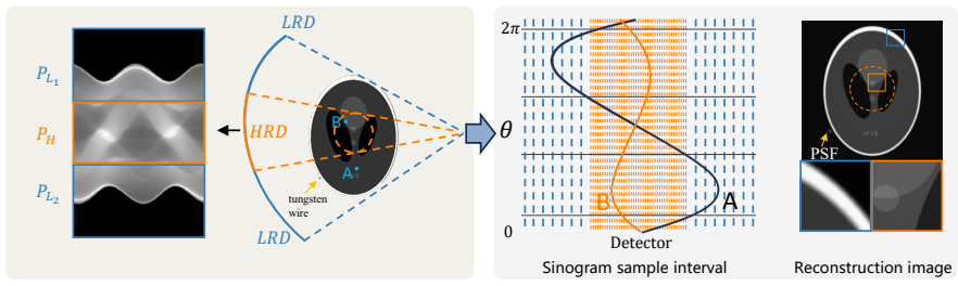
The FIGCT architecture records the majority of projections at low resolution and a small central or offset region at high resolution; the DPFSR framework then uses these local high-resolution measurements as seeds that are inserted into intermediate clean-image estimates during the reverse diffusion process in both the projection domain and the image domain, thereby guiding the generation of a globally consistent high-resolution volume.
What carries the argument
The seeded reverse diffusion step inside DPFSR that inserts local high-resolution measurements into clean-image estimates in both projection and image domains to enforce data consistency.
If this is right
- CT systems can achieve high-resolution imaging over the full field of view while acquiring the majority of data at low resolution, reducing detector cost and data volume.
- The high-resolution region can be placed flexibly inside the field of view without requiring a uniform high-resolution detector array.
- Super-resolution performance improves when high-resolution data are supplied directly in both projection and image domains rather than only at the final image stage.
- Existing diffusion-based reconstruction pipelines can be extended to hybrid-resolution inputs by adding the seeded clean-image update step.
Where Pith is reading between the lines
- If the seeded diffusion approach generalizes, similar hybrid-resolution acquisition could be applied to other modalities such as cone-beam CT or digital tomosynthesis where detector cost is a limiting factor.
- The method implicitly assumes that the low-resolution measurements outside the high-resolution region remain consistent with the high-resolution region; any misalignment in the scanner geometry would therefore need separate correction.
- A practical next test would be to vary the size and position of the high-resolution patch and measure the resulting trade-off between reconstruction quality and data overhead.
Load-bearing premise
Local high-resolution measurements can be inserted into the diffusion process without creating inconsistencies or new artifacts outside the measured high-resolution region.
What would settle it
Reconstruct a full high-resolution ground-truth scan of the same swine lung specimen under both FIGCT geometry and a uniform high-resolution detector array, then measure whether the DPFSR output matches the uniform high-resolution scan in both the seeded region and the surrounding field of view to within the reported PSNR/SSIM margins.
Figures


read the original abstract
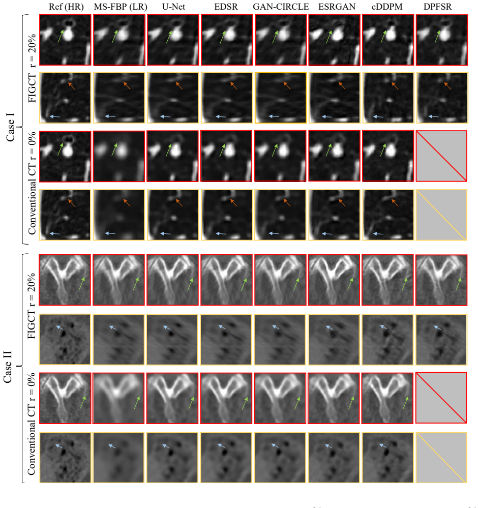
For X-ray computed tomography (CT), a smaller detector pixel size generally leads to higher scanner spatial resolution, but inevitably increases system cost as well as data overhead in acquisition and processing. To achieve high-resolution (HR) CT imaging in a more resource-efficient manner, we propose a Foveated-Imaging Geometry CT (FIGCT) architecture, which integrates local HR data into an acquisition scheme dominated by low-resolution (LR) measurements. We further develop a Diffusion Probabilistic FIGCT Super-Resolution Reconstruction (DPFSR) framework to generate global HR CT images over the full field of view (FOV). The concept of FIGCT is first established, and its typical configurations are characterized according to the arrangement of HR data. Two key indices, namely the HR data fraction (HDF) and the LR-to-HR detector pixel size ratio (LHR), are introduced to describe the FIGCT geometry. The proposed DPFSR incorporates local HR information into intermediate clean-image estimates in both the projection and image domains during the reverse diffusion process. This additional step not only guides HR image generation from LR data, but also improves data consistency between the clean-image estimates and the originally measured data. Preliminary numerical simulation results on FIGCT show that the proposed architecture provides high-precision CT images within the region of interest (ROI) corresponding to the HR data, while the spatial resolution deteriorates rapidly outside the ROI. With DPFSR, global HR reconstruction is achieved on the AAPM Grand Challenge dataset and swine lung CT data, outperforming existing SR methods in terms of Learned Perceptual Image Patch Similarity (LPIPS), PSNR, and SSIM.
Editorial analysis
A structured set of objections, weighed in public.
Referee Report
Summary. The manuscript introduces a Foveated-Imaging Geometry CT (FIGCT) architecture that acquires mostly low-resolution (LR) measurements supplemented by local high-resolution (HR) data in a region of interest (ROI), along with the Diffusion Probabilistic FIGCT Super-Resolution Reconstruction (DPFSR) framework. DPFSR injects local HR information into intermediate clean-image estimates during the reverse diffusion process in both projection and image domains to enforce data consistency. The paper claims that standard FIGCT resolution deteriorates rapidly outside the ROI, whereas DPFSR achieves global HR reconstruction on the AAPM Grand Challenge dataset and swine lung CT data, outperforming existing super-resolution methods according to LPIPS, PSNR, and SSIM.
Significance. If the central claim of globally consistent HR reconstruction that respects all LR measurements holds, the work could enable cost- and data-efficient high-resolution CT by reducing detector pixel count while using diffusion priors to fill in detail outside the foveated region. The dual-domain seeding approach is a potentially useful extension of diffusion-based SR to geometry-constrained problems in medical imaging.
major comments (2)
- [Abstract] Abstract: The claim that DPFSR produces 'global HR reconstruction' that outperforms SR baselines is load-bearing, yet the text provides no description of any verification that the final images, when re-projected under the LR geometry, match the acquired LR sinogram outside the ROI within noise. Without this check, the reported LPIPS/PSNR/SSIM gains cannot be distinguished from hallucinated detail that violates data fidelity.
- [Abstract] Abstract: The outperformance is stated on two datasets without error bars, without an ablation isolating the contribution of the dual-domain seeding step, and with results labeled only as 'preliminary numerical simulation results.' These omissions make the quantitative superiority claim impossible to assess for statistical or methodological robustness.
minor comments (1)
- [Abstract] The definitions of the two key indices (HR data fraction HDF and LR-to-HR detector pixel size ratio LHR) are introduced but their precise mathematical formulations and how they enter the reconstruction pipeline are not shown in the provided text.
Simulated Author's Rebuttal
We thank the referee for the constructive comments. We respond point-by-point below and will revise the manuscript to address the concerns raised.
read point-by-point responses
-
Referee: [Abstract] Abstract: The claim that DPFSR produces 'global HR reconstruction' that outperforms SR baselines is load-bearing, yet the text provides no description of any verification that the final images, when re-projected under the LR geometry, match the acquired LR sinogram outside the ROI within noise. Without this check, the reported LPIPS/PSNR/SSIM gains cannot be distinguished from hallucinated detail that violates data fidelity.
Authors: We acknowledge that an explicit re-projection check comparing the final HR images against the acquired LR sinogram outside the ROI (within noise) would strengthen the data-fidelity claim. The DPFSR framework already incorporates projection-domain seeding during the reverse process to enforce consistency with measured data, but we agree this additional verification step was not described. We will add quantitative sinogram-domain fidelity metrics and corresponding analysis to the revised manuscript. revision: yes
-
Referee: [Abstract] Abstract: The outperformance is stated on two datasets without error bars, without an ablation isolating the contribution of the dual-domain seeding step, and with results labeled only as 'preliminary numerical simulation results.' These omissions make the quantitative superiority claim impossible to assess for statistical or methodological robustness.
Authors: The results are labeled preliminary because they are numerical simulations. To improve statistical and methodological robustness we will add error bars from repeated runs, include an ablation isolating the dual-domain seeding contribution, and update the abstract and results sections accordingly. revision: yes
Circularity Check
No circularity: DPFSR derivation is self-contained and independent of its inputs
full rationale
The paper defines a new FIGCT geometry via HDF and LHR indices and a DPFSR diffusion process that injects local HR data into reverse steps in projection and image domains. No equations are shown that define any output quantity in terms of itself, no fitted parameters are relabeled as predictions, and no self-citations supply load-bearing uniqueness theorems or ansatzes. Results are obtained from external AAPM and swine datasets with standard metrics, so the central reconstruction claim does not reduce to its own inputs by construction.
Axiom & Free-Parameter Ledger
axioms (1)
- domain assumption Diffusion models can be conditioned on local high-resolution measurements to enforce data consistency across the full FOV.
invented entities (2)
-
FIGCT architecture
no independent evidence
-
DPFSR framework
no independent evidence
Reference graph
Works this paper leans on
-
[1]
Medical Imaging 2022: Physics of Medical Imaging , volume=
High-resolution CT reconstruction from Zoom-In Partial Scans (ZIPS) with simultaneous estimation of inter-scan ROI motion , author=. Medical Imaging 2022: Physics of Medical Imaging , volume=. 2022 , organization=
2022
-
[2]
Physics in Medicine & Biology , abstract =
Zhang, Zheng and Chen, Buxin and Xia, Dan and Sidky, Emil Y and Pan, Xiaochuan , title =. Physics in Medicine & Biology , abstract =. 2025 , month =. doi:10.1088/1361-6560/ada7be , url =
-
[3]
Medical Imaging 2025: Physics of Medical Imaging , editor =
WenXin Mo and Hewei Gao , title =. Medical Imaging 2025: Physics of Medical Imaging , editor =. 2025 , doi =
2025
-
[4]
Medical physics , volume=
Empirical optimization of energy bin weights for compressing measurements with realistic photon counting x-ray detectors , author=. Medical physics , volume=. 2024 , publisher=
2024
-
[5]
Pontone, Gianluca and Bertella, Erika and Mushtaq, Saima and Loguercio, Monica and Cortinovis, Sarah and Baggiano, Andrea and Conte, Edoardo and Annoni, Andrea and Formenti, Alberto and Beltrama, Virginia and Guaricci, Andrea Igoren and Andreini, Daniele , title =. Radiology , volume =. 2014 , doi =. https://doi.org/10.1148/radiol.13130909 , abstract =
-
[6]
Improved Training of Wasserstein GANs , url =
Gulrajani, Ishaan and Ahmed, Faruk and Arjovsky, Martin and Dumoulin, Vincent and Courville, Aaron C , booktitle =. Improved Training of Wasserstein GANs , url =
-
[7]
In: 2018 IEEE/CVF Con- ference on Computer Vision and Pattern Recognition
Zhang, Richard and Isola, Phillip and Efros, Alexei A. and Shechtman, Eli and Wang, Oliver , month = jun, year =. The. 2018. doi:10.1109/CVPR.2018.00068 , abstract =
-
[8]
2016 Low-Dose CT Grand Challenge , year =
Cynthia McCollough , publisher =. 2016 Low-Dose CT Grand Challenge , year =. doi:10.21227/4yqw-2364 , url =
-
[9]
and Hadjiiski, Lubomir and Tourassi, Georgia D
Armato III, Samuel G. and Hadjiiski, Lubomir and Tourassi, Georgia D. and Drukker, Karen and Giger, Maryellen L. and Li, Feng and Redmond, George and Farahani, Keyvan and Kirby, Justin S. and Clarke, Laurence P. , title =. 2015 , publisher =. doi:10.7937/K9/TCIA.2015.UZLSU3FL , url =
-
[10]
Journal of Digital Imaging , author =
The. Journal of Digital Imaging , author =. 2013 , pages =. doi:10.1007/s10278-013-9622-7 , abstract =
-
[11]
MAGIC: Manifold and Graph Integrative Convolutional Network for Low-Dose CT Reconstruction , year=
Xia, Wenjun and Lu, Zexin and Huang, Yongqiang and Shi, Zuoqiang and Liu, Yan and Chen, Hu and Chen, Yang and Zhou, Jiliu and Zhang, Yi , journal=. MAGIC: Manifold and Graph Integrative Convolutional Network for Low-Dose CT Reconstruction , year=
-
[12]
Hallucination
Tivnan, Matthew and Yoon, Siyeop and Chen, Zhennong and Li, Xiang and Wu, Dufan and Li, Quanzheng , editor =. Hallucination. Medical. 2024 , pages =
2024
-
[13]
Liu, Jiaming and Anirudh, Rushil and Thiagarajan, Jayaraman J. and He, Stewart and Mohan, K. Aditya and Kamilov, Ulugbek S. and Kim, Hyojin , month = oct, year =. 2023. doi:10.1109/ICCV51070.2023.00963 , abstract =
-
[14]
European Journal of Radiology , author =
General principles of. European Journal of Radiology , author =. 2003 , pages =. doi:10.1016/S0720-048X(02)00358-3 , abstract =
-
[15]
Katsuyuki Taguchi and George S. K. Fung and Qiulin Tang and Jochen Cammin , title =. Medical Imaging 2013: Physics of Medical Imaging , editor =. 2013 , doi =
2013
-
[16]
Lugmayr, Andreas and Danelljan, Martin and Romero, Andres and Yu, Fisher and Timofte, Radu and Van Gool, Luc , month = jun, year =. 2022. doi:10.1109/CVPR52688.2022.01117 , language =
-
[17]
SRDiff: Single image super-resolution with diffusion probabilistic models , journal =
Haoying Li and Yifan Yang and Meng Chang and Shiqi Chen and Huajun Feng and Zhihai Xu and Qi Li and Yueting Chen , keywords =. SRDiff: Single image super-resolution with diffusion probabilistic models , journal =. 2022 , issn =. doi:https://doi.org/10.1016/j.neucom.2022.01.029 , url =
-
[18]
Physics in Medicine & Biology , abstract =
Sharma, Kriti Sen and Holzner, Christian and Vasilescu, Dragoş M and Jin, Xin and Narayanan, Shree and Agah, Masoud and Hoffman, Eric A and Yu, Hengyong and Wang, Ge , title =. Physics in Medicine & Biology , abstract =. 2013 , month =. doi:10.1088/0031-9155/58/12/4297 , url =
-
[19]
Yu, Haichao and Liu, Ding and Shi, Honghui and Yu, Hanchao and Wang, Zhangyang and Wang, Xinchao and Cross, Brent and Bramler, Matthew and Huang, Thomas S. , month = sep, year =. Computed tomography super-resolution using convolutional neural networks , url =. 2017. doi:10.1109/icip.2017.8297022 , abstract =
-
[20]
Physics in Medicine & Biology , author =
Interior tomography in microscopic. Physics in Medicine & Biology , author =. 2018 , note =. doi:10.1088/1361-6560/aab46f , language =
-
[21]
Rays , volume=
High resolution spiral computed tomography of the pancreas , author=. Rays , volume=
-
[22]
and Wu, Pengwei and Mahesh, Mahadevappa and Siewerdsen, Jeffrey H
Hernandez, Andrew M. and Wu, Pengwei and Mahesh, Mahadevappa and Siewerdsen, Jeffrey H. and Boone, John M. , title =. Medical Physics , volume =. doi:https://doi.org/10.1002/mp.14789 , url =. https://aapm.onlinelibrary.wiley.com/doi/pdf/10.1002/mp.14789 , abstract =
-
[23]
IEEE Transactions on Medical Imaging , author =
Spatial-resolution enhancement in computed tomography , volume =. IEEE Transactions on Medical Imaging , author =. 2005 , note =. doi:10.1109/tmi.2004.840846 , abstract =
-
[24]
Physics in Medicine & Biology , author =
End-to-end deep learning for interior tomography with low-dose x-ray. Physics in Medicine & Biology , author =. 2022 , note =. doi:10.1088/1361-6560/ac6560 , abstract =
-
[25]
Computers in Biology and Medicine , volume=
Self-supervised CT super-resolution with hybrid model , author=. Computers in Biology and Medicine , volume=. 2021 , publisher=
2021
-
[26]
Computers in Biology and Medicine , volume=
CT image super-resolution reconstruction based on global hybrid attention , author=. Computers in Biology and Medicine , volume=. 2022 , publisher=
2022
-
[27]
Physics in Medicine & Biology , volume=
Super-resolution dual-layer CBCT imaging with model-guided deep learning , author=. Physics in Medicine & Biology , volume=. 2024 , publisher=
2024
-
[28]
Measurement Science and Technology , abstract =
Zhao, Qingxian and Li, Jing and Li, Yi and Luo, Shouhua , title =. Measurement Science and Technology , abstract =. 2023 , month =. doi:10.1088/1361-6501/acd0ca , url =
-
[29]
High-Fidelity Modeling of Shift-Variant Focal-Spot Blur for High-Resolution CT , author=
-
[30]
Physics in Medicine & Biology , volume=
Model-based iterative reconstruction for flat-panel cone-beam CT with focal spot blur, detector blur, and correlated noise , author=. Physics in Medicine & Biology , volume=. 2015 , publisher=
2015
-
[31]
2021 IEEE International Conference on Medical Imaging Physics and Engineering (ICMIPE) , pages=
Preliminary studies on Dual-energy CT image super-resolution based on dual-dictionary learning , author=. 2021 IEEE International Conference on Medical Imaging Physics and Engineering (ICMIPE) , pages=. 2021 , organization=
2021
-
[32]
Juan Li and Jin Wu and Huiping Deng and Jin Liu , keywords =. A self-learning image super-resolution method via sparse representation and non-local similarity , journal =. 2016 , note =. doi:https://doi.org/10.1016/j.neucom.2015.07.139 , url =
-
[33]
Proceedings of the IEEE conference on computer vision and pattern recognition workshops , pages=
Enhanced deep residual networks for single image super-resolution , author=. Proceedings of the IEEE conference on computer vision and pattern recognition workshops , pages=
-
[34]
International Conference on Medical image computing and computer-assisted intervention , pages=
U-net: Convolutional networks for biomedical image segmentation , author=. International Conference on Medical image computing and computer-assisted intervention , pages=. 2015 , organization=
2015
-
[35]
Proceedings of the European conference on computer vision (ECCV) workshops , pages=
Esrgan: Enhanced super-resolution generative adversarial networks , author=. Proceedings of the European conference on computer vision (ECCV) workshops , pages=
-
[36]
Super resolution in CT
Yan, Ziye and Li, Jianwu and Lu, Yao and Yan, Hongxia and Zhao, Yanfeng. Super resolution in CT. International Journal of Imaging Systems and Technology. 2015
2015
-
[37]
Journal of Medical Imaging , volume=
Impact of deep learning-based image super-resolution on binary signal detection , author=. Journal of Medical Imaging , volume=. 2021 , publisher=
2021
-
[38]
Impact of using sinogram domain data in the super‐resolution of. Medical Physics , author =. 2024 , note =. doi:10.1002/mp.16807 , abstract =
-
[39]
Investigative radiology , volume=
150- m spatial resolution using photon-counting detector computed tomography technology: technical performance and first patient images , author=. Investigative radiology , volume=. 2018 , publisher=
2018
-
[40]
Computed tomography super-resolution using deep convolutional neural network
Junyoung Park and Donghwi Hwang and Kyeong Yun Kim and Seung Kwan Kang and Yu Kyeong Kim and Jae Sung Lee. Computed tomography super-resolution using deep convolutional neural network. Physics in Medicine & Biology. 2018
2018
-
[41]
and Saha, Punam K
You, Chenyu and Li, Guang and Zhang, Yi and Zhang, Xiaoliu and Shan, Hongming and Li, Mengzhou and Ju, Shenghong and Zhao, Zhen and Zhang, Zhuiyang and Cong, Wenxiang and Vannier, Michael W. and Saha, Punam K. and Hoffman, Eric A. and Wang, Ge. CT Super-Resolution GAN Constrained by the Identical, Residual, and Cycle Learning Ensemble (GAN-CIRCLE). IEEE T...
2020
-
[42]
Denoising Diffusion Probabilistic Models , url =
Ho, Jonathan and Jain, Ajay and Abbeel, Pieter , booktitle =. Denoising Diffusion Probabilistic Models , url =
-
[43]
Medical Physics , volume=
SU-E-I-73: Clinical Evaluation of CT Image Reconstructed Using Interior Tomography , author=. Medical Physics , volume=. 2014 , publisher=
2014
-
[44]
and Norouzi, Mohammad , journal=
Saharia, Chitwan and Ho, Jonathan and Chan, William and Salimans, Tim and Fleet, David J. and Norouzi, Mohammad , journal=. Image Super-Resolution via Iterative Refinement , year=
-
[45]
2024 , url=
Photon-counting CT using a Conditional Diffusion Model for Super-resolution and Texture-preservation , author=. 2024 , url=
2024
-
[46]
MEDICAL PHYSICS , volume=
A novel coplanar multi-modality tomographic imaging for image guidance in radiotherapy using hybrid radiation detector , author=. MEDICAL PHYSICS , volume=. 2020 , organization=
2020
-
[47]
Proceedings of the IEEE conference on computer vision and pattern recognition , pages=
The unreasonable effectiveness of deep features as a perceptual metric , author=. Proceedings of the IEEE conference on computer vision and pattern recognition , pages=
-
[48]
Pontone, E
G. Pontone, E. Bertella, S. Mushtaq, M. Loguercio, S. Cortinovis, A. Baggiano, E. Conte, A. Annoni, A. Formenti, V. Beltrama, A. I. Guaricci, and D. Andreini, Coronary Artery Disease: Diagnostic Accuracy of CT Coronary Angiography—A Comparison of High and Standard Spatial Resolution Scanning, Radiology 271 , 688--694 (2014), PMID: 24520943
2014
-
[49]
Y. Yang, S. Wang, D. Pal, Z. Yin, N. J. Pelc, and A. S. Wang, Empirical optimization of energy bin weights for compressing measurements with realistic photon counting x-ray detectors, Medical physics 51 , 224--238 (2024)
2024
-
[50]
A. M. Hernandez, P. Wu, M. Mahesh, J. H. Siewerdsen, and J. M. Boone, Location and direction dependence in the 3D MTF for a high-resolution CT system, Medical Physics 48 , 2760--2771 (2021)
2021
-
[51]
Xiaochuan Pan , Lifeng Yu , and Chien-Min Kao , Spatial-resolution enhancement in computed tomography, IEEE Transactions on Medical Imaging 24 , 246--253 (2005), Publisher: Institute of Electrical and Electronics Engineers (IEEE)
2005
-
[52]
Q. Zhao, J. Li, Y. Li, and S. Luo, Super-resolution model-based iterative reconstruction for lens-coupled micro-CT imaging, Measurement Science and Technology 34 , 085401 (2023)
2023
-
[53]
Haneda, B
E. Haneda, B. De Man, B. Claus, and L. Fu, High-resolution CT reconstruction from Zoom-In Partial Scans (ZIPS) with simultaneous estimation of inter-scan ROI motion, in Medical Imaging 2022: Physics of Medical Imaging , volume 12031, pages 646--653, SPIE, 2022
2022
-
[54]
Prokop, General principles of MDCT , European Journal of Radiology 45 , S4--S10 (2003)
M. Prokop, General principles of MDCT , European Journal of Radiology 45 , S4--S10 (2003)
2003
-
[55]
Steven Tilley, W
I. Steven Tilley, W. Zbijewski, and J. W. Stayman, High-Fidelity Modeling of Shift-Variant Focal-Spot Blur for High-Resolution CT, Int’l Mtg. Fully 3D Image Recon. in Radiology and Nuc. Med
-
[56]
Tilley, J
S. Tilley, J. H. Siewerdsen, and J. W. Stayman, Model-based iterative reconstruction for flat-panel cone-beam CT with focal spot blur, detector blur, and correlated noise, Physics in Medicine & Biology 61 , 296 (2015)
2015
-
[57]
Zhong, A
X. Zhong, A. Cai, N. Liang, X. Yu, L. Li, and B. Yan, Preliminary studies on Dual-energy CT image super-resolution based on dual-dictionary learning, in 2021 IEEE International Conference on Medical Imaging Physics and Engineering (ICMIPE) , pages 1--4, IEEE, 2021
2021
-
[58]
J. Li, J. Wu, H. Deng, and J. Liu, A self-learning image super-resolution method via sparse representation and non-local similarity, Neurocomputing 184 , 196--206 (2016), RoLoD: Robust Local Descriptors for Computer Vision 2014
2016
-
[59]
Zhang, S
Z. Zhang, S. Yu, W. Qin, X. Liang, Y. Xie, and G. Cao, Self-supervised CT super-resolution with hybrid model, Computers in Biology and Medicine 138 , 104775 (2021)
2021
-
[60]
J. Chi, Z. Sun, H. Wang, P. Lyu, X. Yu, and C. Wu, CT image super-resolution reconstruction based on global hybrid attention, Computers in Biology and Medicine 150 , 106112 (2022)
2022
-
[61]
J. Zhu, T. Su, X. Zhang, H. Cui, Y. Tan, H. Zheng, D. Liang, J. Guo, and Y. Ge, Super-resolution dual-layer CBCT imaging with model-guided deep learning, Physics in Medicine & Biology 69 , 015016 (2024)
2024
-
[62]
H. Yu, D. Liu, H. Shi, H. Yu, Z. Wang, X. Wang, B. Cross, M. Bramler, and T. S. Huang, Computed tomography super-resolution using convolutional neural networks, in 2017 IEEE International Conference on Image Processing ( ICIP ) , pages 3944--3948, Beijing, 2017, IEEE
2017
-
[63]
J. Park, D. Hwang, K. Y. Kim, S. K. Kang, Y. K. Kim, and J. S. Lee, Computed tomography super-resolution using deep convolutional neural network., Physics in Medicine & Biology 63 , 145011 (2018)
2018
-
[64]
J. Ho, A. Jain, and P. Abbeel, Denoising Diffusion Probabilistic Models, in Advances in Neural Information Processing Systems , edited by H. Larochelle, M. Ranzato, R. Hadsell, M. Balcan, and H. Lin, volume 33, pages 6840--6851, Curran Associates, Inc., 2020
2020
-
[65]
Saharia, J
C. Saharia, J. Ho, W. Chan, T. Salimans, D. J. Fleet, and M. Norouzi, Image Super-Resolution via Iterative Refinement, IEEE Transactions on Pattern Analysis and Machine Intelligence 45 , 4713--4726 (2023)
2023
-
[66]
H. Li, Y. Yang, M. Chang, S. Chen, H. Feng, Z. Xu, Q. Li, and Y. Chen, SRDiff: Single image super-resolution with diffusion probabilistic models, Neurocomputing 479 , 47--59 (2022)
2022
-
[67]
J. Liu, R. Anirudh, J. J. Thiagarajan, S. He, K. A. Mohan, U. S. Kamilov, and H. Kim, DOLCE : A Model - Based Probabilistic Diffusion Framework for Limited - Angle CT Reconstruction , in 2023 IEEE / CVF International Conference on Computer Vision ( ICCV ) , pages 10464--10474, Paris, France, 2023, IEEE
2023
-
[68]
Zhang, V
X. Zhang, V. A. Kelkar, J. Granstedt, H. Li, and M. A. Anastasio, Impact of deep learning-based image super-resolution on binary signal detection, Journal of Medical Imaging 8 , 065501--065501 (2021)
2021
-
[69]
Tivnan, S
M. Tivnan, S. Yoon, Z. Chen, X. Li, D. Wu, and Q. Li, Hallucination Index : An Image Quality Metric for Generative Reconstruction Models , in Medical Image Computing and Computer Assisted Intervention – MICCAI 2024 , edited by M. G. Linguraru, Q. Dou, A. Feragen, S. Giannarou, B. Glocker, K. Lekadir, and J. A. Schnabel, pages 449--458, Cham, 2024, Springe...
2024
-
[70]
C. You, G. Li, Y. Zhang, X. Zhang, H. Shan, M. Li, S. Ju, Z. Zhao, Z. Zhang, W. Cong, M. W. Vannier, P. K. Saha, E. A. Hoffman, and G. Wang, CT Super-Resolution GAN Constrained by the Identical, Residual, and Cycle Learning Ensemble (GAN-CIRCLE), IEEE Transactions on Medical Imaging 39 , 188--203 (2020)
2020
-
[71]
S. Luo, T. Shen, Y. Sun, J. Li, G. Li, and X. Tang, Interior tomography in microscopic CT with image reconstruction constrained by full field of view scan at low spatial resolution, Physics in Medicine & Biology 63 , 075006 (2018), Publisher: IOP Publishing
2018
-
[72]
Gao and Y
H. Gao and Y. Xia, A novel coplanar multi-modality tomographic imaging for image guidance in radiotherapy using hybrid radiation detector, in MEDICAL PHYSICS , volume 47, pages E570--E570, WILEY 111 RIVER ST, HOBOKEN 07030-5774, NJ USA, 2020
2020
-
[73]
Zhang, B
Z. Zhang, B. Chen, D. Xia, E. Y. Sidky, and X. Pan, Accurate image reconstruction within and beyond the field-of-view of CT system from data with truncation, Physics in Medicine & Biology 70 , 035005 (2025)
2025
-
[74]
Lugmayr, M
A. Lugmayr, M. Danelljan, A. Romero, F. Yu, R. Timofte, and L. Van Gool, RePaint : Inpainting using Denoising Diffusion Probabilistic Models , in 2022 IEEE / CVF Conference on Computer Vision and Pattern Recognition ( CVPR ) , pages 11451--11461, New Orleans, LA, USA, 2022, IEEE
2022
-
[75]
W. Xia, Z. Lu, Y. Huang, Z. Shi, Y. Liu, H. Chen, Y. Chen, J. Zhou, and Y. Zhang, MAGIC: Manifold and Graph Integrative Convolutional Network for Low-Dose CT Reconstruction, IEEE Transactions on Medical Imaging 40 , 3459--3472 (2021)
2021
-
[76]
Zhang, P
R. Zhang, P. Isola, A. A. Efros, E. Shechtman, and O. Wang, The Unreasonable Effectiveness of Deep Features as a Perceptual Metric , in 2018 IEEE / CVF Conference on Computer Vision and Pattern Recognition , pages 586--595, Salt Lake City, UT, 2018, IEEE
2018
-
[77]
McCollough, 2016 Low-Dose CT Grand Challenge, 2022
C. McCollough, 2016 Low-Dose CT Grand Challenge, 2022
2016
-
[78]
Ronneberger, P
O. Ronneberger, P. Fischer, and T. Brox, U-net: Convolutional networks for biomedical image segmentation, in International Conference on Medical image computing and computer-assisted intervention , pages 234--241, Springer, 2015
2015
-
[79]
B. Lim, S. Son, H. Kim, S. Nah, and K. Mu Lee, Enhanced deep residual networks for single image super-resolution, in Proceedings of the IEEE conference on computer vision and pattern recognition workshops , pages 136--144, 2017
2017
-
[80]
X. Wang, K. Yu, S. Wu, J. Gu, Y. Liu, C. Dong, Y. Qiao, and C. Change Loy, Esrgan: Enhanced super-resolution generative adversarial networks, in Proceedings of the European conference on computer vision (ECCV) workshops , pages 0--0, 2018
2018
discussion (0)
Sign in with ORCID, Apple, or X to comment. Anyone can read and Pith papers without signing in.