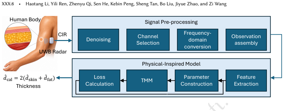
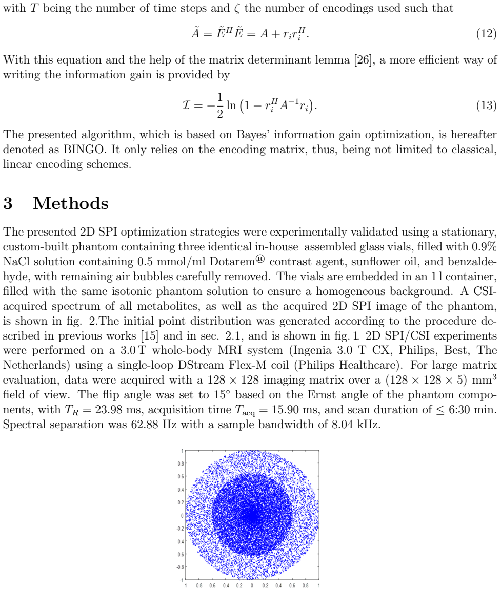
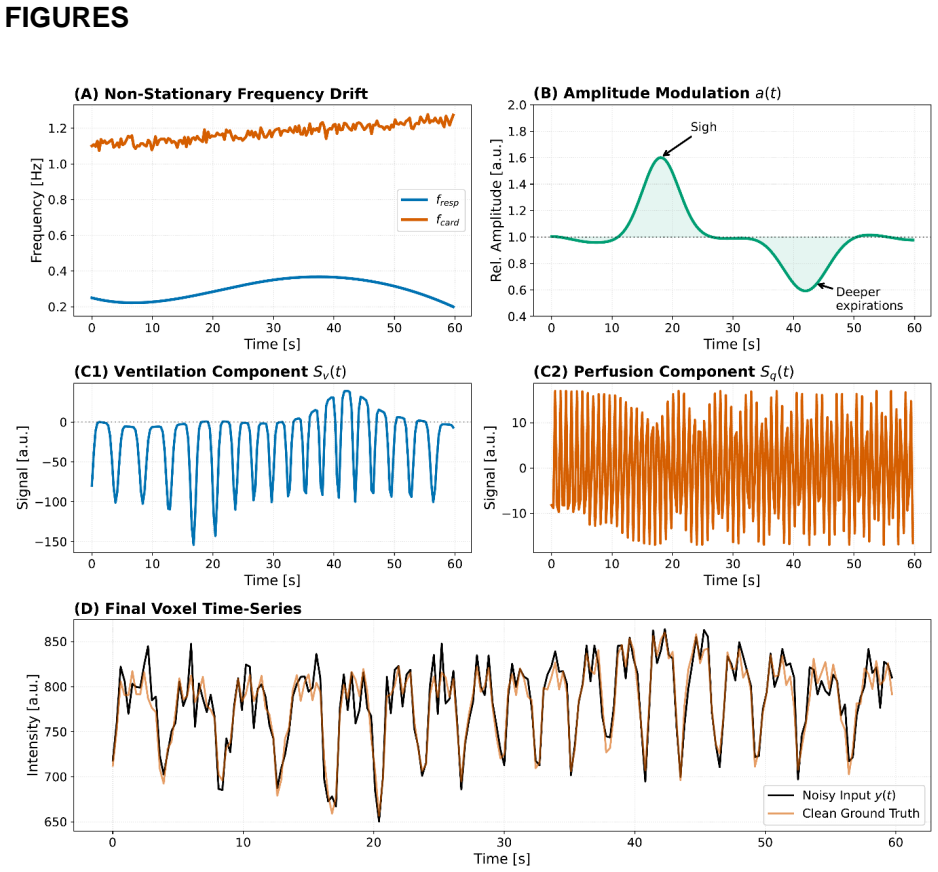
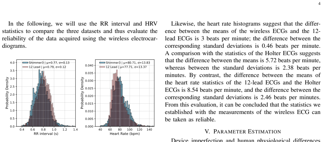
pith.machine review for the scientific record.sign in
physics.med-ph
Medical Physics
Radiation therapy. Radiation dosimetry. Biomedical imaging modelling. Reconstruction, processing, and analysis. Biomedical system modelling and analysis. Health physics. New imaging or therapy modalities.
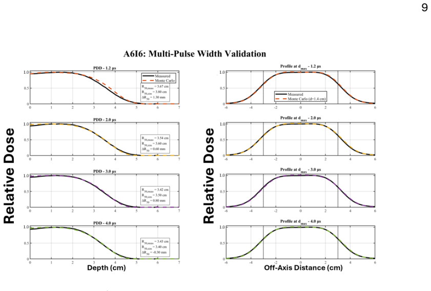
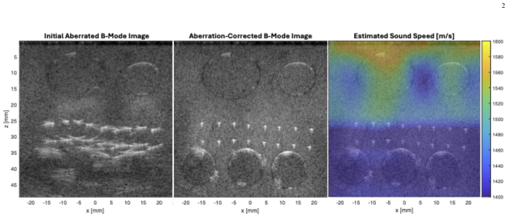
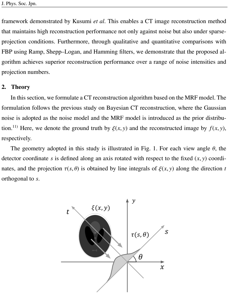
X-ray computed tomography (CT) reveals the materials' internal structures non-destructively from a tilt series of projected images. Filtered back projection (FBP) is a widely-adopted reconstruction algorithm in CT owing to its small computational cost. Under low-dose or sparse-view conditions, however, FBP often amplifies noise, severely degrading the reconstructed images. In this study, we evaluated the performance of a Bayesian CT reconstruction algorithm based on the Markov random field model under such adverse conditions. Through simulations, we demonstrated that the proposed algorithm shows higher reconstruction performance than FBP under both low-dose and sparse-view conditions. The hyperparameters are estimated by minimizing the Bayesian free energy, enabling adaptive reconstruction that reflects the noise characteristics of the observed projection data. These results suggest that the proposed algorithm can broaden the applicability of CT to dose-sensitive applications and time-constrained measurements, where only limited observed projection data are available.
This work proposes Attractor-Vascular Coupling Theory (AVCT), a mathematical framework showing that cardiac attractor geometry encodes blood pressure (BP) information sufficient for AAMI-standard estimation, and validates the theory through a calibrated cuffless BP model using photoplethysmography (PPG). AVCT is grounded in Cardiac Stability Theory and operationalized using Takens delay embedding and attractor morphology extraction. Two theorems, one proposition, and one corollary formally justify the use of PPG attractor features for BP estimation and predict the feature-importance hierarchy. A LightGBM model trained on pulse transit time (PTT) and Cardiac Stability Index (CSI) attractor features under single-point calibration was evaluated using strict leave-one-subject-out cross-validation (LOSO-CV) on 46 subjects from BIDMC ICU (n = 9) and VitalDB surgical data (n = 37), comprising 29,684 windows. The model achieved systolic BP (SBP) mean absolute error (MAE) of 2.05 mmHg and diastolic BP (DBP) MAE of 1.67 mmHg, with correlations r = 0.990 and r = 0.991, satisfying the AAMI/IEEE SP10 requirement of MAE below 5 mmHg. Median per-subject MAE was 1.87/1.54 mmHg, and 70%/76% of subjects individually satisfied AAMI criteria. A PPG-only ablation using nine smartphone attractor features matched the ECG+PPG model within 0.05 mmHg, demonstrating that clinical-grade BP tracking is achievable using only a smartphone camera while surpassing prior generalized LOSO-CV results using fewer sensors. All four AVCT predictions were quantitatively confirmed, with 91.5% error reduction from uncalibrated to calibrated estimation (epsilon_cal = 0.915). Unlike post-hoc explainable AI methods, AVCT predicts features satisfying the architectural faithfulness criterion of the Explainable-AI Trustworthiness (EAT) framework and grounding BP estimation in nonlinear dynamical systems theory.
Proof-of-concept tilts compact magnet and rat together, yielding usable scans while motion occurs and opening vestibular studies.
abstractclick to expand
Current magnetic resonance imaging (MRI) requires the subject to remain stationary to limit motion artifacts and avoid unwanted field-induced brain stimulation. However, imaging during large-scale motion could enable studies in which motion itself is central. One example is the study of brain networks involved in vestibular function, which senses head motion. Here, we demonstrate Moving MRI (mMRI), a system that enables imaging during large-scale motion by moving the subject and scanner together to minimize relative motion. We implemented a proof-of-concept platform using a compact, cryogen-free superconducting magnet mounted on a pneumatically actuated tilt mechanism that moves the magnet, gradients, and RF coil as a unit during scanning. Phantom and in vivo rat brain scans were acquired during repetitive tilting. We characterized artifacts arising from tilt-induced field shifts and residual subject-scanner motion, and partially reduced these effects. mMRI enables imaging during large-scale movement and may broaden access to naturalistic vestibular paradigms while providing a foundation for future human systems.
Combined acquisition gives stable estimates of tissue properties and vascular fraction in calf muscle, unlike separate diffusion-only scans.
abstractclick to expand
Quantifying muscle tissue properties is crucial for understanding pathophysiological changes occurring in skeletal muscle (SM). In particular, T2 relaxation and diffusion MRI (dMRI) are promising techniques. However, typical methods measure T2 and diffusion separately, making them less specific to microstructure than emerging combined diffusion-relaxation techniques. Here we demonstrate a combined diffusion-relaxation MRI approach for disentangling T2 and diffusivity properties in SM. A diffusion-relaxation acquisition was implemented on a 3 T scanner, combining six b-values and four echo times within a 12-min single-slice protocol. Five healthy participants were enrolled. Data were analysed with six microstructural diffusion and diffusion-relaxation models. Mean parameter values were extracted from manually segmented calf muscles. Models neglecting T2 relaxation showed strong TE dependence: mean diffusivity (MD) decreased by up to 47\%, fractional anisotropy (FA) increased by up to 75\%, and vascular fraction fv increased by up to 297\% when TE increased from 50 to 90 ms. Diffusion-relaxation models produced TE-independent estimates. Tissue and vascular relaxation times ranged 31-36 ms T2t and 66-86 ms T2v, respectively. Simulations confirmed improved accuracy for fv estimation (r=0.95; RMSE=0.03) and reduced TE-related bias. Combined diffusion-relaxation MRI provides robust, TE-independent estimates of muscle microstructural and perfusion-related biomarkers. The quantitative improvements observed - particularly in the estimation of fv - show its potential to provide non-invasive biomarkers for the assessment of muscle physiology, exercise adaptation, rehabilitation, and neuromuscular pathology.
Purpose: Diffusion MRI has shown promise for breast cancer screening, lesion characterization,and treatment response monitoring without contrast agents, but further translation is constraint by the gradient performance of conventional systems. The aim of this work is to develop a single-axis high performance bilateral plug-and-play breast gradient insert to enable strong-gradient diffusion MRI. Methods: An in-house breast gradient insert and bed-tabletop was constructed entirely from commercially available materials, providing a cost-effective solution compatible with existing MRI systems. Its wiring pattern was optimized for torque and force balancing, power dissipation, and target field performance. Evaluation included gradient field characterization, peripheral nerve stimulation simulation verification, and temperature and eddy current assessment. The setup was used for imaging of a diffusion phantom based on soy lecithin across a range of b-values. Results: Gradient efficiency reached 2.8 mT/m/A, enabling local strengths up to 1850 mT/m (660 A). No peripheral nerve stimulation was observed during tests on five healthy volunteers. Eddy currents were successfully characterized employed in standard correction methods. Imaging showed the feasibility of $b = 10 000 s/mm^2$ acquisitions at TE = 78 ms versus 161 ms with scanner gradients. Conclusion: This work demonstrates a dedicated bilateral breast gradient insert for safe and feasible strong-gradient breast diffusion MRI, and represents a first step toward dedicated hardware for breast cancer detection and characterization without contrast agents.
White matter hyperintensities (WMH) are bright regions on T2-weighted magnetic resonance imaging (MRI) scans and are associated with cerebrovascular pathology and neurodegeneration, including myelin loss. While Luxol Fast Blue histopathology provides visualization of myelin integrity, quantitative analysis requires measuring Optical Density as a proxy for myelin concentration. However, differences in laboratory protocols and tissue processing introduce staining variability that acts as systematic noise, obscuring the biological signal and preventing consistent comparison across histology runs. To address this, we developed an automated pipeline that identifies reference (non-pathologic) regions in whole-slide images to compute normalized Optical Density heatmaps. We validated this approach through two complementary evaluations: (1) comparison against expert ratings of myelin loss severity, and (2) cross-modal spatial comparison with co-registered 7T ex vivo MRI for voxel-wise evaluation within white matter regions. The pipeline's reference selection showed strong concordance with expert-identified reference regions, and normalized Optical Density demonstrated a substantially stronger correlation with MRI signal intensity than raw measurements. This correlation persisted within WMH, confirming that the pipeline captures continuous myelin pathology rather than merely the presence or absence of myelin loss contrast. By mitigating staining artifacts, this pipeline provides a robust, validated framework for quantitative cross-modal comparison, establishing a critical methodological foundation for future translation to in vivo myelin mapping and biomarker discovery.
Body fat percentage and its spatial distribution are clinically important health indicators. However, existing measurement methods often impose a tradeoff between accuracy and accessibility. Clinical-grade techniques, such as Dual-Energy X-ray Absorptiometry (DEXA) and hydrostatic weighing, provide accurate measurements but require specialized equipment and trained operators, making them difficult to access and unsuitable for everyday use. In contrast, consumer-level methods, such as Bioelectrical Impedance Analysis (BIA) smart scales and skinfold calipers, are more accessible but typically provide only coarse-grained estimates, are prone to user error, or require intrusive physical contact. In this work, we present UWB-Fat, the first system that leverages commodity ultra-wideband (UWB) radar to enable non-intrusive, accessible, and accurate caliper-equivalent skinfold thickness estimation, serving as a convenient replacement for the skinfold caliper. UWB-Fat collects UWB signal at specified body sites non-intrusively without operator assistance. It extracts body-composition-related features from UWB signals by exploiting dielectric contrasts among skin, fat, and muscle tissues. Then, it uses a physics-inspired model to estimate site-specific skinfold thickness. We evaluate UWB-Fat on 15 participants, achieving a root mean square error of 0.63~mm for pooled-site subcutaneous fat thickness. These results highlight the potential of UWB-Fat to support low-cost, self-administered, and everyday body fat monitoring.
Deep learning-based organs/structures-at-risk(OARs) auto-contouring models can improve radiotherapy workflows, but models trained on adult data often underperform in pediatric patients. Developing robust pediatric-specific models is hindered by data scarcity and fragmentation across centers. Federated learning (FL) enables privacy-preserving collaborative training without the need for data sharing. We evaluated the feasibility and performance of FL for developing pediatric-specific OAR segmentation models across two European medical centers. Computed tomography (CT) images from pediatric patients from Utrecht and Heidelberg with a renal tumor or abdominal neuroblastoma were retrospectively collected and locally processed. An nnU-Net-based framework segmented 19 OARs using local and FL schemes. FL was implemented with secure weight exchange on a cloud storage across institutional firewalls. Performance was assessed using the Dice similarity coefficient (DSC), 95th percentile Hausdorff distance, and mean surface distance. Robustness to patient orientation, false-positive segmentation of surgically removed kidneys, and failure cases were identified. A total of 310 postoperative CTs from 272 patients (105 renal tumors, 167 neuroblastomas) were included. Local models performed well on their respective center data but showed significantly reduced cross-center performance for four to seven of the nine evaluated OARs (DSC). In contrast, the FL model matched local performance for at least seven of nine OARs and achieved the best cross-center results across three metrics, with DSC gains of 0.003-0.007 over local models. FL also maintained stable performance across patient orientations and reduced false-positive kidney segmentations. Real-world FL improves cross-center robustness of CT-based OAR segmentation models in pediatric upper abdominal tumors.
Simplifies to physically interpretable differential relations under alignment, enabling consistency checks and geometric calibration in CT.
abstractclick to expand
The John equation serves as the mathematical foundation of the X-ray transform, describing the intrinsic compatibility conditions that projection data must satisfy. In this paper, within three-dimensional (3D) Euclidean space, an innovative mixed parameterization scheme is adopted: the source point is represented using cylindrical coordinates a=(s cos{\theta},s sin{\theta},z_0), and the ray direction is represented using spherical coordinates d=\{rho}(-cos\{beta}sin{\alpha},cos\{beta}cos{\alpha},sin\{beta}). The specific form of the John equation under this geometric parameterization is systematically derived. Through detailed partial differential operator transformations, application of -1 homogeneity, and algebraic simplification, a complete system of constraint equations is obtained. In particular, under the special configurations where the ray direction is perpendicular to the radial direction of the source point in the horizontal plane (i.e., the so-called alignment condition:{\alpha} = {\theta}) and the ray has no tilt (\{beta} = 0), the constraint equations simplify to differential relations with clear physical meanings. This paper not only establishes a bridge between abstract mathematical theory and concrete imaging geometry, but also provides rigorous mathematical tools for data consistency verification, geometric parameter calibration, and incomplete-data reconstruction in 3D Computed Tomography (CT) systems. The research results are of great significance for advancing the mathematical theory and practical applications of CT imaging.
Cylindrical-spherical parameterization produces differential relations useful for verifying projection consistency in CT.
abstractclick to expand
The John equation serves as the mathematical foundation of the X-ray transform, describing the intrinsic compatibility conditions that projection data must satisfy. In this paper, within three-dimensional (3D) Euclidean space, an innovative mixed parameterization scheme is adopted: the source point is represented using cylindrical coordinates a=(s cos{\theta},s sin{\theta},z_0), and the ray direction is represented using spherical coordinates d=\{rho}(-cos\{beta}sin{\alpha},cos\{beta}cos{\alpha},sin\{beta}). The specific form of the John equation under this geometric parameterization is systematically derived. Through detailed partial differential operator transformations, application of -1 homogeneity, and algebraic simplification, a complete system of constraint equations is obtained. In particular, under the special configurations where the ray direction is perpendicular to the radial direction of the source point in the horizontal plane (i.e., the so-called alignment condition:{\alpha} = {\theta}) and the ray has no tilt (\{beta} = 0), the constraint equations simplify to differential relations with clear physical meanings. This paper not only establishes a bridge between abstract mathematical theory and concrete imaging geometry, but also provides rigorous mathematical tools for data consistency verification, geometric parameter calibration, and incomplete-data reconstruction in 3D Computed Tomography (CT) systems. The research results are of great significance for advancing the mathematical theory and practical applications of CT imaging.
The oil-encapsulated alkali metal system outperforms urokinase via mechanical, chemical, and thermal actions without bleeding risks.
abstractclick to expand
Thrombotic vascular diseases contribute to significant global mortality, yet current therapeutic strategies face persistent challenges including bleeding risks, suboptimal efficiency, and procedural complexity. Here, we report a micro-explosive thermochemical thrombolysis (METCT) therapy via injectable liquid alkali metal (LAM) encapsulated in dimethyl silicone (LAM@oil), which enables prompt, efficient and safe vascular recanalization within an ultrafast timeframe (< 90 seconds). This LAM@oil system effectively disrupts thrombus tissue through a synergistic triple-action mechanism: Mechanical micro-explosions forces, alkaline ablation due to highly localized exothermic chemical reactions, and thermal thrombolysis mediated by elevated temperature. Upon thrombolysis completion, the non-toxic reaction byproducts (sodium and potassium ions) exhibit physiologically biocompatible and metabolizable effects. Critically, the LAM@oil demonstrates significantly higher thrombolytic efficacy compared to clinically available thrombolytic drugs (residual thrombus area percent 10.87%+-7.16% for LAM@oil vs. 80.86%+-13.32% for urokinase), with no associated bleeding risks. This strategy opens a byproduct-free, cost-effective, and high-efficiency alternative to conventional thrombolytics, holding big potential for clinical translation in acute thrombosis management.
Commercial treatment planning systems for electron FLASH radiotherapy are unavailable, and the dosimetric precision required for ultra-high dose rate delivery makes Monte Carlo (MC) simulation the gold standard approach. This work establishes a methodology for generating pulse-width-specific phase space (PHSP) files for the Mobetron UHDR system (9 MeV), accounting for systematic beam quality shifts caused by RF waveguide loading across pulse widths of 1.2-4.0 microsecond. Using GAMOS 6.2.0, source parameters were iteratively refined against experimental targets: mean energy was optimized by matching phantom-measured R50 in the fall-off region, while energy spread was refined using surface dose and build-up gradients. Relationships derived from a mid-range 6 cm aperture were applied across all clinical configurations (2.5-10 cm) to test the aperture-independence of beam loading effects. Mean energy decreased exponentially from 9.58 to 9.04 MeV (R^2=0.99) with increasing pulse width, while energy spread increased quadratically (R^2=0.99), with a strong negative correlation (r=-0.98). Cross-aperture validation confirmed that energy shifts are independent of downstream collimation. The geometric mean pulse width (2.28 microsecond) was evaluated as a universal clinical reference, yielding 9.32 MeV mean energy. Across experimental extremes, R50 deviations were within 1.3 mm and critical depth-dose parameters remained within 2.0 mm, meeting AAPM TG-106 tolerances. Validated regression models enable beam parameter prediction at arbitrary pulse widths, and the universal reference reduces computational burden by 75% while maintaining clinical accuracy.
Dose rates from 0.01 to 100 Gy/s and grid patterns are now available for cell studies on an existing medical accelerator.
abstractclick to expand
Advanced radiotherapy approaches such as FLASH irradiation and spatially fractionated radiotherapy (SFRT) show potential to improve the therapeutic ratio, yet their biological mechanisms and optimal delivery parameters remain uncertain. Progress requires accessible proton research platforms with flexible temporal and spatial dose delivery. We report on the adaptation of the Beam Transfer Line (BTL) of the Bern Medical Cyclotron (BMC) for radiobiology research with FLASH and proton minibeam capabilities. The BMC is optimized for the production of radionuclides for medical imaging, and is able to extract currents up to 150 $ \mathrm{\mu A}$. The 18 MeV proton beam was passively shaped using collimators, scattering foils, and extended drift space to generate irradiation fields. A dosimetric framework was implemented using an in-beam ionization chamber and radiochromic film with LET-dependent corrections. Beam uniformity and SFRT profiles with various grid spacings were evaluated at realistic target distances. The developed beamline enables stable delivery under controlled conditions in both conventional and FLASH regimes, spanning dose rates from 0.01 to 100 Gy/s. Dose uniformity within a 20 mm radius was below 8\%. Film measurements confirmed the need for LET-dependent corrections and indicated that quantitative dosimetry in in-vitro setups is achievable with appropriate LET corrections. The low proton energy (15.54(12) MeV extracted into air, 8.14(28) MeV delivered to cells in flask) facilitates compact SFRT implementation with well-resolved minibeams. The adapted BMC provides a flexible and accessible platform for systematic pre-clinical proton radiobiology studies under varied dose-rate and spatial delivery conditions. This supports optimization of emerging modalities such as proton FLASH and SFRT and helps bridge accelerator technology and radiobiology.
Wave propagation through complex poroelastic multilayered media is difficult to model and invert because pronounced heterogeneity, scattering, mode conversion and fluid-solid coupling jointly distort acoustic signals during propagation. Here we present Physics-Constrained Learning for Complex Multilayered Media (PCL-CMM), a general framework that integrates Biot's poroelastic theory with the elastic wave equation to bridge the gap between physically rigorous wave modelling and data-driven learning. PCL-CMM constructs a high-fidelity digital twin that dynamically computes an effective acoustic stiffness tensor for forward wave modelling and incorporates the resulting physical constraint as a loss term to regularize the training of deep neural networks. We demonstrate PCL-CMM on transcranial photoacoustic imaging, where skull-induced acoustic distortions severely degrade image formation. Across simulations and ex vivo experiments, PCL-CMM effectively compensates for these distortions and improves SSIM by more than 0.06 compared with purely data-driven neural networks. This work establishes a physics-constrained learning framework for acoustic wave modelling in complex poroelastic multilayered media.
Monte Carlo simulations with real anatomy and beam data show how breast volume, density, and skin thickness change dose, supporting safer 3D
abstractclick to expand
This study develops and validates a patient-specific Monte Carlo (MC) dosimetry framework for propagation-based phase-contrast breast CT (BCT) at the Imaging and Medical Beamline (IMBL), ANSTO Australian Synchrotron, for accurate mean glandular dose (MGD) estimation. BCT provides 3D imaging without breast compression, improving comfort and visualization of internal structures for cancer detection. Propagation-based phase contrast improves soft-tissue contrast at equal or lower dose than conventional systems. Accurate dosimetry remains essential for safety and optimisation. Most MC-based MGD studies use non-patient-specific phantoms that ignore anatomical variability, while existing patient-specific methods lack a unified framework. Here, a voxel-based MC framework using EGSnrc was implemented to compute MGD in realistic anthropomorphic breast phantoms derived from synchrotron BCT images. IMBL beam characteristics were used as source inputs. Homogeneous phantoms were also generated to compute air Kerma to MGD conversion coefficients (DgN) for comparison with heterogeneous models. Simulations covered breast height, skin thickness, and photon energies (28 to 38 keV). Results show MGD depends strongly on anatomy and energy. Higher glandular density reduces MGD, while larger breast volume increases dose. A 2 mm increase in skin thickness reduces MGD by 10%. Differences between heterogeneous and homogeneous phantoms show variations in DgN, highlighting the need for anatomical realism. The framework provides a robust basis for patient-specific dosimetry in synchrotron phase-contrast BCT, enabling precise MGD estimation and supporting safe, optimised clinical imaging. This supports improved protocol design and contributes to standardised patient-specific dosimetry for clinical translation across varying breast anatomies and imaging conditions within synchrotron BCT applications.
Magnetite aggregates provide better neutron shielding than conventional concrete, with shorter attenuation lengths for spectra typical in…
abstractclick to expand
The neutron shielding properties of high-density concrete and magnetite aggregates were evaluated using both experimental measurements and Monte Carlo simulations. Because these materials are commonly used in medical accelerator facilities, it is essential to characterize their behavior under neutron radiation to ensure adequate shielding performance. Our experimental results show good agreement with the Monte Carlo calculations, confirming the reliability of the simulation approach. The attenuated neutron doses for various shielding thicknesses were determined for each aggregate type based on simulation and then compared as dose ratios. The findings indicate that magnetite provides superior neutron shielding, exhibiting a shorter attenuation length than conventional concrete for the same barrier thickness. The neutron attenuation characteristics of both concrete and magnetite were studied for typical neutron spectra encountered in clinical proton-therapy accelerators, including treatment rooms, primary, secondary barriers, and mazes. These results can support the optimization of radiation-shielding designs in medical and research facilities.
The lack of analytical models describing diffusion time dependence at intermediate time scales in complex tissue microstructure limits the accurate quantification of extracellular diffusivity and tissue microstructure. We introduce TRACED, a biophysical model that incorporates diffusion time dependence in cell distributions to quantify pathologically-relevant properties in solid tumors. Neural networks were trained on Monte Carlo diffusion simulations using sphere distribution-based geometries to enable the rapid computation of time-dependent diffusion MRI signals in cell populations of variable cell size. Model sensitivity and fit performance were assessed via simulation. Diffusion data from eight mixed-grade glioma patients was fitted using the TRACED model. Data fitting was performed using a novel physics-informed transfer learning pipeline, Sim2PINN. In two patients, cell size measurements were compared directly with image-localized histology. Simulation results indicate improved parameter estimation compared to the simple two-compartment model. TRACED enabled the simultaneous in vivo quantification of intracellular volume fraction, cell size distribution, extracellular intrinsic diffusivity, and tortuosity in glioma patients. Neural network implementations of diffusion time-dependence and tortuosity showed behavior consistent with coarse-graining and effective medium theory, respectively. Future work will explore the clinical utility of TRACED parameters in additional patients.
Applicator-specific phase space (PHSP) files recorded at the aperture exit reduce Monte Carlo dose calculation time by 30-50% for electron FLASH radiotherapy. However, positioning PHSP scoring planes coincident with the applicator-air interface introduces boundary sampling artifacts. This study characterizes these artifacts in Geant4-based simulations and demonstrates their mitigation. PHSP files were generated using GAMOS 6.2.0 for a 9 MeV Mobetron UHDR model across twelve clinical aperture configurations (2.5-10 cm diameter). Three scoring plane positions were evaluated relative to the physical aperture exit: coincident with the interface, 0.1 mm downstream, and 1 mm downstream. Scoring at the exact interface produced proximal R50 shifts of up to 2.2 mm and Distance-to-Agreement (DTA) values of 4-6 mm, exceeding clinical acceptance criteria. Artifact severity scaled inversely with aperture diameter, with the smallest configurations most severely affected. A 0.1 mm offset partially restored the primary electron energy spectrum but failed to recover the bremsstrahlung tail. A 1 mm offset fully resolved all artifacts, achieving mean DTA values within 2.0 mm, equivalent to or better than linac-exit references. These artifacts arise from the degenerate behavior of the fUseSafety step-limitation algorithm when safety equals zero at exact material boundaries, producing incomplete secondary electron equilibration and suppressed bremsstrahlung production. Angular distribution analysis revealed a near-forward particle pileup in the 0 mm PHSP and a deficit of large-angle secondaries recovered by the 1 mm offset. A 1 mm downstream offset fully mitigates these artifacts while introducing negligible perturbation to primary beam characteristics. This requirement applies to any Geant4-based framework (including TOPAS and GATE) scoring PHSP files at material exit surfaces.
Transmembrane water permeability, which regulates cellular water exchange and is influenced by water channels such as aquaporin-4 (AQP4), has been implicated in glioma progression and may affect tumour infiltration and treatment response. Non-invasive mapping of water exchange may therefore provide biomarkers of glioma pathology. This study investigates the feasibility of characterizing water exchange in gliomas using diffusion MRI with free gradient waveforms, known as the Restriction-Exchange (ResEx) approach, which enables exchange quantification independent of restricted diffusion effects. Thirteen patients with histologically confirmed gliomas (ten glioblastomas, three astrocytomas) underwent preoperative MRI at 3T using a custom ResEx protocol. Multiple diffusion-weighted acquisitions with selective exchange sensitivity were performed to estimate voxel-wise maps of the apparent diffusion coefficient (ADC), diffusion kurtosis, and water exchange rate. ResEx-derived maps revealed heterogeneous spatial patterns across and within tumours. Elevated exchange rates were commonly observed in enhancing tumour margins, potentially reflecting smaller cells, increased membrane permeability or AQP4 upregulation. In some cases, elevated exchange extended into non-enhancing peritumoural regions. Exchange values in oedema were slightly higher than in healthy tissue, suggesting potential infiltration or membrane disruption. Diffusion MRI with free gradient waveforms permits non-invasive mapping of water exchange in gliomas and reveals physiological information not captured by standard imaging. Exchange rate mapping may offer novel biomarkers of tumour aggressiveness, infiltration, and treatment response, and holds promise for surgical and radiotherapy planning.
A differentiable beamforming framework optimizes aberration correction using angular coherence, improving transcranial ULM and fUS imaging…
abstractclick to expand
Skull-induced aberrations remain a major drawback of transcranial ultrasound localization microscopy (ULM), degrading sensitivity and spatial accuracy through microbubble mislocalization, false detections, and imaging artifacts, such as disconnected or duplicated vessels. Here, we present a differentiable beamforming framework for automatic aberration correction in transcranial Doppler and ULM. Our approach uses spatially distributed delay-based parameterization of the aberration that is optimized in a closed-loop manner using angular coherence as an objective function. We demonstrate robust improvements of transcranial ULM, in vivo, with enhanced resolution of both mouse and nonhuman primate (NHP) brains. We also extended differentiable beamforming to functional measurements, with improvements in the sensitivity of transcranial functional ultrasound (fUS) and ULM based hemodynamic quantification. Extending this approach to 3D transcranial ULM imaging in NHPs, we show efficient correction of skull induced aberrations and removal of artifacts, such as vessel duplications. By providing a fully automated and generalizable solution for aberration correction, this work lowers a major technical barrier to transcranial ultrasound imaging, enabling broader adoption of non-invasive, super-resolution and functional neuroimaging across laboratories and across species.
Grating interferometry is a promising diagnostic technique that enables simultaneous acquisition of three complementary, synergistic X-ray images: transmission, differential phase, and dark-field. Its key advantage over other setups is its ability to use large pixels and, hence, large-area detectors, as well as its compatibility with low-coherence, compact X-ray sources, both of which are key factors for human-scale imaging. It has already demonstrated strong potential for chest imaging applications, including the diagnosis of pulmonary emphysema, fibrosis, and cancer. To retrieve transmission, differential phase, and dark-field images from data, an algorithm is required to separate the distinct mechanisms contributing to measured contrast. Since its realization, this image-retrieval step has remained fundamentally unchanged. In this work, we develop a novel transmission- and dark-field retrieval algorithm for grating-interferometry derived from the X-ray Fokker-Planck equation. To demonstrate and validate our Fokker-Planck algorithm, we apply it to experimental measurements of a test sample and to data from a mouse chest acquired with varying exposure times and added Poisson noise. The retrieved images were qualitatively and quantitatively compared with those retrieved using a conventional sinusoidal-fitting approach. Across both samples, the Fokker--Planck method produced images consistent with conventional retrieval, with a comparable signal-to-noise ratio. Notably, our Fokker-Planck method suppresses artefacts arising in the conventional approach under grating perturbations (e.g., structural defects like scratches) and reduced flux or visibility, yielding smoother and more reproducible images. Additionally, we demonstrate that our Fokker-Planck method has an advantage over the conventional dark-field retrieval method for fast sample imaging with short exposure times and high noise.
Purpose: As interest in CEST-MRI grows, particularly in the preclinical setting, the necessity for standardized and easy-to-use acquisition and data analysis pipelines has become apparent. While vendors have increasingly introduced support for CEST acquisitions on both clinical and preclinical hardware, image post-processing and analysis pipelines remain siloed based on privately developed code. We aim to develop an easy-to-use, open-source graphical user interface toolbox for preclinical CEST-MRI data analysis (Preclinical CEST-MRI Analysis Tool; Pre-CAT), supporting multiple acquisition types, organ systems, and CEST contrast mechanisms. Methods: Pre-CAT was developed in Python and utilizes the Numpy, Scipy, and Matplotlib libraries for data analysis and plotting. Inbuilt data processing steps include image loading, reconstruction, post-processing, and segmentation. Pre-CAT also supports data analysis for QUESP, CEST-MRF, and field mapping experiments using consensus protocols and methods. Pre-CAT's web interface and GUI were developed using Streamlit, an open-source Python framework. Pre-CAT is hosted and accessible online and can be downloaded for local installation to complete the data analysis pipeline in roughly one minute using modern hardware. Results: Pre-CAT analysis pipelines for Z-spectroscopy, CEST-MRF, and quantitative CEST (QUESP/QUEST) are demonstrated. Conclusion: With the introduction of Pre-CAT, we aim to standardize the preclinical CEST-MRI data analysis pipeline, fostering collaboration across research sites and reducing methodological redundancy. Pre-CAT is open-source and relatively modular, encouraging the addition of new methods and protocols.
The ramp filter kernel and cutoff frequency are fundamental parameters of the Feldkamp-Davis-Kress (FDK) algorithm that determine the resolution and noise characteristics of the reconstructed image. Despite their importance, systematic evaluations of their combined effect on task-based image quality in preclinical micro-CT are scarce, and many studies do not report the filter configuration used. We reconstruct identical data from a GE eXplore CT 120 scanner using four filter kernels (ramp, Shepp-Logan, cosine, Hamming) at four cutoff frequencies (1.0, 0.8, 0.6, and $0.379\times$ Nyquist, matched to the detector-to-voxel size ratio) and evaluate each of the sixteen configurations using the modulation transfer function (MTF), noise power spectrum (NPS), and non-prewhitening detectability index (NPW $d'$). Qualitative assessment is performed on a mouse lung specimen. Across the sixteen configurations, $\mathrm{MTF}_{10}$ ranges from 0.93 to 2.35 lp/mm, integrated NPS from 75,670 to 13,259 $\mathrm{HU}^2$, and the Rose criterion crossing diameter from 2.86 to 0.93 mm at $\Delta C = 500$ HU and from 7.74 to 3.62 mm at 100 HU. This note presents the data as a concise visual and quantitative reference for groups selecting FDK filter parameters for preclinical cone-beam CT.
A model using patient dose maps and blood dynamics quantifies how particle beams limit immune-cell damage compared with conventional beams.
abstractclick to expand
Treatment-related lymphopenia is a frequent and clinically significant consequence of cancer therapy that can compromise immune-mediated tumor control and worsen patient outcomes. Despite its importance, no mechanistic framework exists to accurately predict the severity of lymphopenia from patient-specific data. Here, we present a biokinetic model that quantitatively describes lymphocyte depletion and recovery during and after radiotherapy, integrating radiation dose-volume distributions, blood circulation dynamics, and distinct kinetics of fast- and slow-recovering lymphocyte populations. The model was calibrated and validated using 56 independent clinical datasets encompassing various tumor sites and treatment modalities. It reproduces observed lymphocyte counts and enables prediction of individual severity of lymphopenia from baseline or early-treatment counts. Applying this framework, we demonstrate that particle therapy reduces lymphocyte depletion by ~30% compared with photon therapy, providing a quantitative explanation for its observed immune-sparing benefit. By linking radiation physics, immune kinetics, and clinical outcomes, our model establishes a mechanistically grounded predictive approach for anticipating systemic immune toxicity. Beyond radiotherapy, this framework offers a generalizable strategy for integrating early biological markers into treatment optimization, advancing personalized and immune-preserving cancer therapy.
In conventional radiotherapy, the probability of controlling tumor growth is quantified using Tumor Control Probability (TCP) models. Instead, the probability of experiencing a side effect after the irradiation of healthy tissues and organs is typically assessed using the concept of Normal Tissue Complication Probability (NTCP), an additional crucial metric for evaluating and comparing treatment plans.
This work is dedicated to the development, implementation, and application of a general mechanistic model to describe the effects of particle therapy (PT) on different tissue organizations beyond Poissonian assumptions, extending the Generalized Stochastic Microdosimetric Model (GSM2), i.e., a stochastic radiobiological model that describes the time evolution of DNA lesions in a cell nucleus according to microdosimetric principles, to the study of macroscopic biological systems. Specifically, we extend the biological stage of radiation damage of the GSM2 model to larger spatial and temporal scales, involving cell populations with a specific geometric and functional architecture.
The model's single-cell resolution allows it to account for energy deposition and tissue heterogeneity, considering different organ volume effects, cell type distributions, and oxygen gradients for different radiation qualities of the beam, that is, type, energy, and LET of radiation, and various fractionation schemes. We show the interplay between physical and environmental parameters on the induction of side effects on healthy tissues, for different radiation qualities and fractionation schemes, and we highlight the impact of biochemical heterogeneities in the target environment, for tumor response.
Sound speed heterogeneities can create aberrations in B-mode ultrasound images by inducing tissue-dependent delays and diffractive effects that conventional beamforming does not incorporate. By using the Fourier split-step method to simulate pressure fields in heterogenous sound speed media, reverse-time migration (RTM) can reconstruct the B-mode image by cross-correlating transmitted and received pressure fields. As a result, RTM is differentiable with respect to sound speed. This enables the reconstruction of the sound speed profile that minimizes the aberration in the B-mode image. In seismic imaging, this form of diffraction tomography, known as wave-equation migration velocity analysis, can roughly be understood as a type of full-waveform inversion (FWI) that acts in the image domain rather than errors in the received channel data. This is the first work applying WEMVA to medical pulse-echo ultrasound imaging. Phantom experiments show dramatic improvements in image quality with measured improvements in point target resolution from 1.22$\pm$1.01 to 0.32$\pm$0.07 mm and lesion contrast from 3.05 to 4.39 dB.
Spatially fractionated radiation therapy (SFRT) planning requires three coordinated tasks: generation of high-dose sphere structures, position-aware optimization, and peak-valley dose ratio evaluation. We present MAAS-SFRThelper, a shared-source Eclipse Scripting Application Programming Interface (ESAPI) plugin that integrates structure generation, geometric-aware optimization, and peak-valley dose ratio evaluation for SFRT into a single workflow inside Varian's Eclipse treatment planning system. The plugin exposes five task-oriented tabs sharing common services for sphere extraction and objective creation. The SphereLattice tab generates sphere lattices using five placement patterns. The Optimization tab searches over candidate lattice positions using a four-metric geometric surrogate score and triggers VMAT optimization and dose calculation. The Evaluation tab implements four analysis modes; its three-dimensional peak-valley classification recovers sphere centers from the lattice structure through a geometric extraction pipeline rather than relying on dose thresholds. We validated all functionality on digital phantoms against analytic ground truth. The plugin is distributed as source code under the Varian Limited Use Software License Agreement. Source code and documentation are publicly available on GitHub.
Proton therapy has been rapidly advancing due to its excellent conformal index, but its relatively low relative biological effect (RBE) has somewhat limited its therapeutic efficacy for certain tumors. To address this, we previously proposed a nitrogen-targeting Proton-Carbon-Alpha-Therapy (Proton-CAT) enhancement method. In this letter, we present combined multi-scale DNA damage simulations and in vitro cell experiments, further investigating the mechanism of the Proton-CAT. It has been show that $^{15}$N enrichment significantly enhances complex DNA damage induced by high linear energy transfer(LET) particles within tumor regions. Under 30\% $^{15}$N conditions, $\alpha$ and $^{12}$C particle induced DSB++ increased by 175.19\% and 52.94\%, respectively. Furthermore, in vitro cell experiments using $^{15}$N-glutamine ($^{15}$N-Glu) as the $^{15}$N carrier indicated that high concentrations of $^{15}$N-Glu did not bring about significant cytotoxicity. Following 2 Gy irradiation, the cell viability in the 500 $\mu$g/mL $^{15}$N-Glu treated group exhibited a net reduction of about 15.41\% compared to the control group.This indicates that the enhanced effect of Proton-CAT primarily stems from increased complex DNA damage. This work provides a theoretical basis and multi-scale research framework for the development of the Proton-CAT.
Objective: This study aims to investigate the influence of organ architecture (specifically the distinction between serial and parallel tissue) on the protective FLASH effect when organs are irradiated with inhomogeneous dose distributions.
Approach: An in silico modeling framework was developed using two distinct methods to calculate the effective FLASH dose: the first method utilized a biophysical model of radiolytic oxygen depletion (ROD); the second employed a phenomenological logistic function where the effective FLASH dose is a function of local dose and dose rate. Both models assume that the underlying mechanism behind the FLASH effect is local. Normal Tissue Complication Probability (NTCP) for heterogeneous dose distributions was calculated using the Lyman-Kutcher-Burman (LKB) model and the generalized equivalent uniform dose, varying the volume effect parameter n from 1.0 (parallel) to below 0.01 (serial) to explore different architectures.
Results: Both the ROD and phenomenological models showed FLASH sparing compared to conventional radiotherapy. Also, the sparing increased with decreasing $n$ (the sparing is more important for serial organs). For example, for a specific calculation, when the NTCP for conventional radiotherapy was 0.2 (set value) the corresponding NTCP for FLASH delivery ranged from 0.14 for n=1 to 0.11 for n=0.1.
Significance: Our results indicate that if the underlying mechanism/s behind the FLASH effect is/are local, the toxicity sparing associated to FLASH-RT can be dependent on the architecture of the irradiated organ/tissue, being more important for serial organs, which are more sensitive to large local doses than to average doses.
Dual-energy computed tomography (DECT) enables material-specific imaging through acquisitions at two different X-ray energy spectra. Material decomposition from DECT data is an ill-posed inverse problem that is highly sensitive to noise amplification. Conventional methods face challenges regarding accuracy and computational efficiency. We present a novel physics-informed deep learning (DL) framework for DECT material decomposition that eliminates the requirement for ground-truth material images during training. Our approach incorporates a polychromatic forward model into the training pipeline, enabling the network to learn the decomposition mapping by minimizing discrepancies in the projection domain. We validate our method on the AAPM DL-Spectral CT Challenge dataset, comparing performance against three state-of-the-art methods. In the projection domain, our method achieves the lowest root mean squared error (RMSE) across test datasets. For virtual monoenergetic images (VMIs) at 30 keV, 50 keV, and 70 keV, the approach consistently outperforms all conventional methods in both RMSE and structural similarity index (SSIM). These results demonstrate the potential of DL for accurate material decomposition in DECT without requiring labeled training data.
Demonstrated speeds suppress breathing artifacts while enabling the throughput needed for facilities to reach financial viability.
abstractclick to expand
Proton therapy exploits the finite range of charged particles in tissue to achieve dose distributions no photon based modality can replicate. Yet the modality reaches fewer than 1 percent of patients who might benefit a gap rooted in cost and complexity rather than clinical evidence. This Review reframes proton therapy adoption as a physics problem. Two fundamental bottlenecks are identified: cost, arising from scaling laws governing accelerator design, beam transport, and radiation shielding; and motion, arising from the spatiotemporal mismatch between sequential pencil beam scanning and respiratory tumour displacement. We trace how successive compact architectures from gantry-integrated energy selection to gantry mounted accelerators and upright fixed beam systems have progressively reduced facility scale toward LINAC like simplicity and cost-effectiveness. An economic physics framework incorporating fixed and variable operating costs demonstrates that delivery speed has greater leverage on cost per patient than capital cost reduction alone. Field delivery times of approximately 10 seconds now demonstrated across fundamentally different architectures simultaneously suppress the interplay effect and enable the patient throughput required for financial viability. The same physics that resolves the motion problem drives the economic case for broad adoption. Emerging directions, including proton arc therapy, FLASH irradiation, and adaptive delivery define the path toward global democratization of the modality.
Measuring gating delays and syncing with internal signals allows reliable control of electron pulse sequences for accurate preclinical dose
abstractclick to expand
Background: Clinical linear accelerators are an accessible platform for preclinical research on the biological effects of ultra rapid electron irradiation (FLASH). However, they are not inherently designed for the accurate pulse control required for experiments using a small number of relatively high-dose pulses, and available methods for beam control such as respiratory gating can be error prone owing to system latency. Here we experimentally characterize the temporal latency of the respiratory gating system for controlling beam-on and beam-off at the individual linac pulse level. Methods and Materials: We used programmable controller boards and a relay circuit to monitor and control delivery of specific numbers of pulses through the built-in monitor chamber and respiratory gating system of a Varian Trilogy linac. We implemented two methods an adaptive method using only the delivered pulse signal, and a synchronization method additionally using the linac internal pulse-timing signal and characterized their performance for standard and customized pulse sequences. Results: Characterizing the latency parameters permitted choosing optimal timing parameters that maximized the rate of successfully delivering the desired number of pulses using both adaptive and synchronization methods. Conclusions: We demonstrated that accounting for latency and/or using the ability to read the prior information on expected pulse timing can provide high accuracy in delivering specified numbers of pulses. This reliability is critical for accurate dose delivery in preclinical FLASH research of single fraction and especially fractionated dosing regimens. The ability to generate custom pulse sequences enables more detailed exploration of the temporal dependence of biological FLASH effects.
Quantitative post-treatment activity imaging is essential for personalised dosimetry after Yttrium-90 selective internal radiation therapy (SIRT). Yttrium-90 PET offers high spatial resolution but is extremely low-count, whereas bremsstrahlung SPECT has higher count statistics but is degraded by blur, scatter, and septal penetration. Since both modalities image the same microsphere distribution, joint synergistic reconstruction can exploit their physical coupling, with CT attenuation correction (CTAC) providing additional anatomical guidance.
We propose weighted directional total nuclear variation (w-dTNV), a joint variational regulariser for coupled PET/SPECT reconstruction with CTAC-guided anisotropy. w-dTNV penalises the nuclear norm of a dual-modality Jacobian, promoting co-located, geometry-consistent edges without forcing intensity correlation. Directionality is derived from the CTAC attenuation map $\mu$ and applied to PET/SPECT gradients, allowing efficient per-voxel spectral computations. PET/SPECT scale disparity is mitigated using data-driven modality normalisation from preliminary reconstructions.
We evaluate w-dTNV on a NEMA IEC phantom with 20 bootstrapped PET noise realisations and on 9 post-SIRT patients with 45 lesions, against dTV, w-TNV, and sequential hybrid kernel expectation maximisation (SHKEM). In the phantom, w-dTNV improves recovery coefficients over dTV and w-TNV, and improves recovery over SHKEM for the smallest spheres. In patients, w-dTNV gives higher tumour-to-background ratios than SHKEM at comparable background activity. These results suggest that CTAC-guided synergistic variational coupling improves lesion recovery and clinical lesion contrast, offering a practical route towards more stable post-SIRT Yttrium-90 activity estimates for personalised dosimetry.
Radiotherapy with Very High Energy Electron (VHEE) beams is being extensively investigated for the treatment of deep-seated tumours, even in view of novel protocols based on the so-called FLASH effect. Laser WakeField Acceleration (LWFA) provides a compact and affordable accelerator technology for VHEE electron beams, featuring ultra-high instantaneous dose rates and holding the promise to provide Ultra-High (average) Dose Rates (UHDRs) needed to activate the FLASH effect, with major efforts ongoing worldwide to fulfill this promise. Therapeutic doses are already at reach, using pencil beams produced via LWFA. These beams typically exhibit significant energy spread, and small transverse size. These features are rather different from those of other beams considered so far in radiotherapy studies. In view of a rapid clinical translation of LWFA-VHEE beams it is therefore of paramount importance to assess the role of these properties in the dose delivery to the patient. Here we present a study carried out via start-to-end (PIC and Monte Carlo) simulations, of the main dosimetric features of a realistic laser-driven VHEE pencil beam targeted on a brain tumor. The entire tumor coverage is achieved by a scanning procedure; the dose pattern resulting from tessellation, i.e. the overlapping of adjacent beamlets, and the role of energy spread are thoroughly discussed. Dose Volume Histograms are presented, and their quality is discussed. The impact of the FLASH effect is also considered, introducing a degree of healthy tissue sparing in the modelling. Finally, the foreseen technological path toward the achievement of FLASH dose rates with LWFA-VHEE beams is briefly outlined.
Short-scan FDK reconstruction is widely used in preclinical cone-beam micro-CT because it reduces scan time and radiation dose, and because the large volume sizes typical of micro-CT make iterative methods impractical for routine use. Short scans, however, introduce non-uniform data redundancy that must be corrected by Parker weighting to avoid directional shading artefacts. This note provides a visual and quantitative summary of Parker weighting as implemented for the eXplore CT 120 scanner. We illustrate the weight maps in the detector and sinogram domains, demonstrate the shading artefacts that arise without correction on both an image quality phantom and an in vivo mouse lung, and show via MTF, NPS, and detectability analysis that Parker weighting corrects HU inaccuracies without degrading image quality. No new methods are introduced; the aim is to serve as a concise practical reference for groups implementing or evaluating short-scan FDK pipelines.
Low-field MRI is increasingly considered accessible for imaging owing to its lower cost, simpler infrastructure requirements, and potential for mobile and point-of-care deployment. A central challenge is achieving clinically useful field strength and homogeneity while keeping the magnet lightweight and maintaining patient accessibility. This work presents the design and magnetostatic simulation of a pole-less, 0.2 T, C-type bipolar magnet comprising two cylindrical N52 permanent magnets and four concentric rings that replace traditional pole pieces to enhance field homogeneity and reduce weight in bipolar magnet designs. Geometric parameters, including each magnet ring thickness, height, angular anchorage, spacing between magnets, and the magnets' vertical offset relative to the horizontal yokes, were manually investigated to improve magnetic field homogeneity in a 20 cm DSV. Simulations were performed in CST Studio Suite, yielding a peak field of 0.2 T, with a peak inhomogeneity of 1.43 mT across the 20 cm DSV and a total weight of 590 kg. A pole piece design with comparable dimensions, used as a benchmark for inhomogeneity and weight, was designed and simulated. It yielded a peak field of 0.15 T and a weight of 890 kg, with a 0.7 mT inhomogeneity over a 20 cm DSV. This study demonstrates the feasibility of replacing the traditional pole pieces with magnet rings to reduce weight while enhancing patient access with the C-magnet structure in yoked MRI systems.
Preclinical micro-CT reconstruction involves large projection sizes and volumes that make iterative methods costly - 5x to 50x slower than analytic alternatives on modern GPUs. Whether this cost is justified depends on the imaging task, yet head-to-head comparisons using task-based metrics on identical preclinical data are lacking. We benchmark four reconstruction methods on identical acquisitions from an eXplore CT 120 micro-CT scanner (Trifoil Imaging, USA): an open-source Feldkamp-Davis-Kress (FDK) pipeline, the proprietary vendor software, and two iterative toolboxes at default settings - ASTRA SIRT and TIGRE OS-SART. Using the modulation transfer function (MTF), noise power spectrum (NPS), and non-prewhitening detectability index (NPW d'), we show that single-metric rankings are misleading: the vendor software achieves the highest spatial resolution ($\mathrm{MTF}_{10} = 2.96$ lp/mm) but fails to reach the Rose criterion ($d'=3$) for 100 HU contrast objects on a half-scan acquisition. ASTRA SIRT, at 5x the computation time of FDK, provides the best low-contrast detectability, while TIGRE OS-SART at 50x the cost offers no additional benefit and exhibits instability across scan protocols. For high-contrast tasks, all methods perform comparably. We release our FDK pipeline as open-source software, providing a fast, transparent, and integrable reconstruction tool for the preclinical micro-CT community.
Objective: Triaxial accelerometers (TAAs) are widely used in homecare medicine. This study investigates whether TAA signals recorded at the fingertip encode respiratory information, particularly instantaneous respiratory rate (IRR) and respiratory effort, during sleep.
Method: We propose an antiderivative-based nonlinear transformation to convert TAA signals into a respiratory surrogate, termed TAA-resp. To quantify the embedded respiratory-induced motion, a modern time-frequency analysis tool is applied to derive an index, referred to as the respiratory motion index (RMI). The proposed TAA-resp and RMI are validated on a dataset comprising 39 full-night recordings with simultaneous polysomnography (PSG) and a fingertip TAA measurements. Criteria for labeling TAA-resp signal quality as good, moderate, or poor are established, and expert annotations are obtained.
Result: On average, TAA-resp over 22.2% $\pm$ 15.6% of full-night recordings encodes high-quality respiratory information, reaching up to 58.9% in some cases. TAA-resp shows stronger correlation with thoracic and abdominal motion than with airflow, indicating predominant capture of respiratory effort. High-quality TAA-resp offers an accurate IRR estimate with root mean square error $0.027 \pm 0.022$ Hz. RMI is higher for high-quality segments and lower for poor-quality segments, and its distribution aligns with physiology, with higher values during REM, N2, and N3 sleep and in the absence of apnea or hypopnea events. In leave-one-subject-out cross-validation, RMI predicts quality labels with 0.74 sensitivity and 0.75 specificity.
Conclusion: Fingertip-mounted TAAs encode meaningful respiratory information. Leveraging this underutilized signal may enhance home-based sleep monitoring in channel-limited settings.
Purpose: Widespread adoption and methodological advancement of Magnetic Resonance Fingerprinting (MRF) are limited by the lack of unified, reproducible implementation frameworks and fragmented open-source tools. To address these barriers, we introduce OpenMRF - a comprehensive Pulseq-based solution - designed to enable consistent, reproducible, and transferable MRF research across vendors, sites, and field strengths.
Methods: OpenMRF integrates modular Pulseq-based sequence design, Bloch-simulation-based dictionary creation directly from .seq files, and iterative low-rank subspace reconstruction. The framework was evaluated through digital phantom simulations, a multi-site ISMRM/NIST phantom study on Siemens MRI systems at 0.55 T, 1.5 T, and 3 T as well as GE and United Imaging 3 T platforms, and representative in vivo acquisitions in the liver (0.55 T), myocardium (1.5 T), and brain (3 T).
Results: Simulations demonstrated high mapping accuracy in an ISMRM/NIST-like digital phantom, with low-rank reconstruction yielding deviations of 0.03+/-0.32 % (T1) and 0.12+/-1.94 % (T2). The multi-site phantom study yielded relaxation times consistent with reference values at all field strengths, with mean deviations of -0.1+/-2.9 % (T1), -1.5+/-8.7 % (T2), and -4.0+/-7.2 % (T1rho). In vivo acquisitions produced high-quality parameter maps across platforms and field strengths.
Conclusion: OpenMRF provides a robust, open-source, end-to-end Pulseq-based solution for MRF that enables reproducible sequence implementation, physics-accurate dictionary simulation, and advanced reconstruction across vendors and field strengths. By providing a unified platform for method development, comparison, and multi-site validation, OpenMRF aims to accelerate reproducible and harmonized quantitative MRI research within the community.
Drug delivery to the brain is limited by the blood-brain barrier (BBB). We developed a capacitive micromachined ultrasonic transducer (CMUT)-based transcranial focused ultrasound system capable of both delivering therapy via BBB opening and monitoring microbubble activity across a broad frequency range. The performance of the geometrically focused half-ring array consisting of five transmitters and one receiving element was first assessed through simulations and in-vitro acoustic measurements with microbubbles. Use of phase-inversion (PI) during transmission effectively suppressed CMUT-generated harmonics and enhanced broadband detection of microbubble emissions. In rats, the same system achieved spatially localized BBB opening, confirmed by T1-weighted magnetic resonance imaging. BBB permeability mapping using dynamic contrast-enhanced magnetic resonance imaging (Ktrans) scaled with pressure. Time-resolved acoustic spectra captured microbubble arrival and decay kinetics, and 7-20dB enhancement in the effective dynamic range is observed with PI processing of acoustic emission signals. Together, these findings establish an integrated CMUT platform for combined therapeutic and sensing applications for BBB opening in small animal models, providing a foundation for future real-time, frequency-agile, closed-loop control of ultrasound-mediated drug delivery to the brain.
Row column addressed (RCA) transducers present a promising solution for ultrafast volumetric imaging with a reduced channel count and a large field of view. However, RCA-based 3D imaging is fundamentally limited by severe sidelobe artifacts and a low signal-to-noise ratio (SNR), primarily due to weak transmit focusing inherent in RCA based ultrafast imaging strategies. To overcome these challenges, we propose a cross fusion and correlation (CFAC) method that leverages the incoherence of sidelobe artifacts and noise across datasets acquired using orthogonal apertures and multiple steering angle sets. The performance of the proposed method was validated through simulations, in vitro imaging of a multi-purpose ultrasound phantom, and in vivo experiments, and benchmarked against four established techniques: orthogonal plane wave (OPW) imaging, XDoppler method, row-column-specific frame-multiply-and-sum beamforming (RC-FMAS), and coherent factor (CF) imaging. Simulation results demonstrated that CFAC reduced sidelobe levels by 42.0 dB, 38.9 dB, 28.3 dB, and 25.5 dB compared to OPW, XDoppler, RC-FMAS, and CF, respectively. In phantom experiments, CFAC improved the CNR by up to 17.5 dB. Furthermore, in vivo imaging of a rat kidney showed that CFAC enables visualization of a significantly more detailed microvascular network, achieving a CNR improvement of over 25 dB against all benchmarked methods. In conclusion, the proposed CFAC method effectively suppresses sidelobe artifacts and noise in RCA-based imaging under low-SNR conditions, enabling high-contrast 3D visualization while preserving the high frame rate capabilities of ultrafast ultrasound imaging.
OptoCENTAL uses digital, solid and liquid models to validate NIRS and DOT systems for real-time pregnancy assessment.
abstractclick to expand
Optical imaging and spectroscopy solutions, such as near-infrared spectroscopy (NIRS) and diffuse optical tomography (DOT), have the potential to provide compact, bedside monitoring of the placenta in the clinic, thanks to recent advancements in miniaturisation and wireless wearability. This would provide neonatologist with continuous assessment of the pregnancy status in real-time, as well as tools to possibly predict delivery outcomes. We present here OptoCENTAL, a standardized platform based on multiple optical phantoms, from digital, through solid to liquid, for a comprehensive bench-testing, characterisation and validation of any photonics solution and instrumentation that aims at in vivo, clinical monitoring of the human placenta. Results: Exemplary applications of the OptoCENTAL platform on different types of optical systems, from wearable, continuous-wave devices to broadband and time-domain NIRS systems, demonstrate the flexibility of its procedures to be implemented with any setup, allowing users to compare performances across different solutions. The results also show the capability of OptoCENTAL to provide quantitative assessment of the major features required by any photonic solution for providing effective and efficient monitoring of the placenta, including basic instrument performances, quantification of monitoring accuracy, as well as depth sensitivity. OptoCENTAL represent the first-of-a-kind effort in standardising bench-testing and validation of optical imaging and spectroscopy methods in the framework of placental clinical applications, further advancing the translation of such modalities into the hospitals, as well as towards future certification and commercialisation of such technologies.
Continuous monitoring of physiological signals is essential for the early detection of health problems. A measurement system that ensures high sensitivity, accuracy, and user comfort is needed. In this study, we designed and optimized a flexible piezoresistive yarn (FPY) sensor to achieve a high sensitivity and wide working range for detecting physiological signals. The representative sensor design was constructed by applying an FPY bonding pattern, utilizing tightly arranged triangular patterns and using minimal FPY. The prototype sensor operates in two measurement modes, strain and pressure, and was evaluated for measuring neck motion, finger bending, respiratory signals, and arterial blood pressure (ABP) waveforms. A qualitative evaluation, performed by comparing the characteristics of the measurement results of each physiological signal with those from related studies, indicates a high similarity in its morphological characteristics. Then, a quantitative evaluation through baseline drift analysis demonstrates that the FPY sensor displays high measurement stability. The ABP waveform measurement shows the most stable baseline, with a mean absolute error (MAE) of $0.0051 \pm 0.0029$ in terms of baseline drift, using normalized values from 0 to 1. Based on our results, the prototype sensor can be used as an innovative solution for physiological signal monitoring and can be further enhanced for personalized healthcare and sports applications.
An explicit positronium (Ps) source model was implemented in Geant4 to provide direct event-level control over annihilation channel selection, decay timing, and photon emission topology. The implementation supports direct annihilation, para-positronium (p-Ps), and ortho-positronium (o-Ps) branches with user-defined fractions, explicit routing of o-Ps events to two-photon (2-gamma) or three-photon (3-gamma) decay, exponential or fixed delay sampling, optional prompt-photon emission, and optional positron-range displacement. Event-level truth metadata were retained to support downstream validation and analysis. The implementation was evaluated in controlled Geant4 studies using native reference configurations, explicit branch-fraction sweeps, lifetime sweeps, timing benchmarks, and a frozen point-source downstream test harness. Observed 2-gamma and 3-gamma fractions followed the requested control parameters with the expected linear behavior, and measured mean delays reproduced the prescribed lifetime settings with near one-to-one agreement. Computational cost scaled linearly with event count, with modest overhead relative to native Geant4 operation. A minimal downstream validation framework was used to verify branch-consistent handling of pure and mixed datasets, including expected method-source compatibility and recovery of valid events under unified 2-gamma and 3-gamma routing. These results establish a practical and internally consistent code framework for explicit positronium modeling in Geant4 and provide a simple pathway for PET researchers to incorporate controlled Ps generation into existing simulation pipelines.
Vision Transformers (ViTs) have shown strong empirical performance on high-dimensional medical imaging data, yet their behavior under survival objectives and the interpretability of their attention mechanisms remain poorly understood. Under shallow ViTs, we design controlled experiments showing that token-level attention dynamics can recover outcome-relevant regions and that attention-based thresholding enables effective token pruning, improving both interpretability and predictive performance. We also study pretrained deep ViTs for survival analysis and propose a radiomics-guided hybrid model that integrates pixel-based embeddings with interpretable radiomic features through a multimodal Cox framework and contrastive alignment. Applied to a COVID-19 chest X-ray cohort with a composite ICU admission or mortality endpoint, the proposed approach achieves competitive discrimination while providing clinically meaningful attention maps and feature-group importance.
Traditional X-ray computed tomography (CT) scanning strategies typically select projection angles uniformly and allocate dose equally. In practice, however, CT scans often need to be fast, radiation-efficient, and adaptive. Sparse-view tomography addresses these requirements by reducing both the number of angles and the total dose budget. Under such constraints, angle selection and dose allocation should be information-driven, with more dose assigned to informative directions. To this end, we propose a dose-aware acquisition and reconstruction framework that combines a PWLS-PnP reconstruction backbone with an RL-based strategy for adaptive angle selection, explicitly accounting for angle-dependent photon statistics. Numerical experiments show that the proposed approach improves overall reconstruction quality and enhances defect detectability compared with conventional strategies, particularly when only a small number of projections or a constrained dose budget is available.
Purpose: Quasi-random Sobol-based sampling schemes exhibit deterministic structural artifacts when aggressively undersampled, particularly at low encoding densities required for accelerated 2D SPI/CSI. To address these limitations, two advanced undersampling strategies are investigated to mitigate deterministic behavior, improving image quality for time-constrained applications such as hyperpolarized MRI.
Methods: An optimized Sobol sequence-derived point distribution with Heaviside-type density gradient center oversampling served as the initial sampling pattern. Undersampling was performed using two point-reduction algorithms: radius-adaptive stochastic undersampling (RAST), which applies a geometric, radius-dependent minimum-distance criterion, and Bayesian Information Gain Optimization (BINGO), that removes points based on their information gain to the reconstructed image. Phantom experiments were conducted on a 3 T clinical MRI system using up to 16-fold undersampling. Image quality was quantified using a performance score derived from RMSE, SSIM, and HFEN.
Results: Both RAST and BINGO outperformed deterministic undersampling across all metrics. RAST achieved highest and most robust performance, with improvements up to 238% in the averaged metric score, while BINGO yielded improvements of 133% across matrix resolutions.
Conclusion: The proposed strategies effectively reduce the number of encoding points in low-discrepancy 2D SPI point distributions while maintaining image quality under strong acceleration. RAST provides superior metric performance, whereas BINGO offers broad applicability, including suitability for non-linear encoding fields. These approaches support rapid acquisition workflows required for real-time and hyperpolarized applications.
Accurate assessment of corneal mechanical properties is critical for understanding ocular biomechanics, predicting refractive surgery outcomes, and optimizing cross-linking (CXL) treatments. Conventional uniaxial tensile test is limited by non-physiological boundary conditions and simplified stress distributions. Inflation testing more closely reproduces the in vivo stress state but has traditionally lacked full-field deformation mapping. In this work, we present an integrated experimental-computational protocol combining inflation testing of freshly enucleated porcine eyes with high-resolution three-dimensional digital image correlation (3D-DIC). Fifteen corneas were analyzed across three cohorts: (i) de-epithelialized controls, (ii) CXL-treated (standard Dresden protocol), and (iii) anterior stromal ablation via femtosecond laser. Samples were subjected to controlled intraocular pressure (IOP) elevations up to 40 mmHg. The 3D-DIC approach provided dense, pointwise displacement and strain maps across the anterior surface, successfully quantifying the localized stiffening effects of CXL and the increased compliance induced by stromal ablation. These full-field kinematic data were integrated into a membrane-theory finite element framework to resolve principal in-plane strains, that were used for subsequent inverse modeling to derive anisotropic hyperelastic parameters of porcine corneal tissue. Overall, the method establishes an end-to-end route from physiologic loading to full-field strain mapping and constitutive parameter identification, enabling quantitative evaluation of treatment-induced biomechanical changes in the cornea.
This manuscript introduces a novel method to assess tissue oxygen concentration via the quantum entanglement (QE) of photons originating from positronium which is produced within the patient's body during positron emission tomography. We also investigate the possibility of assessing hypoxia by simultaneously detecting positronium lifetime and the positronium decay rate ratio.
We introduce two distinct quantum sensing approaches. Method 1 utilizes the correlation between oxygen concentration and ortho-positronium (o-Ps) decay rates, relying on the simultaneous measurement of the mean o-Ps lifetime ($\tau_{\mathrm{oPs}}$) and the $3\gamma$-to-$2\gamma$ annihilation rate ratio of o-Ps ($R_{\mathrm{oPs-3\gamma/2\gamma}}$). Method 2 introduces a novel hypothesis: that the degree of QE is sensitive to the relative contribution of annihilation mechanisms (pick-off vs. conversion), which in turn depends on oxygen concentration. We derive a formula for partial pressure of oxygen ($p\mathrm{O}_2$) as a function of $R_{\mathrm{oPs-3\gamma/2\gamma}}$ and $\tau_{\mathrm{oPs}}$ and estimate the measurement accuracy required for these parameters - and for the degree of QE - to sense in-vivo oxygen pressure in the range between hypoxic and physoxic conditions.
Theoretical models and quantitative estimates for $R_{\mathrm{oPs-3\gamma/2\gamma}}$, $\tau_{\mathrm{oPs}}$ and for the degree of QE ($C_{\mathrm{QE}}$ ) as a function of $p\mathrm{O}_2$ are provided for water, isopropanol, cyclohexane, isooctane, and adipose tissue. In particular, applying the formulas derived under the working hypothesis that in pick-off process the photons are not entangled, we estimated that for $p\mathrm{O}_2 = 0$, the degree of quantum entanglement $C_{\mathrm{QE}}$ is equal to 0.890 for adipose, 0.886 for isopropanol, 0.867 for water, 0.818 for cyclohexane, and 0.784 for isooctane.
The LEOPs (Light-ERG-Oscillatory Potentials) dataset provides light-adapted (LA) electroretinogram (ERG) and Oscillatory Potentials (OPs) waveforms for typically developing Control, Autism Spectrum Disorder (ASD) and ASD + Attention Deficit Hyperactivity Disorder (ADHD) childhood and adolescent populations. The ERGs were recorded in the Right And Left eyes with skin electrodes using the handheld RETeval device at two sites in Australia and the United Kingdom. The LEOPs dataset includes 5309 single flash ERG and 4434 OPs waveforms as well as images selected from each participant showing the position of the skin electrode. The LEOPs dataset is constructed from recordings using a 9 step randomized flash series from $-0.37$ to $1.20$~$Td.s$, a 2 step at 113 and 446 $Td.s$ flash strengths (2500 Control, 1730 ASD and 451 ASD + ADHD samples), as well as the $85$~$Td.s$ (Light Adapted 3 $cd.s.m^{-2}$ (LA3)) equivalent International Society of Clinical Electrophysiology of Vision (ISCEV) Standard flash with 435 Control, 176 ASD and 37 ASD + ADHD waveform samples. Code for the stimulus is provided along with participant demographics, date and time of testing, and where available diagnostic scores for the ASD and ASD + ADHD groups, alongside iris color, electrode position with image files and time domain values for the ERG and summed values for the OPs. The repository contains excel file, exported JSON files on the patient level that are more suitable for machine learning tasks, images of electrode position for each recording and the protocol files for use with the RETeval.
Purpose: To develop a robust deep learning framework for non-contrast-enhanced functional lung MRI, overcoming the limitations of spectral decomposition in the presence of physiological non-stationarity.
Methods: We introduce VQ-Wave (Ventilation/Q-perfusion Waveform-based Assessment of Variable Evolutions), a physics-driven spatio-temporal inception neural network trained on synthetic signal models to estimate ventilation and perfusion parameters. By processing local spatial context alongside temporal evolution, the network learns to decouple physiological signals from noise. The training generator simulated non-stationary dynamics, including amplitude modulations, frequency drifts, and noise. Performance was validated against matrix pencil (MP) decomposition using numerical phantoms and in-vivo lung MRI acquired in four healthy volunteers and two children with cystic fibrosis (CF) at 1.5T.
Results: In numerical benchmarks, VQ-Wave demonstrated superior robustness to non-stationarity, maintaining low global and regional error rates where MP exhibited stochastic instability due to spectral leakage. In-vivo, VQ-Wave accurately captured functional defects in patients with CF yielding ventilation and perfusion maps with high quantitative stability (mean variation < 12%) even when scan time was reduced from 45s to 15s. Conversely, under irregular physiology and short scan lengths, MP decomposition severely degraded, exhibiting systematic amplitude instability, overestimation bias, and regional signal dropouts.
Conclusion: VQ-Wave offers a robust, physics-driven neural network-based alternative to spectral decomposition. By effectively handling physiological irregularity and noise, it enables reliable functional lung imaging with substantially shortened acquisition protocols.
Distributions match simulations at three energies and four positions, showing sensitivity to beam energy for potential real-time checks.
abstractclick to expand
Prompt Gamma Timing (PGT) is a promising technique for in vivo range verification in particle therapy, exploiting the time-of-flight between primary particles and prompt gamma rays emitted by nuclear interactions. PGT distribution is highly sensitive to beam energy and target density, which, under controlled detector positioning, enables real-time monitoring of particle range, detection of morphological changes, and support for adaptive treatment strategies. This study investigates for the first time the application of PGT in carbon ion therapy. Measurements were performed using a dedicated detection system composed of a silicon strip sensor for primary ion timing and a LaBr3(Ce) read out by a SiPM for secondary radiation. Carbon ion beams with energies of 166.41, 268.86, and 398.84 MeV/u irradiated a homogeneous 30.0 cm PMMA target at CNAO. The secondary radiation detector was positioned at four off-beam positions to assess the robustness of the PGT technique. Simulations based on Geant4 were conducted for all configurations to evaluate agreement and predictive capability. A bin-by-bin comparison of experimental and simulated PGT intensities demonstrated strong agreement within the 95% confidence interval, with no incompatible bins at 166.41 MeV/u, at most 1% at 268.86 MeV/u, and up to 8% at 398.84 MeV/u, depending on detector position. Photons were identified as the dominant contribution to the detected signals, particularly for detector positions upstream with respect to the primary particle beam, minimizing signal contamination from neutrons and charged fragments. The validated experimental-simulation framework confirms the capability of the proposed PGT system to resolve energy-dependent differences and highlights its potential for detecting clinically relevant changes in the particle beam range, supporting further development toward real-time monitoring in carbon ion therapy.
{\bf Purpose}: To develop a geometry-governed diffusion framework that explains differential tissue response under FLASH ultra-high dose rate (UHDR) irradiation by explicitly accounting for structural heterogeneity and anomalous transport in biological tissues. {\bf Methods}: We formulate a generalized diffusion--reaction model on fractal substrates to describe molecular transport in heterogeneous media. Tissue architecture is characterized by a fractal (Hausdorff) dimension \(D\), while scale-dependent transport inefficiency and memory effects are captured by a fractional parameter \(\theta\). Analytical solutions for radially symmetric geometries are derived and compared with classical normal (Euclidean) diffusion and a Gaussian reference model under identical physical conditions. Transport behavior is quantified through transient probability distributions and steady-state spatial profiles. {\bf Results}: The model reveals systematic suppression of long-range transport and enhanced localization as tissue structural complexity increases. Increasing \(\theta\) leads to subdiffusive dynamics, reduced effective diffusion lengths, and persistent non-Gaussian concentration profiles, even in the steady state. While increasing \(D\) alone enhances spatial accessibility, fractional dynamics dominate transport behavior when \(\theta>0\), counteracting geometric connectivity. These effects produce a separation between regimes characterized by efficient inter-track overlap and rapid homogenization, and regimes marked by isolated, long-lived reactive domains.
A finger ring with multi-electrode bioimpedance sensing produces conductivity images of digital arteries and trains neural networks to…
abstractclick to expand
Continuous ambulatory monitoring of peripheral vascular perfusion could enable earlier detection of vascular dysfunction in individuals with diabetes mellitus and more timely management of cardiovascular disease. Clinical imaging modalities provide high-fidelity vascular information but are impractical for ambulatory use, whereas most wearable devices are limited to single-modality sensing and do not provide imaging. Electrical bioimpedance has the potential to bridge this gap by enabling rapid spatial and temporal imaging while remaining sensitive to hemodynamic changes. Here, we introduce a wearable ring with 8 electrodes and 32-channel bioimpedance sensing for finger blood flow imaging. In 96 healthy participants measured at rest and during autonomic maneuvers, we resolve conductivity images in the digital arteries associated with pulsatile blood flow and train neural network models for continuous cuffless blood pressure waveform estimation. We demonstrate the feasibility of bioimpedance imaging in a ring form factor, supporting its potential for ambulatory cuffless hemodynamic monitoring.
Purpose: Novel MR sequence developments still today allow generation of new diagnostic tools or novel imaging biomarkers. Programming MRI pulse sequences, however, is time-consuming and requires deep expertise in sequence design, restrictions by hardware constraints and MRI physics; even small modifications often require substantial debugging and validation. LLMs can assist when given structured prompts and error feedback, but many generated sequences still exhibit physical inconsistencies. We present Agent4MR, an agent-based framework that automatically generates and refines PyPulseq sequences using a structured, physics-aware validation report. These agents can perform also autonomous research. Methods: We evaluated Agent4MR on a spin-echo EPI task across three state-of-the-art LLMs and compared it to a context-only baseline (LLM4MR) and to a human developer with the same tools. We tested an MR autoresearch on a fluid-suppressed spin-echo EPI challenge for three different model generations. Results: Across all models, Agent4MR consistently produced artifact-free, physically valid sequences in a single user interaction, reducing the number of required interactions below the human baseline while maintaining correct timing and k-space coverage. Autonomous agents could then improve a sequence to match a given target contrast in an autoresearch approach. Conclusion: An appropriate agentic harness with physics-based validation can turn general-purpose LLMs into reliable MRI sequence developers and may ultimately enable non-experts to refine or innovate MR sequences guided by biological or clinical questions, or let swarms of agents realize sequence programming for them. Keywords: MRI; pulse sequence; PyPulseq; large language models; agents; autoresearch, sequence development.
Purpose: Accurate dose calculation is essential in radiotherapy for precise tumor irradiation while sparing healthy tissue. With the growing adoption of MRI-guided and real-time adaptive radiotherapy, fast and accurate dose calculation on CT and MRI is increasingly needed. The DoseRAD2026 dataset and challenge provide a public benchmark of paired CT and MRI data with beam-level photon and proton Monte Carlo dose distributions for developing and evaluating advanced dose calculation methods. Acquisition and validation methods: The dataset comprises paired CT and MRI from 115 patients (75 training, 40 testing) treated on an MRI-linac for thoracic or abdominal lesions, derived from the SynthRAD2025 dataset. Pre-processing included deformable image registration, air-cavity correction, and resampling. Ground-truth photon (6 MV) and proton dose distributions were computed using open-source Monte Carlo algorithms, yielding 40,500 photon beams and 81,000 proton beamlets. Data format and usage notes: Data are organized into photon and proton subsets with paired CT-MRI images, beam-level dose distributions, and JSON beam configuration files. Files are provided in compressed MetaImage (.mha) format. The dataset is released under CC BY-NC 4.0, with training data available from April 2026 and the test set withheld until March 2030. Potential applications: The dataset supports benchmarking of fast dose calculation methods, including beam-level dose estimation for photon and proton therapy, MRI-based dose calculation in MRI-guided workflows, and real-time adaptive radiotherapy.
In prostate cancer the method matches EQD2 above thresholds while showing clearer biological sparing from fewer larger fractions.
abstractclick to expand
Objectives: Existing voxel-based dose converters transform hypofractionated dose distributions into biologically effective dose (BED) or equivalent dose in 2 Gy fractions (EQD2), but they are not reliably applicable to organ-at-risk (OAR) dose constraints, particularly in low-dose regions, which may lead to dose misinterpretation. This study develops and demonstrates a voxel-based method to convert hypofractionated dose distributions into 2 Gy-equivalent OAR constraints. Methods: To2GyConstraints converter (www.healthy-innovations.com) was applied to a prostate cancer case. The method uses the Linear Quadratic (LQ) model for doses per fraction less than or equal to 7.5 Gy and the Linear Quadratic Linear (LQ-L) model for higher doses. For voxel fraction doses below a threshold defined as the mean between the prescribed hypofractionated fraction dose and 2 Gy, an equivalent number of fractions is calculated. The method then applies an EQDx-type conversion, rather than EQD2, using this calculated fraction number to better reproduce normofractionated dose behavior. Results: For doses above the defined threshold, unlike BED, the To2GyConstraints model produced results consistent with EQD2 and provided clinically realistic dose values comparable to standard dosimetric constraints, thereby offering a clearer demonstration of the radiobiological benefits of hypofractionation in prostate cancer. For doses below the threshold, unlike EQD2, the To2GyConstraints model showed behavior consistent with BED, yielding higher dose estimates when converted to a normofractionation scheme. Conclusions: To2GyConstraints converter shows promising results for radiobiological interpretation of hypofractionation. Further multicenter validation is required. Advances in knowledge: A voxel-based method enabling application of normofractionation OAR constraints to hypofractionated dosimetry after conversion.
Purpose: Recent investigations of radiation-induced contrast enhancements (RICE) in brain tumor patients after proton therapy indicated variability in proton relative biological effectiveness (RBE) and increased radiosensitivity of the periventricular region (PVR). Prior studies, however, were restricted to proton cohorts requiring assumptions on reference radiation. This study assessed proton RBE variability and PVR radiosensitivity using spatially resolved predictive modeling of RICE in a combined photon-proton cohort. Methods and Materials: Predictive models for RICE detected on follow-up magnetic resonance imaging were developed in 152 brain tumor patients treated with photons or protons. Logistic regression was applied at the voxel level to model spatial occurrence and at the patient level to model incidence. A clinical RBE model was derived from voxel-wise comparisons of estimated risk between photon and proton irradiation. Results: In total, 128 RICE of various grades occurred in 64 patients. Voxel-level modeling identified absorbed dose (D), D multiplied by dose-averaged linear energy transfer (LETd) for proton therapy, and PVR as independent predictors of RICE. The model implied a variable proton RBE described by RBE=1+m$\cdot$LETd, with m=0.10 $\mu$m/keV. At the patient level, the equivalent uniform dose (EUDa=8) in the brain based on this RBE achieved the highest predictive performance. Conclusions: RICE was spatially associated with increased LET-dependent proton RBE and elevated PVR radiosensitivity across photon and proton radiotherapy. The cross-modality framework enables clinical assessment of proton RBE without reliance on predefined reference dose-response relationships. Incorporating variable proton RBE and the PVR as an organ at risk may improve risk assessment and mitigation of radiation-induced side effects.
Method recovers accurate effective rates from sparse triple-coincidence data without building the full detector-time system matrix.
abstractclick to expand
Background: Positronium lifetime imaging extends conventional positron emission tomography by using the time interval between positron emission and annihilation as an additional contrast mechanism. Voxel-wise lifetime estimation in fully three-dimensional settings is computationally difficult because the number of feasible detector-time channels grows rapidly, whereas only a small subset is observed in practice. We developed a scalable statistical framework for three-dimensional positronium lifetime estimation based on a time-of-flight-aware partial system matrix restricted to observed detector-time channels, combined with posterior event-to-voxel weighting and a conjugate Gamma--Exponential update for closed-form voxel-wise effective-rate estimation.
Results: Restricting the forward model to observed detector-time channels reduced memory and computational requirements while preserving the Poisson data model for retained detected triple coincidences. In simulated data with 4056 voxels, the analytic Bayesian estimator required 2.76 s versus 74.46 s for 10 L-BFGS-B iterations on the same CPU while accurately recovering the effective-rate map. In a triple-coincidence dataset acquired with a J-PET prototype scanner and a NEMA image-quality phantom, a 234 375-voxel effective-rate map was estimated in approximately 3 s from about $3.64\times10^5$ retained events.
Conclusions: Restricting the system matrix to observed detector-time channels makes fully three-dimensional positronium lifetime estimation computationally practical for sparse triple-coincidence data. The proposed posterior-weighted conjugate update provides a fast and stable single-component surrogate estimator of voxel-wise effective lifetime for large-scale three-dimensional positronium lifetime imaging.
We introduce a novel learning framework for accelerated Monte Carlo (MC) dose calculation termed Energy-Shifting. This approach leverages deep learning to synthesize 6 MV TrueBeam Linear Accelerator (LINAC) dose distributions directly from monoenergetic inputs under identical beam configurations. Unlike conventional denoising techniques, which rely on noisy low-count dose maps that compromise beam profile integrity, our method achieves superior cross-domain generalization on unseen datasets by integrating high-fidelity anatomical textures and source-specific beam similarity into the model's input space. Furthermore, we propose a novel 3D architecture termed TransUNetSE3D, featuring Transformer blocks for global context and Residual Squeeze-and-Excitation (SE) modules for adaptive channel-wise feature recalibration. Hierarchical representations of these blocks are fused into the network's latent space alongside the primary dose-map parameters, allowing physics-aware reconstruction. This hybrid design outperforms existing UNet and Transformer-based benchmarks in both spatial precision and structural preservation, while maintaining the execution speed necessary for real-time use. Our proposed pipeline achieves a Gamma Passing Rate exceeding 98% (3%/3mm) compared to the MC reference, evaluated within the framework of a treatment planning system (TPS) for prostate radiotherapy. These results offer a robust solution for fast volumetric dosimetry in adaptive radiotherapy.
Sub-voxel frequency shifts from dispersed axon models agree with 3T experiments, showing myelin dominates over iron.
abstractclick to expand
Larmor frequency shifts in white matter (WM) vary with fiber orientation due to anisotropic microstructure. Since clinical voxels are significantly larger than these microscopic frequency variations, the measured signal represents a bulk average of local shifts. Accurate estimation of magnetic susceptibility therefore requires accounting for these underlying frequency distributions that exist below the imaging resolution. We evaluated whether Microstructure-informed Quantitative Susceptibility Mapping ({\mu}QSM) can predict orientation-dependent sub-voxel frequency shifts from orientationally dispersed hollow cylinders and spherical inclusions. Diffusion-weighted and multi-gradient-echo images were acquired from ex vivo pig optic nerves at multiple orientations relative to the main magnetic field using a 3T Siemens Connectom scanner. We also analyzed de-ironed optic nerves to try and separate the effects of myelin and iron on susceptibility. The estimated sub-voxel frequency shifts closely matched {\mu}QSM predictions, consistent with mesoscopic field perturbations generated by uniformly magnetized axons. De-ironing had minimal effect on the frequency shifts, indicating negligible iron contribution. {\mu}QSM accurately reproduces the orientation dependence of Larmor frequency shifts in optic nerve WM, providing new insight into their microstructural origin and supporting improved estimation of tissue magnetic susceptibility in Quantitative Susceptibility Mapping.
Cost-effective wireless electrocardiograms (ECGs) enable long-term and scalable monitoring of cardiac patients in their home and work environments. Because they offer greater freedom of movement, they are also suitable for investigating the relationship between cardiac workload and underlying physical exertion. However, this requires that the quality of the generated data meets the standards of clinical devices. The aim of this study is to examine this closely. We therefore analyze data from 54 healthy subjects who performed five physical activities using wireless ECGs outside of clinical settings and without medical supervision. The results are compared with clinically collected data from standard 12-lead ECGs (2493 subjects) and Holter ECGs (29 subjects), with particular attention to the RR interval time series (tachogram) and heart rate variability (HRV). Our study shows significant statistical agreement between the different datasets. We calculated the 95% confidence intervals for the mean RR interval and HRV assuming that (1) the statistics of the 12-lead ECGs could serve as reliable reference, and (2) the statistics of the 12-lead ECGs cannot be taken as reliable reference. The p-values for both conditions (for the RR interval: 0.23 and 0.26 respectively; for HRV: 0.10 and 0.11 respectively) suggest that there is insufficient evidence to reject the hypothesis that significant statistical agreement exists between the different datasets.
Clinical decisions for unruptured intracranial aneurysms depend on detecting growth on follow-up magnetic resonance angiography (MRA). Growth is typically judged from manual 2D diameters on few slices, which vary across clinicians and frequently miss subtle 3D change. Even with 3D segmentations, apparent differences can reflect resolution, segmentation, surface processing, or registration mismatch rather than true growth; most criteria remain heuristic and binary. We show that a Bayesian displacement-based model using the surrounding vessel as an internal reference achieves strong discrimination of aneurysm growth (AUC 0.86-0.87) and improves agreement with expert labels (Cohen's kappa up to 0.66 vs. 0.35 for volumetric criteria), while providing calibrated posterior probabilities with uncertainty bounds. The method registers baseline and follow-up surfaces, computes normal-directed displacements, and summarizes change as the difference between mean aneurysm displacement and mean displacement on the surrounding non-aneurysmal vessel segment. The vessel segment serves as an internal control for imaging and processing variability, assuming negligible structural change over the surveillance interval. We evaluate two cohorts spanning time-of-flight and contrast-enhanced longitudinal MRA studies: a public dataset labeled from neuroradiologist-provided measurements and an institutional dataset labeled by senior and junior raters. Performance is preserved when training on lower-expertise labels, indicating robustness to label variability. Calibrated probabilities may aid clinical decision-making in borderline cases, where high uncertainty can motivate repeat imaging. This framework provides interpretable probabilistic growth assessment from longitudinal MRA, reduces dependence on clinician expertise, and supports cross-center surveillance across scanners and angiography sequences.
For accurate disease characterization using positron emission tomography (PET), it is desirable to image multiple radiotracers in a single scan. Conventional PET methods cannot do this due to the indistinguishable annihilation photons produced by different radiotracers. One approach is to label one radiotracer with a positron+prompt-gamma ($\beta^+\!\!-\!\!\gamma$) isotope producing triple coincidences, and another with a pure positron-emitting ($\beta^+$) isotope producing double coincidences. However, $\beta^+\!\!-\!\!\gamma$ emitters present challenges in correctly identifying the two annihilation photons, or equivalently, assigning the correct line-of-response (LOR) to triple-photon coincidence events. Here, we propose a maximum likelihood estimation (MLE) framework leveraging spatial, timing, and energy information to determine the most probable LOR. Simulation studies validated the method: simulations showed over 96\% and 94\% accuracy for LOR assignment of $\beta^+\!\!-\!\!\gamma$ emitters $^{22}$Na and $^{124}$I point sources, respectively. Furthermore, simulated phantom imaging of $^{22}$Na or $^{124}$I distributions alongside a $\beta^+$ emitter demonstrated that MLE LOR assignment achieved comparable image quality -- measured by contrast recovery coefficient (CRC) and cross-talk ratio (XR) -- to benchmark methods, where the prompt gamma was identified using an energy threshold ($\geq 650$ keV) for $^{22}$Na and as the highest-energy photon for $^{124}$I.
Time-resolved volumetric MR imaging that reconstructs a 3D MRI within sub-seconds to resolve deformable motion is essential for motion-adaptive radiotherapy. Representing patient anatomy and associated motion fields as 3D Gaussians, we developed a spatiotemporal Gaussian representation-based framework (DREME-GSMR), which enables time-resolved dynamic MRI reconstruction from a pre-treatment 3D MR scan without any prior anatomical/motion model. DREME-GSMR represents a reference MRI volume and a corresponding low-rank motion model (as motion-basis components) using 3D Gaussians, and incorporates a dual-path MLP/CNN motion encoder to estimate temporal motion coefficients of the motion model from raw k-space-derived signals. Furthermore, using the solved motion model, DREME-GSMR can infer motion coefficients directly from new online k-space data, allowing subsequent intra-treatment volumetric MR imaging and motion tracking (real-time imaging). A motion-augmentation strategy is further introduced to improve robustness to unseen motion patterns during real-time imaging. DREME-GSMR was evaluated on the XCAT digital phantom, a physical motion phantom, and MR-LINAC datasets acquired from 6 healthy volunteers and 20 patients (with independent sequential scans for cross-evaluation). DREME-GSMR reconstructs MRIs of a ~400ms temporal resolution, with an inference time of ~10ms/volume. In XCAT experiments, DREME-GSMR achieved mean(s.d.) SSIM, tumor center-of-mass-error(COME), and DSC of 0.92(0.01)/0.91(0.02), 0.50(0.15)/0.65(0.19) mm, and 0.92(0.02)/0.92(0.03) for dynamic reconstruction/real-time imaging. For the physical phantom, the mean target COME was 1.19(0.94)/1.40(1.15) mm for dynamic/real-time imaging, while for volunteers and patients, the mean liver COME for real-time imaging was 1.31(0.82) and 0.96(0.64) mm, respectively.
To optimize diffusion MRI acquisition protocols for IMPULSED model at clinical 3T scanner using Bayesian experimental design, enabling accurate cellular-scale parameter estimation under realistic scan time and scanner hardware constraints. Expected Information Gain (EIG) was used as the optimization objective to maximize the information content of acquired measurements for IMPULSED model fitting. Bayesian optimization with Gaussian process surrogates efficiently searched the high-dimensional acquisition parameter space, including pulse types (PGSE, OGSEn1, and OGSEn2), diffusion times, and b-values. Optimized protocols were systematically evaluated against a heuristically designed baseline protocol through simulation studies assessing classification accuracy and parameter estimation performance across SNR levels of 5-40. Robustness to optimization assumptions was examined by varying prior distributions and assumed SNR. In-vivo validation was performed using canine tumor data acquired at 3T. The optimized protocol eliminated OGSEn2 acquisitions, concentrated measurements at high b-values, employing concurrently optimized diffusion timing. Compared to the baseline protocol, the optimized design achieved superior classification accuracy for distinguishing cell populations and reduced parameter estimation error across biologically relevant parameter ranges at various SNRs. Performance advantages were consistent across diverse optimization scenarios, demonstrating robustness to prior knowledge and noise assumptions. In-vivo parameter maps showed substantially improved quality and smoothness. Bayesian optimization substantially improves IMPULSED acquisition design for clinical 3T scanners. This principled, algorithm-agnostic framework enables accurate diffusion MRI cytometry under clinical constraints, with potential applications to tumor characterization and treatment monitoring.
We present an experimental setup and methodology designed to facilitate high-precision thermal measurements required for infrared medical tomography. The approach which is best suited for the study of specialized hardware phantoms comprises a controlled environmental enclosure, infrared detection, internal thermal reference elements, and a comprehensive data acquisition counting chain and protocol. Temporal and spatial corrections applied to sequential thermal images and panoramic projections reduce measurement fluctuations resulting in measurement uncertainty to approximately 25~mK. The capability to resolve weak surface temperature variations, well below 0.1~K, meets the requirement of medical imaging sensitivity. The methodology was validated using wax phantoms with elevated-temperature sources ($\Delta T$ = 1.5 to 10~K). Reconstructed 3D thermal tomographic images of hot spots embedded in hardware phantoms are found to be in quantitative agreement with thermocouple measurements and $\mu CT$ derived source positions. The results demonstrate that the proposed setup and methodology enable high-precision thermal measurements and establish the feasibility of detecting surface temperature variations below 0.1 K, consistent with low-temperature localized internal contrasts ($\Delta T =$ 1-3 K) at subsurface depths of a few centimeters, relevant to biological tissue.
Imperfect correlations mean post-cancellation leaves more residual noise than optimal hardware prevention.
abstractclick to expand
In this Comment, we discuss recent approaches to electromagnetic interference (EMI) mitigation in low-field Magnetic Resonance Imaging (LF-MRI), as presented in arXiv preprints 2509.05955v1, 2406.17804v3, or 2210.06730v2. These and other works explore noise cancellation strategies based on external sensing coils for post-elimination of EMI. We argue that, under realistic conditions, such approaches lead to residual signal contamination that necessarily exceed that obtained with optimal hardware-based pre-elimination.
It processes 597 patient PET/CT studies, matches OpenDose3D at median 0.997 correlation, and finishes each case in 32 minutes.
abstractclick to expand
Purpose: To develop and evaluate DosimeTron, an agentic AI system for automated patient-specific MC internal radiation dosimetry in PET/CT examinations.
Materials and Methods: In this retrospective study, DosimeTron was evaluated on a publicly available PSMA-PET/CT dataset comprising 597 studies from 378 male patients acquired on three scanner models (18-F, n = 369; 68-Ga, n = 228). The system uses GPT-5.2 as its reasoning engine and 23 tools exposed via four Model Context Protocol servers, automating DICOM metadata extraction, image preprocessing, MC simulation, organ segmentation, and dosimetric reporting through natural-language interaction. Agentic performance was assessed using diverse prompt templates spanning single-turn instructions of varying specificity and multi-turn conversational exchanges, monitored via OpenTelemetry traces. Dosimetric accuracy was validated against OpenDose3D across 114 cases and 22 organs using Pearson's r, Lin's concordance correlation coefficient (CCC), and Bland-Altman analysis.
Results: Across all prompt templates and all runs, no execution failures, pipeline errors, or hallucinated outputs were observed. Pearson's r ranged from 0.965 to 1.000 (median 0.997; all p < 0.001) and CCC from 0.963 to 1.000 (median 0.996). Mean absolute percentage difference was below 5% for 19 of 22 organs (median 2.5%). Total per-study processing time (SD) was 32.3 (6.0) minutes.
Conclusion: DosimeTron autonomously executed complex dosimetry pipelines across diverse prompt configurations and achieved high dosimetric agreement with OpenDose3D at clinically acceptable processing times, demonstrating the feasibility of agentic AI for patient-specific Monte Carlo dosimetry in PET/CT.
Breath acetone represents a promising non-invasive biomarker for monitoring fat oxidation during exercise. However, its utility is limited by confounding factors, as well as by the fact that significant changes in concentration occur only hours post-exercise, which makes real-time assessment difficult. We performed an untargeted screening for volatile organic compounds (VOCs) that could serve as markers of fat oxidation beyond acetone, and investigated whether breath measurements taken during exercise could predict post-exercise changes in fat oxidation. Nineteen participants completed two 25-min cycling sessions separated by a brief 5-min rest period. VOC emissions were analysed using proton-transfer-reaction time-of-flight mass spectrometry (PTR-TOF-MS) during exercise and after a 90-min recovery period. Blood $\beta$-hydroxybutyrate (BOHB) concentrations served as the reference marker for fat oxidation. Among 773 relevant analytical features detected in the PTR-TOF-MS measurements, only four signals exhibited strong correlations with BOHB ($\rho$ $\geq$ 0.82, p = 0.0002)-all attributable to acetone or its isotopologues or fragments. End-of-exercise measurements of these signals enabled accurate prediction of participants with substantial post-exercise BOHB changes (F1 score $\geq$ 0.83, accuracy = 0.89). Our study did not reveal any novel breath-based biomarkers of fat oxidation, but it confirmed acetone as the key marker. Moreover, our findings suggest that breath acetone measurements during exercise may already enable basic predictions of post-exercise fat oxidation.
Consumer GPUs can now reconstruct 3D ultrafast ultrasound volumes faster than sound reaches the target depth.
abstractclick to expand
Purpose:
Volumetric ultrafast ultrasound produces massive datasets with high frame rates, dense reconstruction grids, and large channel counts. Beamforming computational demands limit research throughput and prevent real-time applications in emerging modalities such as elastography, functional neuroimaging, and microscopy.
Approach:
We developed mach, an open-source, GPU-accelerated beamformer with a highly optimized delay-and-sum CUDA kernel and an accessible Python interface. mach uses a hybrid delay computation strategy that substantially reduces memory overhead compared to fully precomputed approaches. The CUDA implementation optimizes memory layout for coalesced access and reuses delay computations across frames via shared memory. We benchmarked mach on the PyMUST rotating disk dataset and validated numerical accuracy against existing open-source beamformers.
Results:
mach processes 1.1 trillion points per second on a consumer-grade GPU, achieving $>$10$\times$ faster performance than existing open-source GPU beamformers. On the PyMUST rotating disk benchmark, mach completes reconstruction in 0.23~ms, 6$\times$ faster than the acoustic round-trip time to the imaging depth. Validation against other beamformers confirms numerical accuracy with errors below $-60$~dB for Power Doppler and $-120$~dB for B-mode.
Conclusions:
mach achieves 1.1 trillion points per second throughput, enabling real-time 3D ultrafast ultrasound reconstruction for the first time on consumer-grade hardware. By eliminating the beamforming bottleneck, mach enables real-time applications such as 3D functional neuroimaging, intraoperative guidance, and ultrasound localization microscopy. mach is freely available at https://github.com/Forest-Neurotech/mach
Review details applications in decision support, safety analysis, and patient education that aim to boost efficiency without new risks.
abstractclick to expand
Large language models (LLMs) have emerged as transformative tools in medicine, with strong capabilities in language understanding, reasoning, and structured information extraction. Radiation oncology is particularly well suited for LLM integration due to its data-intensive workflows, reliance on structured guidelines, and documentation burden. This review summarizes recent applications, including domain-specific fine-tuning for decision support, automated nomenclature standardization, registry curation using autonomous LLM agents, and protocol-aware radiotherapy plan evaluation using modular retrieval-augmented generation (RAG). Additional applications include patient safety analysis through incident classification and root cause analysis, electronic health record (EHR)-integrated communication, CT simulation order summarization, daily readiness briefings, and patient education systems. Emerging multimodal approaches enable context-aware contouring, while early studies show LLMs can assist treatment planning by interpreting dosimetric feedback. Together, these advances highlight a shift toward clinically grounded, auditable, and workflow-integrated AI systems that enhance efficiency, safety, and patient engagement.
Single-snapshot 3D capture separates fluorophore concentration from attenuation, enabling quantitative maps for glioma resection.
abstractclick to expand
Conventional 2D fluorescence imaging in glioma surgery cannot separate intrinsic fluorophore strength from attenuation with depth, creating depth-intensity ambiguity that can compromise assessment of residual tumour and fluorescence based grading. This study develops and validates a dual mode light field imaging system that could capture 3D structure and depth corrected fluorescence in a single snapshot by adapting a commercial Lytro Illum camera. A custom 3D printed depth standard was used to optimise main lens focal length and to derive a grayscale - distance linearity from Lytro Desktop depth maps. CdSe/ZnS quantum dot targets and fluorescent brain phantoms were imaged to establish fluorescence intensity distance attenuation models and to recover intrinsic fluorescence. In system optimisation, the increasing FU strengthened grayscale depth linearity and achieved millimetre scale vertical resolution ($R^{2}$ > 0.95) for FU $\ge$ 60 mm. Higher concentration quantum dot wells of the fluorescent target showed consistent attenuation. In fluorescence mode, the deviations of distance estimations across six regions of a fluorescent brain phantom were 0.14 to 2.45% with intensity prediction errors from -11.73% to 6.08% based on the fluorescence intensity-distance model, enabling recovery of intrinsic quantum dot concentrations which are mimicking PpIX characteristics in glioma. This research supports light field imaging as a practical approach for depth resolved quantitative fluorescence and improved intraoperative tumour characterisation.
Ultra-low-field (ULF, <0.1 T) magnetic resonance imaging (MRI) systems offer advantages in cost, portability, and accessibility, but their current utility is still limited by low signal-to-noise ratio (SNR). Deep learning (DL)-based denoising has emerged as a potential strategy to mitigate this limitation. In this work, we present a systematic evaluation of a high-performance DL denoising model trained using the SNRAware framework and applied to 88 mT and 72 mT data. Using a series of controlled experiments, we assessed model performance as a function of spatial resolution, coil impedance matching, readout bandwidth, input noise level, k-space undersampling, anatomy, image contrast, and scanner platform, and compared against analytical denoising algorithms. The model consistently increased the effective SNR of ULF acquisitions, enabling images with nominal spatial resolutions comparable to those commonly used in clinical 3 T protocols. Residual analyses indicated that the model predominantly removed stochastic noise while preserving underlying signal structure. At the same time, the results highlight some constraints: denoising performance remains dependent on the starting SNR of the acquisition, and training-domain mismatch influences behavior under certain artifact conditions. These findings suggest that DL-based denoising can significantly expand the practical capabilities of ULF MRI, while emphasizing potential benefits from hardware-software co-optimization and the need for rigorous clinical validation to determine the diagnostic value of denoised images.