Fully Automated High-Precision Segmentation of Retinal Atrophy and Ellipsoid Zone Thickness in OCT: A Reliable Tool for Real-World GA Monitoring
Pith reviewed 2026-07-01 06:04 UTC · model grok-4.3
The pith
Three deep learning models segment RPE loss and EZ thickness in OCT volumes at sub-pixel precision for GA monitoring.
A machine-rendered reading of the paper's core claim, the machinery that carries it, and where it could break.
Core claim
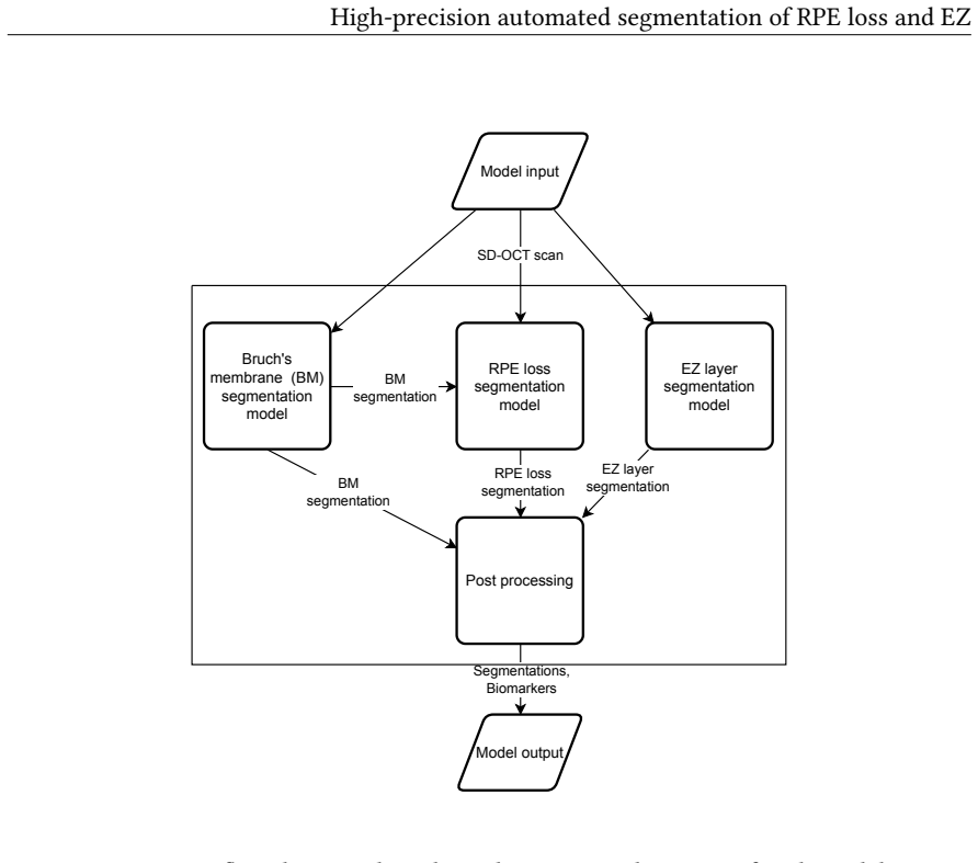
The proposed pipeline uses three specialized semantic segmentation models to delineate RPE loss, EZ boundaries (including interruptions), and Bruch's membrane. Results demonstrated high segmentation accuracy (Dice RPE loss: 0.88, Dice EZ loss: 0.87, Pearson's r > 0.99). Total EZ thickness measurements exhibited a sub-pixel average deviation of 2.15 μm, and segmentation reliability was confirmed by a strong reproducibility score (ICC > 0.98).
What carries the argument
Three specialized semantic segmentation models that separately delineate RPE loss, EZ boundaries including interruptions, and Bruch's membrane.
If this is right
- High Dice scores and sub-pixel thickness accuracy enable detection of small changes in photoreceptor degeneration over short intervals.
- ICC above 0.98 supports replacement of manual grading in clinical trials that require reproducible biomarker endpoints.
- Robustness across lesion sizes and B-scan densities allows consistent measurements even when scan protocols vary.
- Validation across GA, intermediate AMD, neovascular AMD, and healthy eyes supports use throughout the AMD disease spectrum.
- Fully automated output removes inter-reader variability for routine monitoring of treatment response.
Where Pith is reading between the lines
- If the models maintain accuracy on unseen scanner vendors, they could be embedded directly in commercial OCT consoles for immediate biomarker readouts.
- The same segmentation approach might extend to tracking photoreceptor integrity in other macular diseases such as Stargardt or cone-rod dystrophies.
- Longitudinal application could generate automated progression slopes that serve as surrogate endpoints for future GA trials.
- Combining the outputs with existing layer segmentation tools could produce composite indices of outer retinal health without additional manual annotation.
Load-bearing premise
Performance on an independent external set of 43 volumes is sufficient to establish reliability across the full range of real-world GA cases and scanner variations.
What would settle it
A multi-center study on several hundred volumes acquired on different OCT devices showing Dice scores below 0.80 or EZ thickness deviations exceeding 5 μm in any major phenotypic subgroup.
Figures


read the original abstract
Geographic atrophy (GA) secondary to age-related macular degeneration (AMD) requires precise monitoring of relevant structural biomarkers to assess disease stage, progression, and treatment response. This paper presents a fully automated, deep learning-based framework for the high-precision, pixel-wise segmentation of key biomarkers in optical coherence tomography (OCT) imaging: retinal pigment epithelium (RPE) loss, ellipsoid zone (EZ) loss, and EZ thinning. The proposed pipeline uses three specialized semantic segmentation models to delineate RPE loss, EZ boundaries (including interruptions), and Bruch's membrane. To ensure robustness and generalizability, the models were developed on a diverse dataset of 298 SD-OCT volumes representing the full phenotypic spectrum of AMD (GA:222, intermediate AMD: 40, neovascular AMD: 17, healthy: 19) and validated on an independent external dataset (n=43). The comprehensive evaluation was further strengthened using additional datasets to assess repeatability, inter-reader reliability, the impact of B-scan density on measurement accuracy, and subgroup performance stratified by lesion size. Results demonstrated high segmentation accuracy (Dice RPE loss: 0.88, Dice EZ loss: 0.87, Pearson's r > 0.99). Total EZ thickness measurements exhibited a sub-pixel average deviation of 2.15 $\mu m$, and segmentation reliability was confirmed by a strong reproducibility score (ICC > 0.98). By accurately and consistently quantifying outer photoreceptor degeneration and RPE loss, this fully automated framework provides a highly reliable tool for GA assessment in both clinical trials and routine real-world ophthalmic care.
Editorial analysis
A structured set of objections, weighed in public.
Referee Report
Summary. The manuscript presents a fully automated deep learning pipeline employing three specialized semantic segmentation models to delineate RPE loss, EZ boundaries (including interruptions), and Bruch's membrane in SD-OCT volumes for geographic atrophy monitoring in AMD. The models are trained on a diverse set of 298 volumes (GA:222, iAMD:40, nAMD:17, healthy:19) and evaluated on an independent external set of 43 volumes, with additional tests for repeatability, inter-reader reliability, B-scan density effects, and subgroup performance by lesion size. Reported results include Dice scores of 0.88 (RPE loss) and 0.87 (EZ loss), Pearson's r > 0.99, average EZ thickness deviation of 2.15 μm, and ICC > 0.98, positioning the framework as a reliable tool for both clinical trials and real-world care.
Significance. If the generalizability claims hold under more rigorous external scrutiny, the work could deliver a standardized, high-precision automated tool for quantifying outer retinal degeneration biomarkers, reducing inter-observer variability and enabling consistent longitudinal monitoring of GA progression and treatment response.
major comments (1)
- [Abstract / External validation] Abstract and validation description: the external validation cohort (n=43) is substantially smaller than the training set (n=298) and the manuscript provides no explicit evidence that the external cohort matches the training distribution with respect to AMD subtype mix, lesion-size distribution, scanner vendors, or B-scan density. This gap directly undermines the load-bearing claim that the reported Dice scores, sub-pixel thickness error, and ICC > 0.98 establish the pipeline as a 'highly reliable tool for GA assessment in both clinical trials and routine real-world ophthalmic care' across the full phenotypic spectrum.
Simulated Author's Rebuttal
We thank the referee for the detailed review and constructive feedback. We address the major comment on external validation below, and will revise the manuscript accordingly to strengthen the presentation of generalizability.
read point-by-point responses
-
Referee: [Abstract / External validation] Abstract and validation description: the external validation cohort (n=43) is substantially smaller than the training set (n=298) and the manuscript provides no explicit evidence that the external cohort matches the training distribution with respect to AMD subtype mix, lesion-size distribution, scanner vendors, or B-scan density. This gap directly undermines the load-bearing claim that the reported Dice scores, sub-pixel thickness error, and ICC > 0.98 establish the pipeline as a 'highly reliable tool for GA assessment in both clinical trials and routine real-world ophthalmic care' across the full phenotypic spectrum.
Authors: We agree that the external cohort size (n=43) is smaller than the training set and that the manuscript does not currently include an explicit side-by-side comparison of distributions for AMD subtype, lesion size, scanner vendor, or B-scan density. The external set was acquired independently at a different clinical center to assess real-world applicability, but we acknowledge this does not substitute for transparent distribution matching. We will revise the manuscript to add a supplementary table (or expanded methods/results section) comparing these characteristics between training and external cohorts. We will also moderate the abstract and discussion claims to more precisely reflect the validated scope rather than implying coverage of the full phenotypic spectrum without qualification. revision: yes
Circularity Check
No significant circularity; validation is independent
full rationale
The paper trains three semantic segmentation models on a 298-volume dataset and reports direct performance metrics (Dice, Pearson r, ICC, thickness deviation) on a fully independent external 43-volume set plus repeatability cohorts. No equations, fitted parameters, or self-citations are invoked to derive the reported accuracies; the metrics are computed from model outputs versus ground truth without reduction to the training inputs by construction. The derivation chain is therefore self-contained against external benchmarks.
Axiom & Free-Parameter Ledger
Reference graph
Works this paper leans on
-
[1]
Deep. Diagnostics , volume =. doi:10.3390/diagnostics15202580 , urldate =
-
[2]
Ansari, Georg and Sch. Evaluating the. Investigative Ophthalmology & Visual Science , volume =. doi:10.1167/iovs.66.11.34 , abstract =
-
[3]
Birner, Klaudia and Mai, Julia and Boryshchuk, Daniela and Frommlet, Florian and Enzendorfer, Marie Louise and. Exploring. Investigative Ophthalmology & Visual Science , volume =. doi:10.1167/iovs.67.1.22 , urldate =
-
[4]
Acta Ophthalmologica , volume =
Normative Prospective Data on Automatically Quantified Retinal Morphology Correlated to Retinal Function in Healthy Ageing Eyes by Two Microperimetry Devices , author =. Acta Ophthalmologica , volume =. doi:10.1111/aos.17434 , urldate =
-
[5]
and Steiner, Irene and Zarghami, Azin and Sadeghipour, Amir and
Birner, Klaudia and Reiter, Gregor S. and Steiner, Irene and Zarghami, Azin and Sadeghipour, Amir and. Structure-. Investigative Ophthalmology & Visual Science , volume =. doi:10.1167/iovs.66.3.26 , urldate =
-
[6]
Lancet (London, England) , volume =
Statistical Methods for Assessing Agreement between Two Methods of Clinical Measurement , author =. Lancet (London, England) , volume =
-
[7]
Exact Parametric Confidence Intervals for
Carkeet, Andrew , year = 2015, month = mar, journal =. Exact Parametric Confidence Intervals for. doi:10.1097/OPX.0000000000000513 , abstract =
-
[8]
Chakravarthy, Usha and Bailey, Clare C. and Johnston, Robert L. and McKibbin, Martin and Khan, Rehna S. and Mahmood, Sajjad and Downey, Louise and Dhingra, Narendra and Brand, Christopher and Brittain, Christopher J. and Willis, Jeffrey R. and Rabhi, Sarah and Muthutantri, Anushini and Cantrell, Ronald A. , year = 2018, month = jun, journal =. Characteriz...
-
[9]
Coulibaly, Leonard M. and Reiter, Gregor S. and Fuchs, Philipp and Lachinov, Dmitrii and Leingang, Oliver and Vogl, Wolf-Dieter and Bogunovic, Hrvoje and. Progression. Ophthalmology Retina , volume =. doi:10.1016/j.oret.2023.05.004 , urldate =
-
[10]
Computer Methods and Programs in Biomedicine , volume =
Fazekas, Botond and Aresta, Guilherme and Lachinov, Dmitrii and Riedl, Sophie and Mai, Julia and. Computer Methods and Programs in Biomedicine , volume =. doi:10.1016/j.cmpb.2025.108586 , urldate =
-
[11]
Fazekas, Botond and Aresta, Guilherme and Lachinov, Dmitrii and Riedl, Sophie and Mai, Julia and. Medical. doi:10.1007/978-3-031-16452-1_31 , abstract =
-
[12]
Fazekas, Botond and Lachinov, Dmitrii and Aresta, Guilherme and Mai, Julia and. Segmentation of. IEEE Journal of Biomedical and Health Informatics , volume =. doi:10.1109/JBHI.2022.3217962 , urldate =
-
[13]
and Yehoshua, Zohar and Gregori, Giovanni and Penha, Fernando M
Feuer, William J. and Yehoshua, Zohar and Gregori, Giovanni and Penha, Fernando M. and Chew, Emily Y. and Ferris, Frederick L. and Clemons, Traci E. and Lindblad, Anne S. and Rosenfeld, Philip J. , year = 2013, month = jan, journal =. Square. doi:10.1001/jamaophthalmol.2013.572 , urldate =
-
[14]
Investigative Ophthalmology & Visual Science , volume =
Quantifications of. Investigative Ophthalmology & Visual Science , volume =. doi:10.1167/iovs.66.4.65 , urldate =
-
[15]
Gerendas, Bianca S. and Sadeghipour, Amir and Michl, Martin and Goldbach, Felix and Mylonas, Georgios and Gruber, Anastasiia and Alten, Thomas and Leingang, Oliver and Sacu, Stefan and Bogunovic, Hrvoje and. Validation of an. RETINA , volume =. doi:10.1097/IAE.0000000000003557 , urldate =
-
[16]
Heier, Jeffrey S. and Lad, Eleonora M. and Holz, Frank G. and Rosenfeld, Philip J. and Guymer, Robyn H. and Boyer, David and Grossi, Federico and Baumal, Caroline R. and Korobelnik, Jean-Francois and Slakter, Jason S. and Waheed, Nadia K. and Metlapally, Ravi and Pearce, Ian and Steinle, Nathan and Francone, Anibal A. and Hu, Allen and Lally, David R. and...
-
[17]
Holz, Frank G. and Strauss, Erich C. and. Geographic. Ophthalmology , volume =. doi:10.1016/j.ophtha.2013.11.023 , urldate =
-
[18]
Densely connected convolutional networks
Huang, Gao and Liu, Zhuang and Van Der Maaten, Laurens and Weinberger, Kilian Q. , year = 2017, month = jul, pages =. Densely. 2017. doi:10.1109/CVPR.2017.243 , urldate =
-
[19]
Kalra, Gagan and Cetin, Hasan and Whitney, Jon and Yordi, Sari and Cakir, Yavuz and McConville, Conor and Whitmore, Victoria and Bonnay, Michelle and Reese, Jamie L. and Srivastava, Sunil K. and Ehlers, Justis P. , year = 2023, month = mar, journal =. Automated. doi:10.3390/diagnostics13061178 , urldate =
-
[20]
Kermany, Daniel S. and Poon, Wesley and Bawiskar, Anaya and Nehra, Natasha and Davarci, Orhun and Das, Glori and Vasquez, Matthew and Schaal, Shlomit and Raghunathan, Raksha and Wong, Stephen T. C. , year = 2025, month = jun, journal =. Identifying. doi:10.1167/iovs.66.6.55 , urldate =
-
[21]
Koo, Terry K. and Li, Mae Y. , year = 2016, month = jun, journal =. A. doi:10.1016/j.jcm.2016.02.012 , urldate =
-
[22]
and Frank, Sophie and Coulibaly, Leonard M
Kostolna, Klaudia and Reiter, Gregor S. and Frank, Sophie and Coulibaly, Leonard M. and Fuchs, Philipp and R. A. Ophthalmology Science , volume =. doi:10.1016/j.xops.2023.100456 , urldate =
-
[23]
Lachinov, Dmitrii and Seeb. Projective. Medical. doi:10.1007/978-3-030-87193-2_41 , urldate =
-
[24]
and Heitkotter, Heather and Carroll, Joseph , year = 2021, month = oct, journal =
Lee, Karen E. and Heitkotter, Heather and Carroll, Joseph , year = 2021, month = oct, journal =. Challenges. doi:10.1167/tvst.10.12.27 , urldate =
-
[25]
Graefe's Archive for Clinical and Experimental Ophthalmology , volume =
Sequential Structural and Functional Change in Geographic Atrophy on Multimodal Imaging in Non-Exudative Age-Related Macular Degeneration , author =. Graefe's Archive for Clinical and Experimental Ophthalmology , volume =. doi:10.1007/s00417-023-06022-3 , urldate =
-
[26]
Liao, David S. and Grossi, Federico V. and El Mehdi, Delphine and Gerber, Monica R. and Brown, David M. and Heier, Jeffrey S. and Wykoff, Charles C. and Singerman, Lawrence J. and Abraham, Prema and Grassmann, Felix and Nuernberg, Peter and Weber, Bernhard H. F. and Deschatelets, Pascal and Kim, Robert Y. and Chung, Carol Y. and Ribeiro, Ramiro M. and Ham...
-
[27]
Liefers, Bart and Colijn, Johanna M. and. A. Ophthalmology , volume =. doi:10.1016/j.ophtha.2020.02.009 , abstract =
-
[28]
Evaluation of Regression Procedures for Methods Comparison Studies , author =. Clinical Chemistry , volume =. doi:10.1093/clinchem/39.3.424 , urldate =
-
[29]
Dual-Branch Image Projection Network for Geographic Atrophy Segmentation in Retinal
Liu, Xiaoming and Li, Jieyang and Zhang, Ying and Yao, Junping , year = 2025, month = feb, journal =. Dual-Branch Image Projection Network for Geographic Atrophy Segmentation in Retinal. doi:10.1038/s41598-025-90709-6 , urldate =
-
[30]
and Vogl, Wolf-Dieter and Bogunovic, Hrvoje and
Mai, Julia and Lachinov, Dmitrii and Riedl, Sophie and Reiter, Gregor S. and Vogl, Wolf-Dieter and Bogunovic, Hrvoje and. Clinical Validation for Automated Geographic Atrophy Monitoring on. Scientific Reports , volume =. doi:10.1038/s41598-023-34139-2 , urldate =
-
[31]
Mai, Julia and Reiter, Gregor S. and Riedl, Sophie and Vogl, Wolf-Dieter and Sadeghipour, Amir and McKeown, Alex and Foos, Emma and Bogunovic, Hrvoje and. Dynamics of the. Investigative Ophthalmology & Visual Science , volume =. doi:10.1167/iovs.66.11.48 , urldate =
-
[32]
Mai, Julia and Reiter, Gregor S. and Riedl, Sophie and Vogl, Wolf-Dieter and Sadeghipour, Amir and Foos, Emma and McKeown, Alex and Bogunovic, Hrvoje and. Quantitative Comparison of Automated. Scientific Reports , volume =. doi:10.1038/s41598-024-71496-y , urldate =
-
[33]
Mares, Virginia and Reiter, Gregor S. and Gumpinger, Markus and Leigang, Oliver and Bogunovic, Hrvoje and Barthelmes, Daniel and Nehemy, Marcio B. and. Correlation of Retinal Fluid and Photoreceptor and. Acta Ophthalmologica , volume =. doi:10.1111/aos.16799 , urldate =
-
[34]
, year = 2025, month = jun, journal =
Midroni, Julie and Longwell, Jack and Bhambra, Nishaant and Demian, Sueellen and Pecaku, Aurora and Melo, Isabela Martins and Muni, Rajeev H. , year = 2025, month = jun, journal =. Automated Segmentation of Subretinal Fluid from Optical Coherence Tomography:. doi:10.1016/j.xops.2025.100852 , urldate =
-
[35]
and Hu, Zhihong , year = 2020, month = jun, journal =
Mishra, Zubin and Ganegoda, Anushika and Selicha, Jane and Wang, Ziyuan and Sadda, SriniVas R. and Hu, Zhihong , year = 2020, month = jun, journal =. Automated. doi:10.1038/s41598-020-66355-5 , urldate =
-
[36]
Mon. The. Translational Vision Science & Technology , volume =. doi:10.1167/tvst.7.6.40 , urldate =
-
[37]
Morano, Jos. Deep. IEEE Journal of Biomedical and Health Informatics , volume =. doi:10.1109/JBHI.2024.3352970 , urldate =
-
[38]
Morano, Jos. Self-Supervised. Medical. doi:10.1007/978-3-031-43901-8_56 , abstract =
-
[39]
Orlando, Jos. Automated. Scientific Reports , volume =. doi:10.1038/s41598-020-62329-9 , urldate =
-
[40]
Orlando, Jos. U2-. doi:10.48550/ARXIV.1901.07929 , urldate =
-
[41]
Pfau, Maximilian and. Association of Complement. Scientific Reports , volume =. doi:10.1038/s41598-022-22404-9 , urldate =
-
[42]
Pfau, Maximilian and. Progression of. JAMA Ophthalmology , volume =. doi:10.1001/jamaophthalmol.2020.2914 , urldate =
-
[43]
Pramil, Varsha and. A. Ophthalmology Retina , volume =. doi:10.1016/j.oret.2022.08.007 , urldate =
-
[44]
and Mai, Julia and Riedl, Sophie and Birner, Klaudia and Frank, Sophie and Bogunovic, Hrvoje and
Reiter, Gregor S. and Mai, Julia and Riedl, Sophie and Birner, Klaudia and Frank, Sophie and Bogunovic, Hrvoje and. Progress in Retinal and Eye Research , volume =. doi:10.1016/j.preteyeres.2024.101305 , abstract =
-
[45]
Reiter, Gregor S. and Told, Reinhard and Schranz, Markus and Baumann, Lukas and Mylonas, Georgios and Sacu, Stefan and Pollreisz, Andreas and. Subretinal. Investigative Ophthalmology & Visual Science , volume =. doi:10.1167/iovs.61.6.11 , urldate =
-
[46]
and Lachinov, Dmitrii and Grechenig, Christoph and McKeown, Alex and Scheibler, Lukas and Bogunovi
Riedl, Sophie and Vogl, Wolf-Dieter and Mai, Julia and Reiter, Gregor S. and Lachinov, Dmitrii and Grechenig, Christoph and McKeown, Alex and Scheibler, Lukas and Bogunovi. The. Ophthalmology Retina , volume =. doi:10.1016/j.oret.2022.05.030 , urldate =
-
[47]
Journal of Ophthalmology , volume =
Automatic. Journal of Ophthalmology , volume =. doi:10.1155/2020/8204641 , urldate =
-
[48]
Wightman, Ross , year = 2019, publisher =
2019
-
[49]
and Guymer, Robyn and Holz, Frank G
Sadda, Srinivas R. and Guymer, Robyn and Holz, Frank G. and. Consensus. Ophthalmology , volume =. doi:10.1016/j.ophtha.2017.09.028 , urldate =
-
[50]
and Rabe, Christina and Schiffman, Courtney and Yang, Qi and Lee, Aaron Y
Salvi, Anish and Cluceru, Julia and Gao, Simon S. and Rabe, Christina and Schiffman, Courtney and Yang, Qi and Lee, Aaron Y. and Keane, Pearse A. and Sadda, Srinivas R. and Holz, Frank G. and Ferrara, Daniela and Anegondi, Neha , year = 2025, month = mar, journal =. Deep. doi:10.1016/j.xops.2024.100635 , urldate =
-
[51]
Disease. Ophthalmology , volume =. doi:10.1016/j.ophtha.2024.08.017 , urldate =
-
[52]
Investigative Ophthalmology & Visual Science , volume =
Longitudinal Assessment of Progressive Functional Decline in Areas of Intact Retina,. Investigative Ophthalmology & Visual Science , volume =
-
[53]
Seghier, Mohamed L. , year = 2024, journal =. Image. doi:10.1002/ima.23203 , urldate =
-
[54]
and Gim, Nayoon and Blazes, Marian and Lee, Cecilia S
Spaide, Theodore and Rajesh, Anand E. and Gim, Nayoon and Blazes, Marian and Lee, Cecilia S. and Macivannan, Niranchana and Lee, Gary and Lewis, Warren and Salehi, Ali and de Sisternes, Luis and Herrera, Gissel and Shen, Mengxi and Gregori, Giovanni and Rosenfeld, Philip J. and Pramil, Varsha and Waheed, Nadia and Wu, Yue and Zhang, Qinqin and Lee, Aaron ...
-
[55]
Navigating Distribution Shifts in Medical Image Analysis: A Survey
Su, Zixian and Guo, Jingwei and Yang, Xi and Wang, Qiufeng and Coenen, Frans and Huang, Kaizhu , year = 2025, month = aug, number =. Navigating. doi:10.48550/arXiv.2411.05824 , urldate =. arXiv , keywords =:2411.05824 , primaryclass =
work page internal anchor Pith review Pith/arXiv arXiv doi:10.48550/arxiv.2411.05824 2025
-
[56]
Proceedings of the 36th
Tan, Mingxing and Le, Quoc , year = 2019, month = may, pages =. Proceedings of the 36th
2019
-
[57]
Investigative Ophthalmology & Visual Science , volume =
Automated. Investigative Ophthalmology & Visual Science , volume =
-
[58]
and Lachinov, Dmitrii and Bogunovi
Vogl, Wolf-Dieter and Riedl, Sophie and Mai, Julia and Reiter, Gregor S. and Lachinov, Dmitrii and Bogunovi. Predicting. Ophthalmology Retina , volume =. doi:10.1016/j.oret.2022.08.003 , urldate =
-
[59]
Vogl, Wolf-Dieter and Bogunovi. Spatio-Temporal Alterations in Retinal and Choroidal Layers in the Progression of Age-Related Macular Degeneration (. Scientific Reports , volume =. doi:10.1038/s41598-021-85110-y , urldate =
-
[60]
Wightman, Ross , year = 2025, month = jul, doi =
2025
-
[61]
Journal of Medical Imaging , volume =
Family of Boundary Overlap Metrics for the Evaluation of Medical Image Segmentation , author =. Journal of Medical Imaging , volume =. doi:10.1117/1.JMI.5.1.015006 , urldate =
-
[62]
and Cukras, Catherine and Rabe, Christina and Ferrara, Daniela and Spaide, Richard F
Yoshida, Kenta and Anegondi, Neha and Pely, Adam and Zhang, Miao and Debraine, Frederic and Ramesh, Karthik and Steffen, Verena and Gao, Simon S. and Cukras, Catherine and Rabe, Christina and Ferrara, Daniela and Spaide, Richard F. and Sadda, SriniVas R. and Holz, Frank G. and Yang, Qi , year = 2025, month = feb, journal =. Deep. doi:10.1167/tvst.14.2.11 ...
-
[63]
Zekavat, Seyedeh Maryam and Sekimitsu, Sayuri and Ye, Yixuan and Raghu, Vineet and Zhao, Hongyu and Elze, Tobias and Segr. Photoreceptor. Ophthalmology , volume =. doi:10.1016/j.ophtha.2022.02.001 , abstract =
-
[64]
doi:10.1109/TMI.2019.2959609 , urldate =
Zhou, Zongwei and Siddiquee, Md Mahfuzur Rahman and Tajbakhsh, Nima and Liang, Jianming , year = 2020, month = jun, journal =. doi:10.1109/TMI.2019.2959609 , urldate =
discussion (0)
Sign in with ORCID, Apple, or X to comment. Anyone can read and Pith papers without signing in.