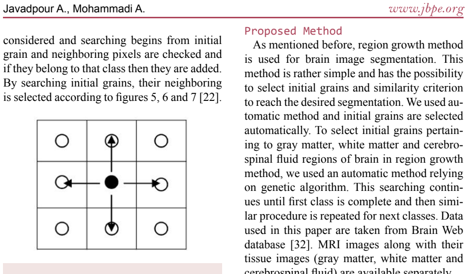
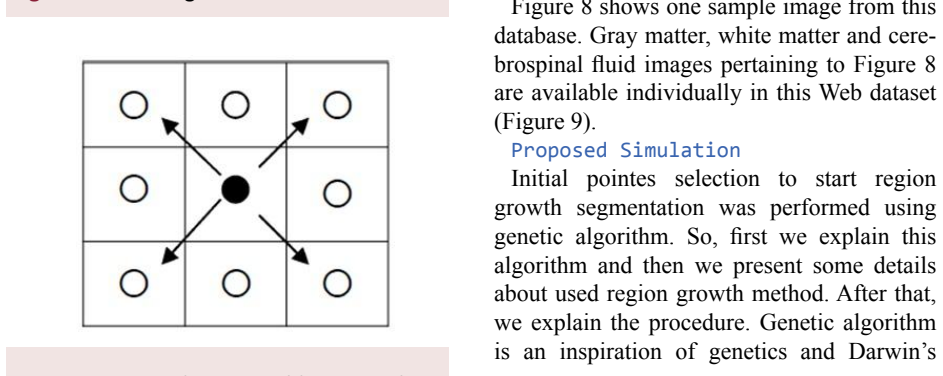
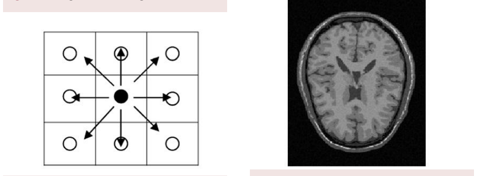
Improving Brain Magnetic Resonance Image MRI Segmentation via a Novel Algorithm based on Genetic and Regional Growth
Pith reviewed 2026-05-24 17:33 UTC · model grok-4.3
The pith
Genetic algorithm automates initial point selection for regional growth to segment brain MRIs with lower error.
A machine-rendered reading of the paper's core claim, the machinery that carries it, and where it could break.
Core claim
By using genetic algorithms to automatically select primary pixels and similarity criteria for the regional growth method, the proposed algorithm improves MRI segmentation accuracy and reduces error compared to manual selection of initial points.
What carries the argument
Genetic algorithm that optimizes initial points and similarity criteria to drive the regional growth segmentation process.
If this is right
- Segmentation error decreases when initial points come from genetic optimization rather than manual choice.
- The method supports more consistent identification of brain tissue changes linked to diseases.
- Automatic selection of similarity criteria improves the validity of the resulting segmentations.
- The combined approach applies directly to existing brain MRI datasets without extra manual setup.
Where Pith is reading between the lines
- The same automation of starting parameters could extend to segmenting other soft-tissue medical scans beyond brain MRIs.
- Comparison against contemporary machine-learning segmentation tools on the same images would test relative performance.
- Wider use might lower reliance on expert radiologists for initial parameter setting in routine scans.
Load-bearing premise
The genetic algorithm's automatic choices of initial points and similarity criteria produce objectively better segmentation than manual selection without needing further tuning for the images tested.
What would settle it
A head-to-head test on new brain MRI images where the genetic algorithm version shows equal or higher segmentation error than conventional regional growth with manual initial points.
Figures


read the original abstract
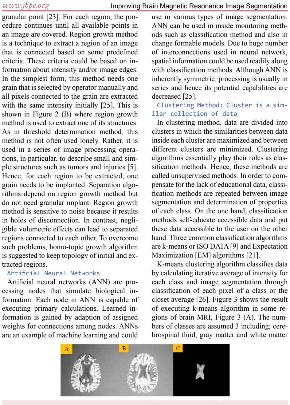
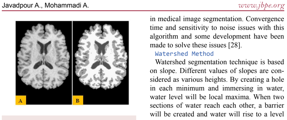
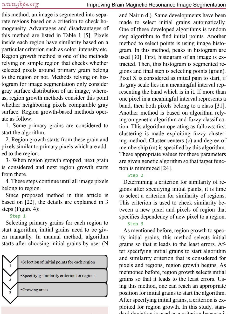
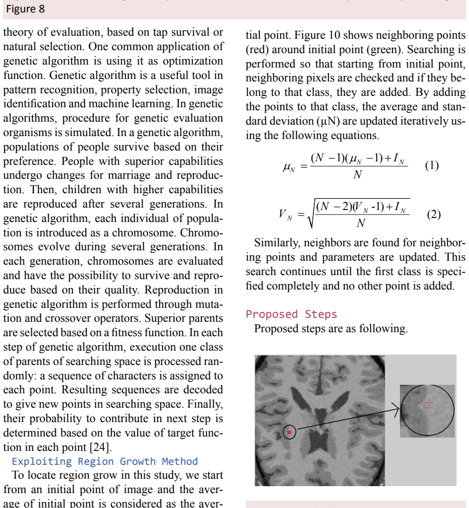
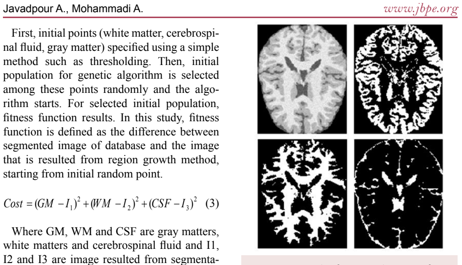
Background: Regarding the importance of right diagnosis in medical applications, various methods have been exploited for processing medical images solar. The method of segmentation is used to analyze anal to miscall structures in medical imaging. Objective: This study describes a new method for brain Magnetic Resonance Image (MRI) segmentation via a novel algorithm based on genetic and regional growth. Methods: Among medical imaging methods, brains MRI segmentation is important due to the high contrast of non-intrusive soft tissue and high spatial resolution. Size variations of brain tissues are often accompanied by various diseases such as Alzheimers disease. As our knowledge about the relationship between various brain diseases and deviation of brain anatomy increases, MRI segmentation is exploited as the first step in early diagnosis. In this paper, the regional growth method and auto-mate selection of initial points by genetic algorithm are used to introduce a new method for MRI segmentation. Primary pixels and similarity criterion are automatically by genetic algorithms to maximize the accuracy and validity in image segmentation. Results: By using genetic algorithms and defining the fixed function of image segmentation, the initial points for the algorithm were found. The proposed algorithms are applied to the images and results are manually selected by regional growth in which the initial points were compared. The results showed that the proposed algorithm could reduce segmentation error effectively. Conclusion: The study concluded that the proposed algorithm could reduce segmentation error effectively and help us to diagnose brain diseases.
Editorial analysis
A structured set of objections, weighed in public.
Referee Report
Summary. The manuscript proposes a hybrid segmentation method for brain MRI that uses a genetic algorithm to automatically select initial seed points and similarity criteria for a subsequent region-growing procedure. The central claim is that this automation yields measurably lower segmentation error than conventional region growing and thereby assists diagnosis of brain diseases such as Alzheimer’s.
Significance. If the claimed error reduction were demonstrated with quantitative, reproducible metrics and independent validation, the approach could supply a practical, parameter-light alternative for automated MRI analysis. No such demonstration is present, so the potential clinical utility cannot be assessed.
major comments (2)
- [Results] Results paragraph: the assertion that 'the results showed that the proposed algorithm could reduce segmentation error effectively' is unsupported by any numerical evidence. No Dice/Jaccard scores, pixel-wise error rates, subject counts, baseline comparisons, or statistical tests appear anywhere in the manuscript.
- [Results] Results paragraph: the evaluation is described as 'results are manually selected by regional growth in which the initial points were compared,' indicating only subjective visual inspection. This directly undermines the central claim of objective superiority.
minor comments (2)
- [Abstract] Abstract contains multiple typographical and grammatical errors ('medical images solar', 'analyze anal to miscall structures', 'auto-mate selection') that must be corrected for readability.
- [Methods] No description is given of the genetic-algorithm fitness function, population size, selection/crossover/mutation operators, or termination criteria, rendering the method non-reproducible.
Simulated Author's Rebuttal
We thank the referee for identifying key deficiencies in the quantitative validation of our proposed method. We agree that the current manuscript lacks the necessary numerical evidence and objective metrics to support its claims, and we will substantially revise the results section in the next version.
read point-by-point responses
-
Referee: [Results] Results paragraph: the assertion that 'the results showed that the proposed algorithm could reduce segmentation error effectively' is unsupported by any numerical evidence. No Dice/Jaccard scores, pixel-wise error rates, subject counts, baseline comparisons, or statistical tests appear anywhere in the manuscript.
Authors: We agree that the manuscript provides no quantitative support for the claim. The revised version will include a full results section reporting Dice coefficients, Jaccard indices, pixel-wise error rates, the number of subjects/images used, direct comparisons against standard region-growing baselines, and statistical significance tests. revision: yes
-
Referee: [Results] Results paragraph: the evaluation is described as 'results are manually selected by regional growth in which the initial points were compared,' indicating only subjective visual inspection. This directly undermines the central claim of objective superiority.
Authors: The manuscript text does describe a manual, subjective comparison process. This is a valid and important criticism. In revision we will replace all subjective visual assessments with the quantitative, reproducible metrics and baseline comparisons noted above. revision: yes
Circularity Check
No circularity in derivation chain
full rationale
The paper proposes an algorithmic method combining genetic algorithms for automatic seed-point and similarity-criterion selection with region-growing segmentation. Its central claim is an empirical assertion that the approach reduces segmentation error, supported only by a qualitative statement that results were 'manually selected' after application to images. No equations, first-principles derivations, or mathematical steps are presented whose outputs reduce to the inputs by construction. No self-citations, uniqueness theorems, or ansatzes are invoked. The absence of quantitative metrics or baselines is a limitation of evidence, not a circular reduction of any claimed derivation to its own fitted values. The paper therefore contains no load-bearing circular steps.
Axiom & Free-Parameter Ledger
Reference graph
Works this paper leans on
-
[1]
Awate SP, Tasdizen T, Foster N, Whitaker RT. Adaptive Markov modeling for mutual-informa- tion-based, unsupervised MRI brain-tissue clas- sification. Med Image Anal. 2006;10:726-39. doi. org/10.1016/j.media.2006.07.002. PubMed PMID: 16919993
-
[2]
Implementing a Smart Method to Eliminate Artifacts of Vital Sig- nals
Javadpour A, Mohammadi A. Implementing a Smart Method to Eliminate Artifacts of Vital Sig- nals. J Biomed Phys Eng. 2015; 5(4): 199–206. PMCID: PMC4681465
work page 2015
-
[3]
Diagnosis of Al- zheimer’s disease from EEG signals: where are we standing? Curr Alzheimer Res
Dauwels J, Vialatte F, Cichocki A. Diagnosis of Al- zheimer’s disease from EEG signals: where are we standing? Curr Alzheimer Res. 2010;7:487-505. doi.org/10.2174/156720510792231720. PubMed PMID: 20455865
-
[4]
Computation in neurons and neural systems: Springer Science & Business Media; 2012
Eeckman F. Computation in neurons and neural systems: Springer Science & Business Media; 2012
work page 2012
-
[5]
Automated Region Growing for Segmentation of Brain Lesion in Diffusion- weighted MRI
Saad NM, Abu-Bakar S, Muda S, Mokji M, Abdullah A, editors. Automated Region Growing for Segmentation of Brain Lesion in Diffusion- weighted MRI. Proceedings of the International MultiConference of Engineers and Computer Scientists; 2012
work page 2012
-
[6]
Banerjee S. The macroeconomics of dementia- -will the world economy get Alzheimer’s disease? Arch Med Res. 2012;43:705-9. doi.org/10.1016/j. arcmed.2012.10.006. PubMed PMID: 23085453
work page doi:10.1016/j 2012
-
[7]
Epidemiology of Alzheimer’s Disease: INTECH Open Access Pub- lisher; 2013
Xu W, Ferrari C, Wang HX. Epidemiology of Alzheimer’s Disease: INTECH Open Access Pub- lisher; 2013. Javadpour A., Mohammadi A. 106 J Biomed Phys Eng 2016; 6(2) www.jbpe.org
work page 2013
-
[8]
Li C, Xu C, Anderson AW, Gore JC, editors. MRI tissue classification and bias field estimation based on coherent local intensity clustering: A unified energy minimization framework. Informa- tion Processing in Medical Imaging: Springer
-
[9]
Segmentation of Brain MRI: INTECH Open Access Publisher; 2012
Xu R, Ohya J, Luo L. Segmentation of Brain MRI: INTECH Open Access Publisher; 2012
work page 2012
-
[10]
Neuroanatomical segmentation in MRI: technological objectives
Worth AJ, Makris N, Caviness Jr VS, Kennedy DN. Neuroanatomical segmentation in MRI: technological objectives. International Journal of Pattern Recognition and Artificial Intel- ligence. 1997;11:1161-87. doi.org/10.1142/ S0218001497000548
work page 1997
-
[11]
Integrated graph cuts for brain MRI segmentation
Song Z, Tustison N, Avants B, Gee JC. Integrated graph cuts for brain MRI segmentation. Medical Image Computing and Computer-Assisted Inter- vention–MICCAI 2006: Springer; 2006. p. 831-8
work page 2006
-
[12]
Ho BC. MRI brain volume abnormalities in young, nonpsychotic relatives of schizophrenia pro- bands are associated with subsequent prodromal symptoms. Schizophr Res. 2007;96:1-13. doi. org/10.1016/j.schres.2007.08.001. PubMed PMID: 17761401. PubMed PMCID: 2222920
-
[13]
Slezak D, Pal S, Kang BH, Gu J, Kuroda H, Kim TH. Signal Processing, Image Processing and Pattern Recognition: International Conference, SIP 2009, Held as Part of the Future Generation Infor- mation Technology Conference, FGIT 2009, Jeju Island, Korea, December 10-12, 2009. Proceed- ings: Springer; 2010
work page 2009
-
[14]
Segmentation of white matter lesions from volumetric MR images
Hojjatoleslami S, Kruggel F, von Cramon D, edi- tors. Segmentation of white matter lesions from volumetric MR images. Medical Image Computing and Computer-Assisted Intervention–MICCAI’99: Springer; 1999. p. 52-61
work page 1999
-
[15]
Magnetic resonance imaging (MRI): considerations and applications in radiotherapy treatment planning
Khoo VS, Dearnaley DP, Finnigan DJ, Padhani A, Tanner SF, Leach MO. Magnetic resonance imaging (MRI): considerations and applications in radiotherapy treatment planning. Radiother Oncol. 1997;42:1-15. doi.org/10.1016/S0167- 8140(96)01866-X. PubMed PMID: 9132820
-
[16]
A Fuzzy Relative of the ISODATA Pro- cess and Its Use in Detecting Compact Well-Sepa- rated Clusters
Dunn JC. A Fuzzy Relative of the ISODATA Pro- cess and Its Use in Detecting Compact Well-Sepa- rated Clusters. Journal of Cybernetics. 1973;3:32-
work page 1973
-
[17]
doi.org/10.1080/01969727308546046
-
[18]
Bezdek JC. Pattern recognition with fuzzy objec- tive function algorithms: Springer Science & Business Media; 2013
work page 2013
-
[19]
The possibilistic c-means algorithm: insights and recommenda- tions
Krishnapuram R, Keller JM. The possibilistic c-means algorithm: insights and recommenda- tions. Fuzzy Systems, IEEE Transactions on. 1996;4:385-93. doi.org/10.1109/91.531779
-
[20]
Improved possibilistic c-means clustering algorithms
Zhang JS, Leung YW. Improved possibilistic c-means clustering algorithms. Fuzzy Systems, IEEE Transactions on. 2004;12:209-17. doi. org/10.1109/TFUZZ.2004.825079
-
[21]
An improved MR image segmentation method based on fuzzy c- means clustering
Lu L, Li M, Zhang X, editors. An improved MR image segmentation method based on fuzzy c- means clustering. Computational Problem-Solving (ICCP), 2012 International Conference on; 2012: IEEE
work page 2012
-
[22]
A fully automated algorithm under modified FCM frame- work for improved brain MR image segmentation
Sikka K, Sinha N, Singh PK, Mishra AK. A fully automated algorithm under modified FCM frame- work for improved brain MR image segmentation. Magn Reson Imaging. 2009;27:994-1004. doi. org/10.1016/j.mri.2009.01.024. PubMed PMID: 19395212
-
[23]
Seed-based region growing study for brain abnormalities segmentation
Khalid NEA, Ibrahim S, Manaf M, Ngah UK, edi- tors. Seed-based region growing study for brain abnormalities segmentation. Information Technol- ogy (ITSim), 2010 International Symposium in; 2010: IEEE. doi.org/10.1109/itsim.2010.5561560
-
[24]
Unifying variational approach and region growing segmentation
Rose J, Grenier T, Revol-Muller C, Odet C, edi- tors. Unifying variational approach and region growing segmentation. European Signal Process- ing Conference; 2010
work page 2010
-
[25]
Genetic based Fuzzy Seeded Region Growing Segmenta- tion for diabetic retinopathy images
Tamilarasi M, Duraiswamy K, editors. Genetic based Fuzzy Seeded Region Growing Segmenta- tion for diabetic retinopathy images. Computer Communication and Informatics (ICCCI), 2013 International Conference on; 2013: IEEE. doi. org/10.1109/iccci.2013.6466117
-
[26]
Brain tumor segmentation from MRI data sets using region growing approach
Węgliński T, Fabijańska A, editors. Brain tumor segmentation from MRI data sets using region growing approach. Perspective Technologies and Methods in MEMS Design (MEMSTECH), 2011 Proceedings of VIIth International Conference on; 2011: IEEE
work page 2011
-
[27]
Survey: interpolation methods in medical image process- ing
Lehmann TM, Gonner C, Spitzer K. Survey: interpolation methods in medical image process- ing. IEEE Trans Med Imaging. 1999;18:1049-75. doi.org/10.1109/42.816070. PubMed PMID: 10661324
-
[28]
A survey of thresholding techniques
Sahoo PK, Soltani S, Wong AK. A survey of thresholding techniques. Computer vision, graph- ics, and image processing. 1988;41:233-60. doi. org/10.1016/0734-189X(88)90022-9
-
[29]
Neural network for enhancement of FCM based brain MRI segmentation
Rostami MT, Ghasemi J, Ghaderi R, editors. Neural network for enhancement of FCM based brain MRI segmentation. Fuzzy Systems (IFSC), 2013 13th Iranian Conference on; 2013: IEEE. doi. org/10.1109/ifsc.2013.6675661
-
[30]
Watersheds in digital spaces: an efficient algorithm based on immersion simu- lations
Vincent L, Soille P. Watersheds in digital spaces: an efficient algorithm based on immersion simu- lations. IEEE Transactions on Pattern Analysis & Machine Intelligence. 1991;(6):583-98. doi. org/10.1109/34.87344
-
[31]
Trémeau A, Tominaga S, Plataniotis KN. Color in image and video processing: most recent Improving Brain Magnetic Resonance Image Segmentation 107 J Biomed Phys Eng 2016; 6(2) www.jbpe.org trends and future research directions. Journal on Image and Video Processing. 2008;2008:7. doi. org/10.1155/2008/581371
-
[32]
Image seg- mentation using automatic seeded region growing and instance-based learning
Gómez O, González JA, Morales EF. Image seg- mentation using automatic seeded region growing and instance-based learning. Progress in pattern recognition, image analysis and applications: Springer; 2007. p. 192-201
work page 2007
-
[33]
Available from: http//brainweb.bic.mni
BrainWeb: 20 Anatomical Models of 20 Normal Brains. Available from: http//brainweb.bic.mni. mcgill.ca/brainweb/anatomic_normal_20
-
[34]
Erfanian A, Mahmoudi B. Real-time ocular artifact suppression using recurrent neural network for electro-encephalogram based brain-computer in- terface. Med Biol Eng Comput. 2005;43:296-305. doi.org/10.1007/BF02345969. PubMed PMID: 15865142
-
[35]
Automatic Sleep Stages Detection Based on EEG Signals Using Combination of Classifiers
Kianzad R, Montazery Kordy H. Automatic Sleep Stages Detection Based on EEG Signals Using Combination of Classifiers. Journal of Electri- cal and Computer Engineering Innovations. 2013;1:99-105
work page 2013
-
[36]
A New Blind Source Separation Method to Remove Artifact in EEG Signals
Chaozhu Z, Siyao L, Abdullah AK, editors. A New Blind Source Separation Method to Remove Artifact in EEG Signals. Instrumentation, Measure- ment, Computer, Communication and Control (IMCCC), 2013 Third International Conference on; 2013: IEEE. doi.org/10.1109/imccc.2013.319
-
[37]
Detection and extraction of the ECG signal parameters
Gholam-Hosseini H, Nazeran H, editors. Detection and extraction of the ECG signal parameters. En- gineering in Medicine and Biology Society, 1998. Proceedings of the 20th Annual International Con- ference of the IEEE; 1998: IEEE. doi.org/10.1109/ iembs.1998.745846. Javadpour A., Mohammadi A. 108
discussion (0)
Sign in with ORCID, Apple, or X to comment. Anyone can read and Pith papers without signing in.