Leveraging Medical Foundation Model Features in Graph Neural Network-Based Retrieval of Breast Histopathology Images
Pith reviewed 2026-05-24 01:07 UTC · model grok-4.3
The pith
Foundation model features in a graph autoencoder improve breast histopathology image retrieval over CNN baselines.
A machine-rendered reading of the paper's core claim, the machinery that carries it, and where it could break.
Core claim
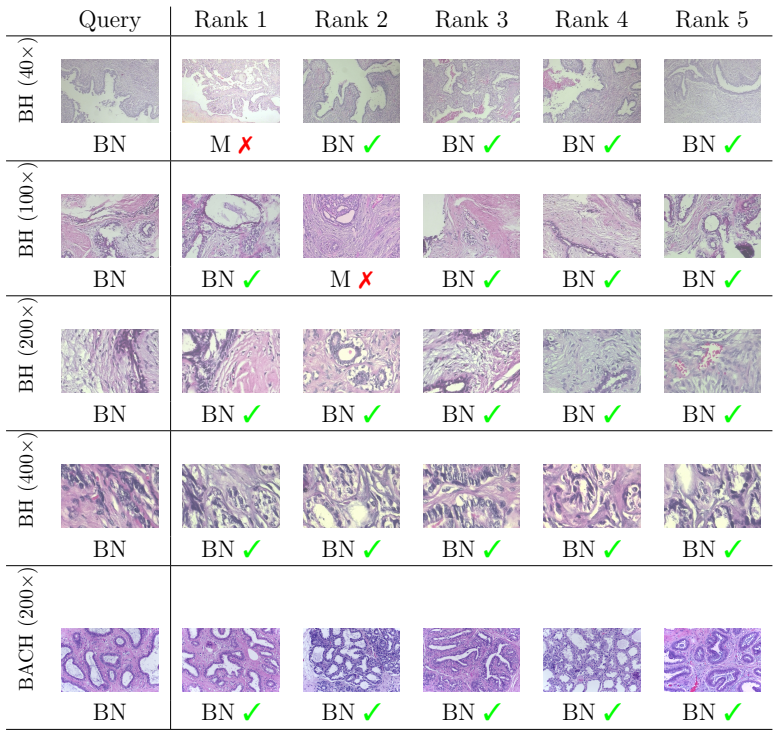
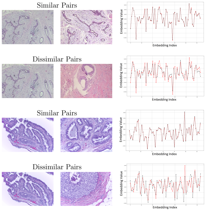
The central claim is that an attention-based adversarially regularized variational graph autoencoder trained on features from foundation models, especially the self-supervised UNI model, produces higher retrieval accuracy than the same architecture trained on features from pre-trained convolutional neural networks, reaching average mAP/mMV of 96.7 percent/91.5 percent on BreakHis and 97.6 percent/94.2 percent on BACH.
What carries the argument
Attention-based adversarially regularized variational graph autoencoder that ingests foundation-model embeddings to encode tissue variability for image retrieval.
Load-bearing premise
The gains measured on two public datasets will continue to appear on new clinical images from different scanners or hospitals.
What would settle it
Evaluation of the same model on an independent, previously unseen breast histopathology dataset where mAP and mMV fall below the CNN-feature baseline.
Figures
read the original abstract
Breast cancer is the most common cancer type in women worldwide. Early detection and appropriate treatment can significantly reduce its impact. While histopathology examinations play a vital role in rapid and accurate diagnosis, they often require experienced medical experts for proper recognition and cancer grading. Automated image retrieval systems have the potential to assist pathologists in identifying cancerous tissues, thereby accelerating the diagnostic process. Nevertheless, proposing an accurate image retrieval model is challenging due to considerable variability among the tissue and cell patterns in histological images. In this work, we leverage the features from foundation models in a novel attention-based adversarially regularized variational graph autoencoder model for breast histological image retrieval. Our results confirm the superior performance of models trained with foundation model features compared to those using pre-trained convolutional neural networks (up to 7.7% and 15.5% for mAP and mMV, respectively), with the pre-trained general-purpose self-supervised model for computational pathology (UNI) delivering the best overall performance. By evaluating two publicly available histology image datasets of breast cancer, our top-performing model, trained with UNI features, achieved average mAP/mMV scores of 96.7%/91.5% and 97.6%/94.2% for the BreakHis and BACH datasets, respectively. Our proposed retrieval model has the potential to be used in clinical settings to enhance diagnostic performance and ultimately benefit patients.
Editorial analysis
A structured set of objections, weighed in public.
Referee Report
Summary. The paper introduces an attention-based adversarially regularized variational graph autoencoder for content-based retrieval of breast histopathology images. It extracts features from medical foundation models (with UNI performing best) and reports that these yield higher retrieval accuracy than pre-trained CNN features, with gains of up to 7.7% mAP and 15.5% mMV; on BreakHis the best model reaches 96.7%/91.5% and on BACH 97.6%/94.2%.
Significance. If the empirical gains prove robust under proper statistical controls and external validation, the work would demonstrate a practical way to combine self-supervised pathology foundation models with graph autoencoders for improved retrieval, which could support diagnostic assistance tools in computational pathology.
major comments (2)
- [Results] Results section: performance is reported solely as point estimates (e.g., 96.7% mAP, 91.5% mMV on BreakHis) with no error bars, standard deviations across random seeds, or statistical significance tests, so it is impossible to determine whether the claimed 7.7% and 15.5% margins over CNN baselines are stable or could arise from training variability.
- [Experiments] Experimental protocol (Methods/Experiments): the manuscript supplies no description of the train/validation/test split strategy (patient-level vs. image-level), number of independent runs, hyperparameter selection procedure, or external-site validation, leaving open the possibility that the reported superiority of UNI features is tied to the specific public datasets and splits used.
minor comments (1)
- [Abstract] The abbreviation mMV is used without an explicit definition in the abstract or early sections; a one-sentence expansion would improve readability.
Simulated Author's Rebuttal
We thank the referee for the constructive feedback on statistical robustness and experimental transparency. We address each major comment below and will revise the manuscript to incorporate the suggested improvements.
read point-by-point responses
-
Referee: [Results] Results section: performance is reported solely as point estimates (e.g., 96.7% mAP, 91.5% mMV on BreakHis) with no error bars, standard deviations across random seeds, or statistical significance tests, so it is impossible to determine whether the claimed 7.7% and 15.5% margins over CNN baselines are stable or could arise from training variability.
Authors: We agree that point estimates alone are insufficient. In the revised manuscript we will report mean performance and standard deviation across five independent runs with different random seeds, add error bars to all tables and figures, and include statistical significance tests (paired t-tests or Wilcoxon signed-rank tests) against the CNN baselines to confirm the reported margins are stable. revision: yes
-
Referee: [Experiments] Experimental protocol (Methods/Experiments): the manuscript supplies no description of the train/validation/test split strategy (patient-level vs. image-level), number of independent runs, hyperparameter selection procedure, or external-site validation, leaving open the possibility that the reported superiority of UNI features is tied to the specific public datasets and splits used.
Authors: We will expand the Methods and Experiments sections with a complete protocol description. Patient-level splits were used (70/15/15 train/val/test) to avoid leakage; hyperparameters were selected via grid search on the validation set; five independent runs were performed. Exact split indices and seeds will be released. External-site validation was outside the current scope and will be noted as a limitation, but the public datasets enable full reproducibility. revision: yes
Circularity Check
No circularity; empirical evaluation on public datasets
full rationale
The paper proposes an attention-based adversarially regularized variational graph autoencoder that ingests features from foundation models (e.g., UNI) or pre-trained CNNs and reports retrieval metrics (mAP, mMV) on the BreakHis and BACH datasets. No equation, prediction, or uniqueness claim reduces by construction to a fitted parameter, self-defined quantity, or self-citation chain. All load-bearing statements are direct experimental outcomes on fixed public benchmarks; the derivation chain is therefore self-contained and does not exhibit any of the enumerated circularity patterns.
Axiom & Free-Parameter Ledger
free parameters (1)
- graph autoencoder hyperparameters
axioms (1)
- domain assumption Features extracted from the pre-trained UNI foundation model are directly suitable as node attributes for the graph autoencoder without further adaptation or domain-specific fine-tuning.
Reference graph
Works this paper leans on
-
[1]
R. Rashmi, K. Prasad, C. B. K. Udupa, Breast histopathological image analysis using image processing techniques for diagnostic puposes: A methodological review., Journal of medical systems 46 (1) (2021) 7.doi: https://doi.org/10.1007/s10916-021-01786-9
-
[2]
M. Arnold, E. Morgan, H. Rumgay, A. Mafra, D. Singh, M. Laver- sanne, J. Vignat, J. R. Gralow, F. Cardoso, S. Siesling, I. Soerjo- mataram, Current and future burden of breast cancer: Global statis- tics for 2020 and 2040, The Breast 66 (2022) 15–23. doi:https: //doi.org/10.1016/j.breast.2022.08.010
-
[3]
A. E. Minarno, K. M. Ghufron, T. S. Sabrila, L. Husniah, F. D. S. Sumadi, CNN based autoencoder application in breast cancer im- age retrieval, in: International Seminar on Intelligent Technology and Its Applications, 2021, pp. 29–34. doi:https://doi.org/10.1109/ ISITIA52817.2021.9502205
-
[4]
H. Burstein, G. Curigliano, S. Loibl, P. Dubsky, M. Gnant, P. Poort- mans, M. Colleoni, C. Denkert, M. Piccart-Gebhart, M. Regan, H.-J. Senn, E. Winer, B. Thurlimann, Estimating the benefits of therapy for early-stage breast cancer: the st. gallen international consensus guide- lines for the primary therapy of early breast cancer 2019, Annals of Oncology ...
work page 2019
-
[5]
Z. Tabatabaei, A. Colomer, K. Engan, J. Oliver, V. Naranjo, Resid- ual block convolutional auto encoder in content-based medical image retrieval, in: IEEE 14th Image, Video, and Multidimensional Signal Processing Workshop, 2022, pp. 1–5. doi:https://doi.org/10.1109/ IVMSP54334.2022.9816325
-
[6]
S. Fuster, F. Khoraminia, U. Kiraz, N. Kanwal, V. Kvikstad, T. Eftestøl, T. C. Zuiverloon, E. A. Janssen, K. Engan, Invasive cancerous area de- tection in non-muscle invasive bladder cancer whole slide images, in: 21 IEEE 14th Image, Video, and Multidimensional Signal Processing Work- shop, 2022, pp. 1–5. doi:https://doi.org/10.1109/IVMSP54334. 2022.9816352
-
[7]
D. Agrawal, A. Agarwal, D. K. Sharma, Content-based image retrieval (cbir): A review, in: P. K. Singh, Y. Singh, J. K. Chhabra, Z. Ill´ es, C. Verma (Eds.), Recent Innovations in Computing, Springer Singa- pore, Singapore, 2022, pp. 439–452. doi:https://doi.org/10.1007/ 978-981-16-8892-8_33
work page 2022
-
[8]
Y. A. Malkov, D. A. Yashunin, Efficient and robust approximate nearest neighbor search using hierarchical navigable small world graphs, IEEE Transactions on Pattern Analysis and Machine Intelligence 42 (4) (2020) 824–836. doi:https://doi.org/10.1109/TPAMI.2018.2889473
-
[9]
L. Z, Z. X, M. H, Z. S, Large-scale retrieval for medical image analytics: A comprehensive review, Medical image analysis 43 (2018) 66–84. doi: https://doi.org/10.1016/J.MEDIA.2017.09.007
-
[10]
J. Silva-Rodr´ ıguez, A. Colomer, M. A. Sales, R. Molina, V. Naranjo, Going deeper through the Gleason scoring scale: An automatic end-to- end system for histology prostate grading and cribriform pattern de- tection, Computer Methods and Programs in Biomedicine 195 (2020). doi:https://doi.org/10.1016/j.cmpb.2020.105637
-
[11]
N. Hegde, J. D. Hipp, Y. Liu, M. Emmert-Buck, E. Reif, D. Smilkov, M. Terry, C. J. Cai, M. B. Amin, C. H. Mermel, P. Q. Nelson, L. H. Peng, G. S. Corrado, M. C. Stumpe, Similar image search for histopathology: SMILY, npj Digital Medicine 2 (1) (2019). doi:https://doi.org/10. 1038/s41746-019-0131-z
work page 2019
-
[12]
R. Yamashita, M. Nishio, R. K. G. Do, K. Togashi, Convolutional neural networks: an overview and application in radiology (2018). doi:https: //doi.org/10.1007/s13244-018-0639-9
- [13]
-
[14]
S. Pan, R. Hu, G. Long, J. Jiang, L. Yao, C. Zhang, Adversarially regularized graph autoencoder for graph embedding, in: IJCAI Interna- 22 tional Joint Conference on Artificial Intelligence, Vol. 2018-July, 2018. doi:https://doi.org/10.24963/ijcai.2018/362
-
[15]
M. Zheng, J. Xu, Y. Shen, C. Tian, J. Li, L. Fei, M. Zong, X. Liu, Attention-based CNNs for Image Classification: A Survey, in: Journal of Physics: Conference Series, Vol. 2171, 2022. doi:https://doi.org/ 10.1088/1742-6596/2171/1/012068
-
[16]
H. Xia, S. Shao, C. Hu, R. Zhang, T. Qiu, F. Xiao, Robust clustering model based on attention mechanism and graph convolutional network, IEEE Transactions on Knowledge and Data Engineering 35 (5) (2023) 5203–5215. doi:https://doi.org/10.1109/TKDE.2022.3150300
- [17]
-
[18]
J. Xiao, Q. Dai, X. Xie, J. Lam, K. W. Kwok, Adversarially reg- ularized graph attention networks for inductive learning on partially labeled graphs, Knowledge-Based Systems 268 (2023). doi:https: //doi.org/10.1016/j.knosys.2023.110456
-
[19]
J. Deng, W. Dong, R. Socher, L.-J. Li, K. Li, L. Fei-Fei, ImageNet: A large-scale hierarchical image database, in: IEEE Conference on Com- puter Vision and Pattern Recognition, 2009, pp. 248–255. doi:https: //doi.org/10.1109/CVPR.2009.5206848
-
[20]
T. N. Kipf, M. Welling, Variational graph auto-encoders, arXiv preprint arXiv:1611.07308 (2016)
work page internal anchor Pith review Pith/arXiv arXiv 2016
- [21]
-
[22]
S. Zhang, Y. Xu, N. Usuyama, H. Xu, J. Bagga, R. Tinn, S. Preston, R. Rao, M. Wei, N. Valluri, et al., BiomedCLIP: a multimodal biomedi- cal foundation model pretrained from fifteen million scientific image-text pairs, arXiv preprint arXiv:2303.00915 (2023). 23
work page internal anchor Pith review Pith/arXiv arXiv 2023
-
[23]
R. J. Chen, T. Ding, M. Y. Lu, D. F. K. Williamson, G. Jaume, A. H. Song, B. Chen, A. Zhang, D. Shao, M. Shaban, M. Williams, L. Old- enburg, L. L. Weishaupt, J. J. Wang, A. Vaidya, L. P. Le, G. Gerber, S. Sahai, W. Williams, F. Mahmood, Towards a general-purpose founda- tion model for computational pathology, Nature Medicine 30 (3) (2024) 850–862. doi:ht...
-
[24]
X. Wang, Y. Du, S. Yang, J. Zhang, M. Wang, J. Zhang, W. Yang, J. Huang, X. Han, RetCCL: Clustering-guided contrastive learning for whole-slide image retrieval, Medical Image Analysis 83 (2023). doi: https://doi.org/10.1016/j.media.2022.102645
-
[25]
F. A. Spanhol, L. S. Oliveira, C. Petitjean, L. Heutte, A Dataset for Breast Cancer Histopathological Image Classification, IEEE Transac- tions on Biomedical Engineering 63 (7) (2016). doi:https://doi.org/ 10.1109/TBME.2015.2496264
-
[26]
G. Aresta, T. Ara´ ujo, S. Kwok, S. S. Chennamsetty, M. Safwan, V. Alex, B. Marami, M. Prastawa, M. Chan, M. Donovan, G. Fernan- dez, J. Zeineh, M. Kohl, C. Walz, F. Ludwig, S. Braunewell, M. Baust, Q. D. Vu, M. N. N. To, E. Kim, J. T. Kwak, S. Galal, V. Sanchez-Freire, N. Brancati, M. Frucci, D. Riccio, Y. Wang, L. Sun, K. Ma, J. Fang, I. Kone, L. Boulma...
-
[27]
N. T. Singh, C. Kaur, A. Chaudhary, S. Goyal, Preprocessing of med- ical images using deep learning: A comprehensive review, in: Interna- tional Conference on Augmented Intelligence and Sustainable Systems, 2023, pp. 521–527. doi:https://doi.org/10.1109/ICAISS58487. 2023.10250462
-
[28]
D. Murcia-G´ omez, I. Rojas-Valenzuela, O. Valenzuela, Impact of im- age preprocessing methods and deep learning models for classifying histopathological breast cancer images, Applied Sciences 12 (22) (2022). doi:https://doi.org/10.3390/app122211375
-
[29]
M. Tan, Q. Le, EfficientNetV2: Smaller models and faster training, in: M. Meila, T. Zhang (Eds.), Proceedings of the 38th International 24 Conference on Machine Learning, Vol. 139 of Proceedings of Machine Learning Research, PMLR, 2021, pp. 10096–10106
work page 2021
-
[30]
Densely connected convolutional networks,
G. Huang, Z. Liu, L. Van Der Maaten, K. Q. Weinberger, Densely connected convolutional networks, in: IEEE Conference on Computer Vision and Pattern Recognition, 2017, pp. 2261–2269. doi:https: //doi.org/10.1109/CVPR.2017.243
-
[31]
C. C. Ukwuoma, M. A. Hossain, J. K. Jackson, G. U. Nneji, H. N. Monday, Z. Qin, Multi-Classification of Breast Cancer Lesions in Histopathological Images Using DEEP Pachi: Multiple Self-Attention Head, Diagnostics 12 (5) (2022). doi:https://doi.org/10.3390/ diagnostics12051152
work page 2022
-
[32]
A. Mahbod, G. Schaefer, R. Ecker, I. Ellinger, Pollen grain micro- scopic image classification using an ensemble of fine-tuned deep convolu- tional neural networks, in: International Conference on Pattern Recog- nition, Springer, 2021, pp. 344–356. doi:https://doi.org/10.1007/ 978-3-030-68763-2_26
work page 2021
-
[33]
D. A. Suju, H. Jose, FLANN: Fast approximate nearest neighbour search algorithm for elucidating human-wildlife conflicts in forest areas, in: 2017 4th International Conference on Signal Processing, Communica- tion and Networking, ICSCN 2017, 2017. doi:https://doi.org/10. 1109/ICSCN.2017.8085676
-
[34]
S. Kalra, H. R. Tizhoosh, C. Choi, S. Shah, P. Diamandis, C. J. Camp- bell, L. Pantanowitz, Yottixel – an image search engine for large archives of histopathology whole slide images, Medical Image Analysis 65 (2020) 101757. doi:https://doi.org/10.1016/J.MEDIA.2020.101757
-
[35]
C. Chen, M. Y. Lu, D. F. Williamson, T. Y. Chen, A. J. Schaumberg, F. Mahmood, Fast and scalable search of whole-slide images via self- supervised deep learning, Nature Biomedical Engineering 6 (12) (2022). doi:https://doi.org/10.1038/s41551-022-00929-8
-
[36]
A. Radford, J. W. Kim, C. Hallacy, A. Ramesh, G. Goh, S. Agarwal, G. Sastry, A. Askell, P. Mishkin, J. Clark, G. Krueger, I. Sutskever, Learning transferable visual models from natural language supervision, in: M. Meila, T. Zhang (Eds.), Proceedings of the 38th International 25 Conference on Machine Learning, Vol. 139 of roceedings of Machine Learning Res...
work page 2021
-
[37]
Walk in the cloud: Learning curves for point clouds shape analysis, pp
M. Caron, H. Touvron, I. Misra, H. Jegou, J. Mairal, P. Bojanowski, A. Joulin, Emerging properties in self-supervised vision transformers, in: IEEE/CVF International Conference on Computer Vision, 2021, pp. 9630–9640. doi:https://doi.org/10.1109/ICCV48922.2021.00951
- [38]
-
[39]
D. Anand, S. Gadiya, A. Sethi, Histographs: graphs in histopathol- ogy, in: J. E. Tomaszewski, A. D. Ward (Eds.), Medical Imaging 2020: Digital Pathology, Vol. 11320, SPIE, 2020, p. 113200O. doi:https: //doi.org/10.1117/12.2550114
-
[40]
D. Ahmedt-Aristizabal, M. A. Armin, S. Denman, C. Fookes, L. Petersson, A survey on graph-based deep learning for computa- tional histopathology, Computerized Medical Imaging and Graphics 95 (2022) 102027. doi:https://doi.org/10.1016/j.compmedimag. 2021.102027
-
[41]
S. Graham, Q. D. Vu, S. E. A. Raza, A. Azam, Y. W. Tsang, J. T. Kwak, N. Rajpoot, Hover-Net: Simultaneous segmentation and classifi- cation of nuclei in multi-tissue histology images, Medical Image Analy- sis 58 (2019) 101563. doi:https://doi.org/10.1016/j.media.2019. 101563
-
[42]
A. Mahbod, G. Schaefer, G. Dorffner, S. Hatamikia, R. Ecker, I. Ellinger, A dual decoder u-net-based model for nuclei instance segmentation in hematoxylin and eosin-stained histological images, Frontiers in Medicine 9 (2022). doi:https://doi.org/10.3389/fmed.2022.978146
-
[43]
D. Ahmedt-Aristizabal, M. A. Armin, S. Denman, C. Fookes, L. Pe- tersson, Graph-based deep learning for medical diagnosis and analy- sis: Past, present and future, Sensors 21 (14) (2021). doi:https: //doi.org/10.3390/s21144758. 26
-
[44]
Y. Zheng, Z. Jiang, H. Zhang, F. Xie, Y. Ma, H. Shi, Y. Zhao, Histopathological Whole Slide Image Analysis Using Context-Based CBIR, IEEE Transactions on Medical Imaging 37 (7) (2018). doi: https://doi.org/10.1109/TMI.2018.2796130
-
[45]
Y. Zheng, B. Jiang, J. Shi, H. Zhang, F. Xie, Encoding histopathological wsis using gnn for scalable diagnostically relevant regions retrieval, in: D. Shen, T. Liu, T. M. Peters, L. H. Staib, C. Essert, S. Zhou, P.-T. Yap, A. Khan (Eds.), Medical Image Computing and Computer Assisted Intervention, Springer International Publishing, Cham, 2019, pp. 550–
work page 2019
-
[46]
doi:https://doi.org/10.1007/978-3-030-32239-7_61
-
[47]
DINOv2: Learning Robust Visual Features without Supervision
M. Oquab, T. Darcet, T. Moutakanni, H. Vo, M. Szafraniec, V. Khali- dov, P. Fernandez, D. Haziza, F. Massa, A. El-Nouby, et al., DINOv2: Learning robust visual features without supervision, arXiv preprint arXiv:2304.07193 (2023)
work page internal anchor Pith review Pith/arXiv arXiv 2023
-
[48]
K. Simonyan, A. Zisserman, Very deep convolutional networks for large- scale image recognition, Computational and Biological Learning Society, 2015, pp. 1–14
work page 2015
-
[49]
M. Sandler, A. Howard, M. Zhu, A. Zhmoginov, L.-C. Chen, Mo- bileNetV2: Inverted residuals and linear bottlenecks, in: Proceedings of the IEEE Conference on Computer Vision and Pattern Recognition,
-
[50]
doi:https://doi.org/10.1109/CVPR.2018.00474
-
[51]
B. Zoph, V. Vasudevan, J. Shlens, Q. V. Le, Learning transferable ar- chitectures for scalable image recognition, in: IEEE/CVF Conference on Computer Vision and Pattern Recognition, 2018, pp. 8697–8710. doi:https://doi.org/10.1109/CVPR.2018.00907
-
[52]
M. Lin, K. Wen, X. Zhu, H. Zhao, X. Sun, Graph Autoencoder with Pre- serving Node Attribute Similarity, Entropy 25 (4) (2023). doi:https: //doi.org/10.3390/e25040567
-
[53]
Z. Gao, Z. Lu, J. Wang, S. Ying, J. Shi, A Convolutional Neural Net- work and Graph Convolutional Network Based Framework for Classifi- cation of Breast Histopathological Images, IEEE Journal of Biomedical and Health Informatics 26 (7) (2022). doi:https://doi.org/10.1109/ JBHI.2022.3153671. 27
-
[54]
M. Aum¨ uller, E. Bernhardsson, A. Faithfull, Ann-benchmarks: A bench- marking tool for approximate nearest neighbor algorithms, in: Similarity Search and Applications: 10th International Conference, SISAP 2017, Munich, Germany, October 4-6, 2017, Proceedings 10, Springer, 2017, pp. 34–49
work page 2017
-
[55]
P. Veliˇ ckovi´ c, G. Cucurull, A. Casanova, A. Romero, P. Lio, Y. Bengio, Graph attention networks, arXiv preprint arXiv:1710.10903 (2017)
work page internal anchor Pith review Pith/arXiv arXiv 2017
-
[56]
Y. Zheng, Z. Jiang, J. Shi, F. Xie, H. Zhang, W. Luo, D. Hu, S. Sun, Z. Jiang, C. Xue, Encoding histopathology whole slide images with location-aware graphs for diagnostically relevant regions retrieval, Med- ical Image Analysis 76 (2022). doi:https://doi.org/10.1016/j. media.2021.102308
work page doi:10.1016/j 2022
-
[57]
E. Parcham, M. Ilbeygi, M. Amini, CBCapsNet: A novel writer- independent offline signature verification model using a cnn-based ar- chitecture and capsule neural networks, Expert Systems with Appli- cations 185 (2021) 115649. doi:https://doi.org/10.1016/j.eswa. 2021.115649
-
[58]
S. Yun, M. Jeong, S. Yoo, S. Lee, S. S. Yi, R. Kim, J. Kang, H. J. Kim, Graph transformer networks: Learning meta-path graphs to improve gnns, Neural Networks 153 (2022) 104–119. doi:https://doi.org/ 10.1016/j.neunet.2022.05.026
-
[59]
J. Johnson, M. Douze, H. J´ egou, Billion-scale similarity search with GPUs, IEEE Transactions on Big Data 7 (3) (2021) 535–547. doi: https://doi.org/10.1109/TBDATA.2019.2921572
- [60]
-
[61]
N. Moshkov, B. Mathe, A. Kertesz-Farkas, R. Hollandi, P. Horvath, Test-time augmentation for deep learning-based cell segmentation on microscopy images, Scientific Reports 10 (1) (2020) 1–7. doi:https: //doi.org/10.1038/s41598-020-61808-3 . 28
-
[62]
A. Mahbod, G. Dorffner, I. Ellinger, R. Woitek, S. Hatamikia, Improv- ing generalization capability of deep learning-based nuclei instance seg- mentation by non-deterministic train time and deterministic test time stain normalization, Computational and Structural Biotechnology Jour- nal 23 (2024) 669–678. doi:https://doi.org/10.1016/j.csbj.2023. 12.042
-
[63]
B. Bancher, A. Mahbod, I. Ellinger, R. Ecker, G. Dorffner, Improving mask r-cnn for nuclei instance segmentation in hematoxylin & eosin- stained histological images, in: MICCAI Workshop on Computational Pathology, Vol. 156, 2021, pp. 20–35. 29
work page 2021
discussion (0)
Sign in with ORCID, Apple, or X to comment. Anyone can read and Pith papers without signing in.