REVIEW 3 major objections 6 minor 36 references
Leveraging Auxiliary Classification for Rib Fracture Segmentation
T0 review · 3 major / 6 minor · reviewed 2026-08-12 · deepseek-v4-flash
Pith's one-line read Adding an auxiliary patch-level classifier with a class activation map at the bottleneck of a 3D U-Net improves rib fracture segmentation on CT by about 1.8% Dice over published baselines.
desk verdict Plausible incremental improvement whose headline numbers are compromised by post-processing thresholds tuned on the evaluation set. read the letter →
The pith
A machine-rendered reading of the paper's core claim, the machinery that carries it, and where it could break.
The reading
What carries the argument
The central object is the CAM module at the bottleneck: global average pooling compresses the encoder output to a vector, a linear layer predicts fracture presence, and the same linear-layer weights are re-expanded and passed through a sigmoid to form a per-channel attention map that is multiplied back into the encoder features before the decoder. This re-weighting focuses the decoder on fracture-like regions, while the epoch-decreasing classification-loss weight theta(tau) lets the auxiliary task guide early learning without dominating later segmentation.
What would settle it
Re-run the experiment with thresholds fixed by cross-validation on the training set only, then evaluate on the original test split; if the with-classifier model no longer beats the no-classifier model by roughly 2.3 Dice points and 2.5-2.9% FROC at levels 2, 4, and 8 (i.e., the gap falls within one standard deviation of the three random initializations), the reported improvement is a threshold-selection artifact rather than a property of the auxiliary classifier.
Extended reading notes
Core claim
The paper's central claim is that an auxiliary classification head at the bottleneck, implemented as a global-average-pooled linear layer whose weights are re-expanded and applied as a channel-wise attention mask through a sigmoid, is what carries the segmentation improvement. The classifier is trained jointly with the segmentation loss (focal plus Dice) using binary cross-entropy on patch labels; the classification loss is weighted by a monotonically decreasing function of epoch so early classification errors do not destabilize later segmentation. In the authors' experiments on RibFrac, the model with this module reaches 64.55% Dice and FROC sensitivities of 0.58, 0.71, 0.79, 0.81, and 0.81 at false-positive levels 0.5, 1, 2, 4, and 8, compared with 62.21% Dice and 0.60, 0.71, 0.75, 0.78, and 0.78 for the same U-Net without the classifier, and it outperforms UNETR, TransUNet, Attention U-Net, and FracNet under matched preprocessing and postprocessing.
Load-bearing premise
The reported advantage over baselines rests on the assumption that the post-processing thresholds (probability 0.6, size 150, bone 300 HU) and the model checkpoint were selected using only validation data, and that the numbers in Table 1 are from a held-out set the authors never tuned on; the paper does not explicitly document which split produced Table 1.
Editorial extensions
If this is right
- Adding the auxiliary classifier raises Dice from 62.21% to 64.55% on the same U-Net backbone, so the gain is attributable to the auxiliary task rather than to model capacity.
- FROC sensitivity improves by 2.5%, 2.9%, and 2.8% at false-positive levels 2, 4, and 8, meaning fewer fractures are missed at operating points relevant for screening.
- The with-classifier model uses only 129 additional parameters yet matches or exceeds much larger baselines such as UNETR, TransUNet, and Attention U-Net under matched preprocessing and postprocessing.
- The epoch-decreasing classification weight lets the model use patch labels early in training and then concentrate on the voxel segmentation objective, which is why the extra task does not degrade final performance.
- Overall, the model reports a 1.846% Dice improvement over the baseline models compared in Table 1.
Reading between the lines
- The same bottleneck classifier could be dropped into other small-lesion segmentation tasks (vertebral fractures, pulmonary nodules, micro-cracks in materials CT) where patch-level positive/negative labels are easier to collect than voxel masks, though the paper only demonstrates rib fractures.
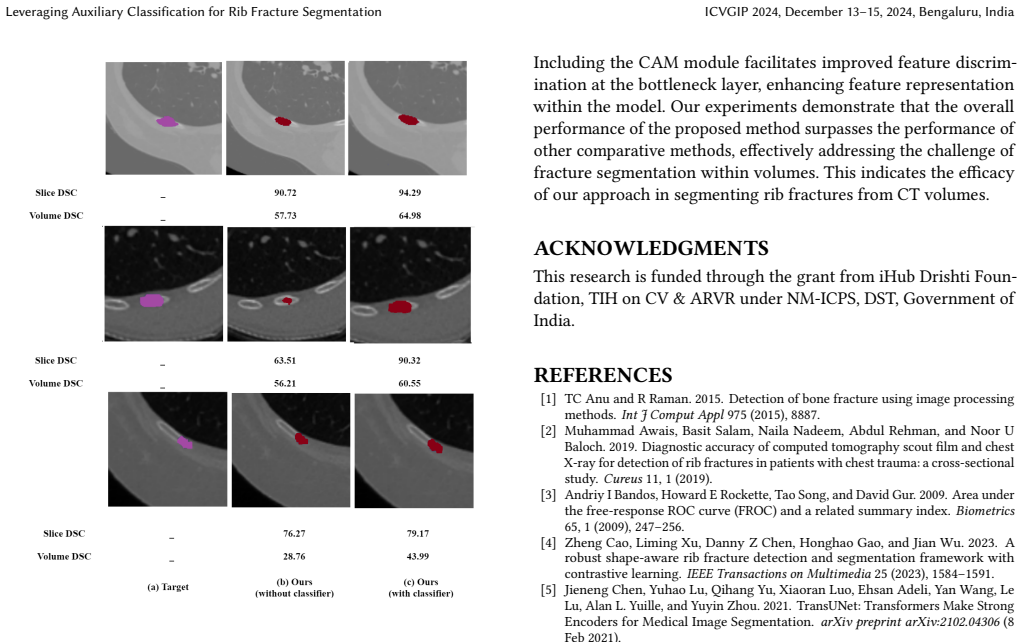
- The CAM maps shown in the paper are a by-product of the mechanism; a quantitative evaluation of their localization accuracy (for example, lesion-centric Dice between CAM and ground truth) would tell whether the classifier is genuinely highlighting fracture regions or merely correlated features.
- Because the no-classifier model wins at FROC level 0.5, the auxiliary task appears to trade away some high-sensitivity operating points; a testable extension is to fuse the two heads at inference or tune the classification-weight schedule to recover that sensitivity without losing the gains at levels 2, 4, and 8.
- The post-processing has three thresholds (probability, size, and bone intensity) that are tuned jointly in the paper, so an ablation isolating each threshold would show how much of the reported gain over baselines comes from the CAM module versus optimal threshold selection.
Signed reviews
Editorial analysis
A structured set of objections, weighed in public.
Referee Report
Summary. The paper proposes an auxiliary patch-level classification task with a class activation map (CAM) module at the bottleneck of a 3D U-Net to improve rib fracture segmentation on CT patches. The model is trained with a combined segmentation loss (focal + Dice) and binary cross-entropy classification loss, with a schedule that down-weights the classification loss over epochs. Post-processing applies probability, connected-component size, and bone-intensity thresholds. Experiments on the RibFrac dataset compare the method against several baselines (UNet, UNETR, TransUNet, Attention U-Net, FracNet) and report Dice similarity coefficient (DSC) and FROC scores. An ablation with and without the auxiliary classifier shows a 2.34-point DSC improvement and small FROC gains, with only 129 additional parameters.
Significance. If the reported gains are robust, the contribution is practically valuable: a nearly parameter-free auxiliary classifier (129 parameters) that yields consistent segmentation improvements over a strong U-Net baseline, and the CAM-based reweighting at the bottleneck is a simple and general mechanism. The ablation with versus without the classifier directly supports the central claim that the auxiliary task helps. However, the evaluation protocol has a load-bearing weakness: the paper does not state which data split is used for the reported results, and the post-processing thresholds appear to be selected on the same split that produces the headline numbers. This makes the absolute DSC/FROC values and the claimed 1.846% improvement over FracNet potentially optimistically biased. The core idea remains defensible and the issue is fixable, so the manuscript warrants major revision rather than rejection.
major comments (3)
- [Sec. 4.2 and Tables 1-2] The paper never states whether Table 1 reports results on the RibFrac validation split (80 CT scans) or the test split (160 CT scans). Section 4.2 only says model selection used minimum validation loss. More seriously, the 'Ours(with classifier)' row in Table 1 exactly matches the best row of the post-processing threshold sweep in Table 2 (probability 0.6, size 150, FROC 0.71/0.79/0.81/0.81/0.58, DSC 64.55). This indicates that the headline numbers are the selected maximum of a threshold search performed on the same data used for the final evaluation. To support the central claim, the authors must either report results on a held-out test split with thresholds fixed a priori, or explicitly describe a separate validation split used for threshold selection and report the corresponding test-set numbers. Without this, the absolute DSC/FROC values and the 1.846% improvement over FracNet may be optimistically biased.
- [Sec. 4.4 and Table 1] The ablation shows FROC improvements at levels 2, 4, and 8 of 0.02 absolute (0.79 vs 0.77, 0.81 vs 0.78, 0.81 vs 0.78), while the reported standard deviations for those metrics are 0.01 for the with-classifier model and up to 0.08 for the without-classifier model. The paper should report whether these differences are statistically significant across the three random initializations (e.g., paired tests or confidence intervals). The DSC improvement of 2.34 points is more compelling, but the FROC gains, which are part of the stated achievements, are marginal and may be within noise.
- [Sec. 3.3 and Table 2] The post-processing protocol fixes the bone threshold at 300 HU but sweeps the probability and size thresholds over a small grid, and the best row is reported. This is the same selection-bias issue as in the first comment. Additionally, the FROC values at the 0.5 false-positive level in Table 2 appear internally inconsistent: for probability threshold 0.4, the 0.5-level FROC is 0.81, equal to the level-8 value, while for other thresholds it drops to 0.57-0.59. The authors should verify these numbers and clarify whether the column labeled '0.5' refers to sensitivity at 0.5 false positives per volume or to a different metric.
minor comments (6)
- [Eq. (3)] Equation (3) uses the notation 𝐺𝑆(Wci) without a formal definition; please clarify that the 're-expansion' operation converts the channel-wise weights back to the spatial dimensions of e3i.
- [Sec. 3.1] The sentence 'we ensure equal patches for both fracture and non-fracture regions' is ambiguous: it could mean an equal number of patches or equal total volume; please rephrase for clarity.
- [Table 1] The table header lists FROC levels as '1 2 4 8 0.5', which is visually confusing because the 0.5 column appears after the 8 column. Please reorder the columns (e.g., 0.5, 1, 2, 4, 8) and add a footnote defining the FROC metric and the meaning of each column.
- [Sec. 4.2] The training description does not mention a learning-rate schedule, batch normalization details, or the exact stopping criterion beyond 'minimum validation loss'; these details are needed for reproducibility.
- [Fig. 5] The CAM visualization is qualitative; consider adding a quantitative measure (e.g., IoU between a thresholded CAM and the ground-truth fracture mask) to support the claim that CAM highlights fracture-related regions.
- [References] References [13] and [30] cite the same RibFrac challenge paper; please merge them to avoid duplication.
Circularity Check
Reported Dice/FROC are the selected best row of a post-processing threshold sweep, making the headline 1.846% gain partly fitted; the auxiliary-classifier mechanism itself is not circular.
-
fitted input called prediction
[Section 4.3/4.4, Tables 1 and 2]
"Table 2 analyzes the proposed model at different probability thresholds and size thresholds, described in the post-processing step, to reduce the false positives, and the bone threshold is kept at 300 HU. The improved performance is observed at size threshold 150 and probability threshold 0.6. [Table 1 row] Ours(with classifier): 0.71±0.03 0.79±0.01 0.81±0.00 0.81±0.00 0.58±0.03 64.55±0.45; [Table 2 row] 0.6 150: 0.71 0.79 0.81 0.81 0.58 64.55."
The exact row selected by the post-processing threshold sweep (0.6, 150) is identical to the row reported as the proposed model's final performance in Table 1. The paper never states that Table 1 was computed on a different split from Table 2, and the matching values indicate the same evaluation data. Thus the headline FROC/DSC numbers are not an independent prediction; they are the selected best of a grid search over probability and size thresholds on the evaluation set. The claimed 'overall dice score enhancement of 1.846% compared to the baseline models' is therefore partly fitted to the data used for reporting, and baselines are not documented as having received the same threshold tuning. This is a fitted post-processing parameter renamed as a predicted result.
full rationale
The only concrete circularity is in the evaluation protocol: the final reported metrics for the proposed model are the same values as the best row of the threshold sweep in Table 2, so the absolute FROC/DSC numbers are selected rather than independently predicted. This makes the numerical advantage over baselines partly an artifact of threshold fitting, especially since the paper does not state that baselines received equivalent threshold tuning. However, the central architectural claim—that an auxiliary patch-level classifier with a CAM module at the bottleneck improves rib fracture segmentation—is not circular by construction. The patch labels are derived from the same ground-truth masks used for the segmentation loss, but that is standard supervised multitask learning rather than a definitional equivalence. The CAM attention is trained with an auxiliary classification loss and evaluated against external RibFrac annotations; the segmentation head is still optimized with segmentation loss. No load-bearing self-citation chain or imported uniqueness theorem is present. The circularity is therefore partial and confined to the reported performance numbers, not to the derivation of the method itself.
Assumptions & free parameters
free parameters (6)
- alpha1, alpha2 (segmentation loss weights)
- gamma (focal loss parameter)
- theta(tau) (classification loss schedule)
- probability threshold =
0.6
- size threshold =
150
- bone threshold (HU) =
300
assumptions (3)
- domain assumption RibFrac ground-truth voxel masks are accurate enough to serve as labels for both the segmentation loss and the derived patch-level classification labels.
- domain assumption CT intensities within the window [-200, 1000] HU, with a bone threshold of 300 HU, capture the relevant bone and fracture signal.
- domain assumption The CAM formulation (GAP followed by linear layer and sigmoid re-weighting) provides a meaningful spatial prior for fracture locations.
Cite this review
Pith. "Pith review of Leveraging Auxiliary Classification for Rib Fracture Segmentation." pith.science (2026). https://pith.science/paper/C7SDZGWJ
@misc{pith2026241109283,
  author       = {Pith},
  title        = {Pith review of: Leveraging Auxiliary Classification for Rib Fracture Segmentation},
  year         = {2026},
  howpublished = {\url{https://pith.science/paper/C7SDZGWJ}},
  note         = {Machine review of arXiv:2411.09283}
}
read the original abstract
Thoracic trauma often results in rib fractures, which demand swift and accurate diagnosis for effective treatment. However, detecting these fractures on rib CT scans poses considerable challenges, involving the analysis of many image slices in sequence. Despite notable advancements in algorithms for automated fracture segmentation, the persisting challenges stem from the diverse shapes and sizes of these fractures. To address these issues, this study introduces a sophisticated deep-learning model with an auxiliary classification task designed to enhance the accuracy of rib fracture segmentation. The auxiliary classification task is crucial in distinguishing between fractured ribs and negative regions, encompassing non-fractured ribs and surrounding tissues, from the patches obtained from CT scans. By leveraging this auxiliary task, the model aims to improve feature representation at the bottleneck layer by highlighting the regions of interest. Experimental results on the RibFrac dataset demonstrate significant improvement in segmentation performance.
Figures


Figures from the paper (3 more)


Reference graph
Works this paper leans on
-
[1]
TC Anu and R Raman. 2015. Detection of bone fracture using image processing methods. Int J Comput Appl 975 (2015), 8887
work page 2015
-
[2]
Muhammad Awais, Basit Salam, Naila Nadeem, Abdul Rehman, and Noor U Baloch. 2019. Diagnostic accuracy of computed tomography scout film and chest X-ray for detection of rib fractures in patients with chest trauma: a cross-sectional study. Cureus 11, 1 (2019)
work page 2019
-
[3]
Andriy I Bandos, Howard E Rockette, Tao Song, and David Gur. 2009. Area under the free-response ROC curve (FROC) and a related summary index. Biometrics 65, 1 (2009), 247–256
work page 2009
-
[4]
Zheng Cao, Liming Xu, Danny Z Chen, Honghao Gao, and Jian Wu. 2023. A robust shape-aware rib fracture detection and segmentation framework with contrastive learning. IEEE Transactions on Multimedia 25 (2023), 1584–1591
work page 2023
-
[5]
Jieneng Chen, Yuhao Lu, Qihang Yu, Xiaoran Luo, Ehsan Adeli, Yan Wang, Le Lu, Alan L. Yuille, and Yuyin Zhou. 2021. TransUNet: Transformers Make Strong Encoders for Medical Image Segmentation. arXiv preprint arXiv:2102.04306 (8 Feb 2021)
arXiv 2021
-
[6]
Abdoulhossein Davoodabadi, Noshin Mosavibioki, Mohammad Mashayekhil, Hamidreza Gilasi, Esmail Abdorrahim Kashi, and Babak Haghpanah. 2022. Corre- lation of rib fracture patterns with abdominal solid organ injury: A retrospective observational cohort study. Chinese Journal of Traumatology 25, 01 (2022), 45–48
work page 2022
-
[7]
Bekir Nihat Dogrul, Ibrahim Kiliccalan, Ekrem Samet Asci, and Selim Can Peker
-
[8]
Alexey Dosovitskiy, Lucas Beyer, Alexander Kolesnikov, Dirk Weissenborn, Xiaohua Zhai, Thomas Unterthiner, Mostafa Dehghani, Matthias Minderer, Georg Heigold, Sylvain Gelly, and Jakob Uszkoreit. 2020. An Image is Worth 16x16 Words: Transformers for Image Recognition at Scale. arXiv preprint arXiv:2010.11929 (22 Oct 2020)
arXiv 2020
Show all 36 references
-
[9]
Ali Hatamizadeh, Yinda Tang, Vishwesh Nath, Donghan Yang, Andriy Myro- nenko, Bennett Landman, Holger R Roth, and Dong Xu. 2022. UNETR: Transform- ers for 3D Medical Image Segmentation. In Proceedings of the IEEE/CVF Winter Conference on Applications of Computer Vision . 574–584
2022
-
[10]
Kaiming He, Xiangyu Zhang, Shaoqing Ren, and Jian Sun. 2015. Delving Deep into Rectifiers: Surpassing Human-Level Performance on ImageNet Classification. In Proceedings of the IEEE International Conference on Computer Vision. 1026–1034
2015
-
[11]
Zilong Huang, Xinggang Wang, Jiasi Wang, Wenyu Liu, and Jingdong Wang
-
[12]
Liang Jin, Jiancheng Yang, Kaiming Kuang, Bingbing Ni, Yiyi Gao, Yingli Sun, Pan Gao, Weiling Ma, Mingyu Tan, Hui Kang, et al. 2020. Deep-learning-assisted detection and segmentation of rib fractures from CT scans: Development and validation of FracNet. EBioMedicine 62 (2020)
2020
-
[13]
Liang Jin, Jiancheng Yang, Kaiming Kuang, Bingbing Ni, Yiyi Gao, Yingli Sun, Pan Gao, Weiling Ma, Mingyu Tan, Hui Kang, Jiajun Chen, and Ming Li. 2020. Deep-Learning-Assisted Detection and Segmentation of Rib Fractures from CT Scans: Development and Validation of FracNet. eBio...
2020
-
[14]
Michelle Kim and James E Moore. 2020. Chest trauma: current recommendations for rib fractures, pneumothorax, and other injuries.Current anesthesiology reports 10 (2020), 61–68
2020
-
[15]
Diederik P Kingma and Max Welling. 2013. Auto-encoding variational bayes. arXiv preprint arXiv:1312.6114 (2013)
2013 arXiv
-
[16]
Kevin Kuo and Amie M Kim. 2019. Rib fracture. (2019)
2019
-
[17]
Jiameng Liu, Zhiming Cui, Yuhang Sun, Caiwen Jiang, Zirong Chen, Hao Yang, Yuyao Zhang, Dijia Wu, and Dinggang Shen. 2021. Multi-scale segmentation network for rib fracture classification from CT images. In Machine Learning in Medical Imaging: 12th International Workshop, MLMI...
2021
-
[18]
A Myronenko. 2018. 3D MRI brain tumor segmentation using autoencoder regularization. Brainlesion Glioma Mult. Scler. Stroke Trauma. Brain Inj.-BrainLes 2019 (2018), 11384
2018
-
[19]
Anastasia Oikonomou and Panos Prassopoulos. 2011. CT imaging of blunt chest trauma. Insights into imaging 2 (2011), 281–295
2011
-
[20]
Ozan Oktay, Jo Schlemper, Loic Le Folgoc, Matthew Lee, Mattias Heinrich, Kazu- nari Misawa, Kensaku Mori, Steven McDonagh, Nils Y Hammerla, Bernhard Kainz, and Ben Glocker. 2018. Attention U-Net: Learning where to look for the pancreas. arXiv preprint arXiv:1804.03999 (11 Apr 2018)
2018 arXiv
-
[21]
Samantha Phan, Diego Torrejon, Jordan Furseth, Erin Mee, and Christine Lus- combe. 2023. Exploiting weak supervision to facilitate segmentation, classifica- tion, and analysis of microplastics (< 100 𝜇m) using Raman microspectroscopy images. Science of the Total Environment 88...
2023
-
[22]
Olaf Ronneberger, Philipp Fischer, and Thomas Brox. 2015. U-net: Convolu- tional networks for biomedical image segmentation. In Medical image computing and computer-assisted intervention–MICCAI 2015: 18th international conference, Munich, Germany, October 5-9, 2015, proceeding...
2015
-
[23]
Holger R Roth, Hirohisa Oda, Xiangrong Zhou, Natsuki Shimizu, Ying Yang, Yuichiro Hayashi, Masahiro Oda, Michitaka Fujiwara, Kazunari Misawa, and Kensaku Mori. 2018. An application of cascaded 3D fully convolutional networks for medical image segmentation. Computerized Medical...
2018
-
[24]
Darshan D Ruikar, KC Santosh, and Ravindra S Hegadi. 2019. Segmentation and analysis of CT images for bone fracture detection and labeling. In Medical Imaging. CRC Press, 130–154
2019
-
[25]
Mark R Sochor, Paul Webber, Brian Bednarski, and Stewart C Wang. 2003. 3D CT imaging versus plain X-ray in diagnosis of rib fractures in lateral impact crashes. In Annual Proceedings/Association for the Advancement of Automotive Medicine , Vol. 47. Association for the Advancem...
2003
-
[26]
Sanghyun Woo, Jongchan Park, Joon-Young Lee, and In So Kweon. 2018. CBAM: Convolutional Block Attention Module. InProceedings of the European Conference on Computer Vision (ECCV) . 3–19
2018
-
[27]
Mingxiang Wu, Zhizhong Chai, Guangwu Qian, Huangjing Lin, Qiong Wang, Liansheng Wang, and Hao Chen. 2021. Development and evaluation of a deep learning algorithm for rib segmentation and fracture detection from multicenter chest CT images. Radiology: Artificial Intelligence 3,...
2021
-
[28]
Wenyu Xing, Zhibin Zhu, Dongni Hou, Yaoting Yue, Fei Dai, Yifang Li, Lin Tong, Yuanlin Song, and Dean Ta. 2022. CM-SegNet: A deep learning-based automatic segmentation approach for medical images by combining convolution and multilayer perceptron. Computers in Biology and Medi...
2022
-
[29]
Jiancheng Yang, Rui Shi, Liang Jin, Xiaoyang Huang, Kaiming Kuang, Donglai Wei, Shixuan Gu, Jianying Liu, Pengfei Liu, Zhizhong Chai, et al . 2024. Deep Rib Fracture Instance Segmentation and Classification from CT on the RibFrac Challenge. arXiv preprint arXiv:2402.09372 (2024)
2024 arXiv
-
[30]
Jiancheng Yang, Rui Shi, Liang Jin, Xiaoyang Huang, Kaiming Kuang, Donglai Wei, Shixuan Gu, Jianying Liu, Pengfei Liu, Zhizhong Chai, Yongjie Xiao, Hao Chen, Liming Xu, Bang Du, Xiangyi Yan, Hao Tang, Adam Alessio, Gregory Holste, Jiapeng Zhang, Xiaoming Wang, Jianye He, Lixua...
2024
-
[31]
Y. Yu, Q. Jin, C. Wang, and Y. Zhang. 2012. Fuzzy clustering-based active contour model with level set evolution for image segmentation. Information Sciences 193 (2012), 82–97. https://doi.org/10.1016/j.ins.2012.01.024
2012 doi
-
[32]
Bolei Zhou, Aditya Khosla, Agata Lapedriza, Aude Oliva, and Antonio Torralba
-
[33]
Zongwei Zhou, Md Mahfuzur Rahman Siddiquee, Nima Tajbakhsh, and Jianming Liang. 2018. Unet++: A nested u-net architecture for medical image segmentation. In Deep Learning in Medical Image Analysis and Multimodal Learning for Clinical Decision Support: 4th International Worksho...
2018
-
[2016]
In Proceedings of the IEEE conference on computer vision and pattern recognition
Learning deep features for discriminative localization. In Proceedings of the IEEE conference on computer vision and pattern recognition . 2921–2929
-
[2018]
In Proceedings of the IEEE conference on computer vision and pattern recognition
Weakly-supervised semantic segmentation network with deep seeded region growing. In Proceedings of the IEEE conference on computer vision and pattern recognition. 7014–7023
-
[2020]
Chinese journal of traumatology 23, 03 (2020), 125–138
Blunt trauma related chest wall and pulmonary injuries: An overview. Chinese journal of traumatology 23, 03 (2020), 125–138
2020
Reviewed August 12, 2026 · model on record in the stance chip above.
Discussion (0). Continue with ORCID to comment.