REVIEW 3 major objections 7 minor 44 references
Intuitive Axial Augmentation Using Polar-Sine-Based Piecewise Distortion for Medical Slice-Wise Segmentation
T0 review · 3 major / 7 minor · reviewed 2026-08-11 · deepseek-v4-flash
Pith's one-line read The paper claims that a polar-sine-based piecewise affine warp of axial CT and MR slices simulates patient posture change during scans and, without extra data, raises segmentation accuracy across multiple model families.
desk verdict New distortion-based augmentation with a plausible clinical motivation, but the empirical case is undercut by validation-set parameter selection and rotation-only baselines. read the letter →
The pith
A machine-rendered reading of the paper's core claim, the machinery that carries it, and where it could break.
The reading
What carries the argument
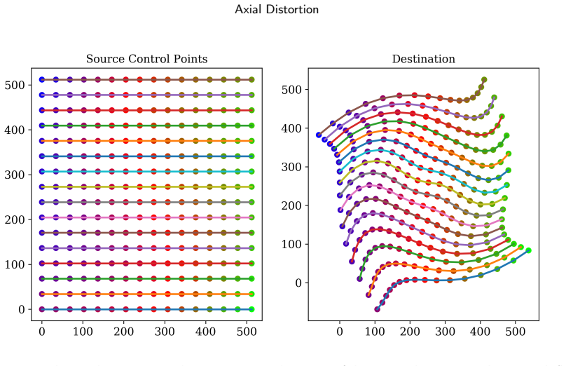
The central object is the polar-sine control-point displacement: a regular grid of control points is converted to polar coordinates around the slice center, and each point's angular coordinate is shifted by a sine function of its radius, $\vartheta_{\mathrm{new}} = \vartheta_{\mathrm{map}} + \frac{\pi}{8} a \sin\!\left(\frac{r_{\mathrm{map}}}{\delta} 2 f \pi\right)$, with per-sample random $a$ and $f$ controlled by $A$ and $\omega$. Because radial distances from the center are preserved, regions near the body surface move more than deep structures, which the authors argue matches how posture changes deform the body. The displaced grid is triangulated and each triangle is warped affinely to produce the augmented slice, with the grid density $\delta$ controlling fidelity. Two supporting mechanisms carry the robustness claims: a geometric model built from DICOM fields (reconstruction diameter, table height, pixel spacing, image position) masks out the scan table, and a similarity-guided search correlates SIFT/ORB matching counts with downstream accuracy to choose $A$ and $\omega$ without neural-network training.
What would settle it
A direct test would compare PSBPD-warped slices against true repeat scans of the same patient: if the SIFT/ORB similarity between a warped slice and the real re-scan is not higher than that between a rotation-only augmented slice and the real re-scan, the physical-realism premise is not supported. A complementary check is to train with PSBPD and with an equally strong but anatomically random warp; if accuracy is matched, the gain is a regularizer rather than realism.
Extended reading notes
Core claim
The discovery the authors are trying to establish is that posture uncertainty during a radiologic scan is a usable source of training variation, and that a polar-sine piecewise affine distortion can synthesize it cheaply. In the displacement rule, each control point keeps its radius but its polar angle becomes $\vartheta_{\mathrm{new}} = \vartheta_{\mathrm{map}} + \frac{\pi}{8} a \sin\!\left(\frac{r_{\mathrm{map}}}{\delta} 2 f \pi\right)$, with amplitude $a$ and frequency $f$ drawn randomly per sample from ranges set by $A$ and $\omega$. The displaced grid is triangulated and applied as a piecewise affine warp to the whole slice, so the paper's evidence is that this warp raises mDice on the private gastric dataset for seven model families and raises class-average Dice on CT-ORG and BraTS over rotation-only augmentation, while preserving enough feature-matching similarity that clinicians would still recognize the anatomy. The authors also claim that the similarity-guided parameter range found without training predicts the intensity range that helps neural networks most, which would spare users expensive end-to-end tuning.
Load-bearing premise
The load-bearing premise is that warping an axial slice along sine-shaped rays produces images that resemble real re-scans of the same patient under posture change; the paper does not test this against actual same-subject multi-posture scans.
Editorial extensions
If this is right
- As a plug-in preprocessing step before normalization, PSBPD improves mDice for ResNet50, ConvNeXt, SegNeXt, MAE, Poolformer, Segformer, and Swin Transformer V2 on the gastric cancer dataset, and for MedNext and SwinUMamba on public CT and MR datasets.
- The method works with as little as one annotated axial slice per CT scan, so it addresses the annotation-cost bottleneck in medical imaging.
- The similarity-guided search identifies a usable intensity range (around $A=6$, $\omega=2$) without training, and the accuracy surface is smooth, so the method is forgiving to parameter choice.
- The scan-table removal step is optional: it helps most models but hurts MAE, because masked reconstruction benefits from the extra pixels.
- Since the augmentation is model-agnostic and the helper modules are non-neural, it can be added to existing training pipelines with minimal computational overhead, measured at sub-millisecond to a few milliseconds per slice.
Reading between the lines
- A direct validity check the paper leaves open is whether a PSBPD-warped slice actually resembles a real re-scan of the same patient under posture change; comparing warped slices with true repeat scans would settle the physical claim and could calibrate the intensity parameters.
- Part of the accuracy gain may come from the warp acting as a strong geometric regularizer rather than from anatomical realism; comparing PSBPD against an equally strong but anatomically random warp, matched in displacement statistics, would separate these explanations.
- The per-slice independent warping ignores slice-to-slice coherence, so sampling one distortion field and applying it consistently across neighboring axial slices is a natural 3D extension that could improve volumetric segmentation.
- The similarity-guided tuning recipe is portable: any geometric augmentation whose pre/post images can be feature-matched could reuse the SIFT/ORB screening to find a safe intensity range, which would make the tuning step cheaper for other augmentations.
Editorial analysis
A structured set of objections, weighed in public.
Referee Report
Summary. The paper proposes Polar-Sine-Based Piecewise Distortion (PSBPD), an augmentation method for axial medical slices that distorts a slice in polar coordinates using a sinusoidal ray mapping, motivated by posture uncertainty during CT/MR scanning. It also introduces a DICOM-metadata-driven scan table removal module and a SIFT/ORB similarity-guided hyperparameter search for the distortion amplitude and frequency. Experiments are reported on the CT-ORG and BraTS public datasets and on a private gastric cancer CT dataset, across convolution-, transformer-, and mamba-based segmentation models, with Dice/Recall/Precision improvements claimed relative to rotation-only or no augmentation.
Significance. If the empirical claims are reliable, PSBPD is a plug-and-play, model-agnostic augmentation that improves slice-wise segmentation in low-annotation settings and has an interpretability rationale that clinicians may find more acceptable than black-box augmentations. The paper has concrete strengths: it provides a public code repository, evaluates on two public benchmarks plus a clinically collected private dataset, and introduces non-adaptive modules with a computational overhead analysis showing that preprocessing is not a training bottleneck. The main limitation is that the evaluation protocol does not clearly separate hyperparameter selection from test reporting and does not provide seed-averaged uncertainty, so the size and even the direction of the reported gains are not yet firmly established.
major comments (3)
- [Section 3.4, Algorithm 1; Tables 4-6] The central claim that the method 'improves accuracy' is not yet substantiated because the evaluation does not state a clean train/validation/test split. Algorithm 1 selects the hyperparameters (A, omega) as k* = argmin_k L_k on D_val, and the manuscript never says explicitly that the Dice values in Tables 4-6 are measured on a test partition that was not used to choose k*. If the reported numbers come from the same D_val used for selection, the improvements are inflated by selection bias. The problem is compounded by the absence of any multiple-seed results or standard deviations. For example, in Table 4 the bladder Dice jumps from 3.21 to 48.55 for MedNext, a change far larger than one would expect from an augmentation effect without seed-averaged error bars. The authors should report an explicit data split, a fixed rule for choosing A and omega, and mean +/- std over at least three random seeds.
- [Section 4.3 and Section 4.5, Fig. 8 and Fig. 11] The similarity-guided parameter search is not validated as a predictor of segmentation accuracy. Section 4.3 reports Pearson correlations of -0.625 and -0.618 between SIFT/ORB matching counts and accuracy and states that similarity begins to plummet at a ~= 3, f ~= 1.5. However, Section 4.5 states that accuracy only starts to decline at A > 7, omega > 3. These two thresholds are not consistent, so it is unclear that the similarity metric identifies the same operating region as the actual accuracy landscape. Moreover, the correlation is computed on the same parameter grid that Algorithm 1 uses to select hyperparameters, so it does not demonstrate predictive value for a new dataset. A concrete fix is to evaluate the similarity-accuracy correlation on a separate grid or a holdout dataset and report the rank correlation between predicted and actual optimal parameters.
- [Section 3.2.2, Eqs. (2)-(4); Section 1] The premise that sinusoidal polar-coordinate distortion simulates genuine posture variation is load-bearing for the paper's interpretability and clinical-acceptance claims, but it is never directly validated. The paper shows that the distorted images are continuous and visually plausible, and that downstream accuracy often improves, but it does not compare the augmented samples to actual repeated scans of the same subject under different postures, nor does it provide a clinical/radiological evaluation of the generated slices. Without such evidence, the claim that the method 'simulates uncertain postures' is an assumption, not an established property. The authors should either provide a validation experiment with paired same-subject scans or soften the interpretability claim accordingly.
minor comments (7)
- [Section 3.2.2, Eq. (3)] The text says 'with theta as the pole' where it should presumably refer to the pole coordinate Theta; Eq. (3) also uses inconsistent notation for array height and width (S_h/S_w vs. H/W).
- [Section 3.4, Algorithm 1] The similarity threshold t ~= 90% is introduced without justification or a sensitivity analysis; it is unclear how the size of K10% changes with t and how sensitive the selected hyperparameters are to this choice.
- [Table 6, Poolformer row] The text says that most frameworks improve 'excluding the Poolformer model,' but Table 6 shows that Poolformer's 'Ours' column is worse than both 'rotate +/-180' and 'w/o Table Removal' on mIoU and mDice; this exception should be acknowledged explicitly and discussed.
- [Section 4.3, Fig. 8] The caption and text refer to 'SIRF' in one place, which appears to be a typo for SIFT; please make the feature-descriptor names consistent.
- [Section 4.2] The statement that 'there are currently two main architectures, convolutional structure and Transformer structure' is outdated given that the paper itself evaluates SwinUMamba, a state-space model; the sentence should be revised to reflect the broader set of modern architectures.
- [Section 3.3, Eq. (12)] The formatting of the conditional 'if ||p - theta2|| > lambda2: p = epsilon' is unclear; please use proper subscripts and explain what happens to pixels outside the valid mask.
- [References and Section 4.4] The text mentions SA-Med2D in Section 4.4 but the dataset description in Section 4.1 lists only CT-ORG and BraTS; please clarify the relationship between SA-Med2D and the CT-ORG results reported in Table 4.
Circularity Check
No significant circularity: the accuracy gains are empirical, the distortion is geometrically defined, and no load-bearing self-citations or in-construction predictions were found.
full rationale
The paper's central claim is that PSBPD improves segmentation accuracy; this is supported by experiments on external benchmarks (CT-ORG, BraTS) and a private dataset across seven architectures. The distortion is defined geometrically via polar-coordinate sine warp (Eqs. 1-9), and the reported accuracy numbers are measured after training, not algebraically derived from the method's definition. Hyperparameter selection in Algorithm 1 uses validation loss to choose A and omega, which is standard model selection; even if the same validation split were reused in the tables, that would be selection bias rather than circularity because the reported Dice is not forced by the argmin. The similarity-guide claim is validated empirically by correlating SIFT/ORB match counts with measured segmentation accuracy (Fig. 8), not by assuming the correlation, so the proxy is independently evidence-backed. The paper contains no load-bearing self-citations: it cites its own GitHub for code and a preprint, but no prior theoretical result is imported to justify the method. The premise that sinusoidal distortion mimics patient posture is a clinical assumption, not a result derived from the target outcome; whether it holds is testable but not circular. The derivation chain is therefore self-contained, and the empirical claims are externally falsifiable.
Assumptions & free parameters
free parameters (4)
- A (distortion amplitude) =
A about 6 (best on validation, Fig. 11)
- omega (angular frequency) =
omega about 2 (best on validation, Fig. 11)
- delta (control point grid density) =
delta >= 16 recommended
- Similarity threshold t =
t about 90%
assumptions (4)
- domain assumption Sinusoidal polar-coordinate distortion of an axial slice generates anatomically plausible images resembling real patient posture variation.
- domain assumption SIFT/ORB feature-match count is a valid proxy for the learnability of augmented samples.
- standard math Delaunay triangulation and piecewise affine mapping preserve the continuity of anatomical structures.
- domain assumption DICOM metadata values (Table Height, Image Position, Pixel Spacing, Reconstruction Diameter) are accurate enough to geometrically locate the CT table.
Cite this review
Pith. "Pith review of Intuitive Axial Augmentation Using Polar-Sine-Based Piecewise Distortion for Medical Slice-Wise Segmentation." pith.science (2026). https://pith.science/paper/SOZN7XKK
@misc{pith2026241203352,
  author       = {Pith},
  title        = {Pith review of: Intuitive Axial Augmentation Using Polar-Sine-Based Piecewise Distortion for Medical Slice-Wise Segmentation},
  year         = {2026},
  howpublished = {\url{https://pith.science/paper/SOZN7XKK}},
  note         = {Machine review of arXiv:2412.03352}
}
read the original abstract
Most data-driven models for medical image analysis rely on universal augmentations to improve accuracy. Experimental evidence has confirmed their effectiveness, but the unclear mechanism underlying them poses a barrier to the widespread acceptance and trust in such methods within the medical community. We revisit and acknowledge the unique characteristics of medical images apart from traditional digital images, and consequently, proposed a medical-specific augmentation algorithm that is more elastic and aligns well with radiology scan procedure. The method performs piecewise affine with sinusoidal distorted ray according to radius on polar coordinates, thus simulating uncertain postures of human lying flat on the scanning table. Our method could generate human visceral distribution without affecting the fundamental relative position on axial plane. Two non-adaptive algorithms, namely Meta-based Scan Table Removal and Similarity-Guided Parameter Search, are introduced to bolster robustness of our augmentation method. In contrast to other methodologies, our method is highlighted for its intuitive design and ease of understanding for medical professionals, thereby enhancing its applicability in clinical scenarios. Experiments show our method improves accuracy with two modality across multiple famous segmentation frameworks without requiring more data samples. Our preview code is available in: https://github.com/MGAMZ/PSBPD.
Figures


Figures from the paper (9 more)


Reference graph
Works this paper leans on
-
[1]
Bali, H., Luitel, A., Upadhyaya, C., 2023. Artifacts among Cone Beam Computed Tomography Images of Patients of Department of Oral Medicine and Radiology in a Tertiary Care Centre: A Descriptive Cross-sectional Study. JNMA J Nepal Med Assoc 61, 18–22. doi:10.31729/jnma.7949
-
[2]
2d object recognition: a comparative analysis of sift, surf and orb feature descriptors
Bansal, M., Kumar, M., Kumar, M., 2021. 2d object recognition: a comparative analysis of sift, surf and orb feature descriptors. Multimedia Tools and Applications 80, 18839–18857. URL: https://doi.org/10.1007/s11042-021-10646-0, doi:10.1007/ s11042-021-10646-0
-
[3]
Deepconvolutionalneuralnetworksforbrainimage analysis on magnetic resonance imaging: a review
Bernal,J.,Kushibar,K.,Asfaw,D.S.,Valverde,S.,Oliver,A.,Martí,R.,Lladó,X.,2019. Deepconvolutionalneuralnetworksforbrainimage analysis on magnetic resonance imaging: a review. Artificial Intelligence in Medicine 95, 64–81. URL:https://www.sciencedirect. com/science/article/pii/S0933365716305206, doi:https://doi.org/10.1016/j.artmed.2018.08.008
-
[4]
Measuring image similarity: an overview of some useful applications
Chalom, E., Asa, E., Biton, E., 2013. Measuring image similarity: an overview of some useful applications. IEEE Instrumentation & Measurement Magazine 16, 24–28. doi:10.1109/MIM.2013.6417053
-
[5]
Chen,Y.W.,Shih,C.T.,Lin,H.H.,Chuang,K.S.,2016.Physicalmodel-basedcontrastenhancementofcomputedtomographyimages:Contrast enhancementofcomputedtomography,in:2016IEEE16thInternationalConferenceonBioinformaticsandBioengineering(BIBE),pp.238–
work page 2016
-
[6]
A review of medical image data augmentation techniques for deep learning applications
Chlap, P., Min, H., Vandenberg, N., Dowling, J., Holloway, L., Haworth, A., 2021. A review of medical image data augmentation techniques for deep learning applications. Journal of medical imaging and radiation oncology 65, 545–563. doi:10.1111/1754-9485.13261. pMID: 34145766
arXiv 2021
-
[7]
Longitudinal image registration with temporally-dependent image similarity measure
Csapo, I., Davis, B., Shi, Y., Sanchez, M., Styner, M., Niethammer, M., 2013. Longitudinal image registration with temporally-dependent image similarity measure. IEEE Transactions on Medical Imaging 32, 1939–1951. doi:10.1109/TMI.2013.2269814
arXiv 2013
-
[8]
Areviewondelaunaytriangulationwithapplicationoncomputervision
Dinas,S.,Bañón,J.,2014. Areviewondelaunaytriangulationwithapplicationoncomputervision. IJCSE-InternationalJournalofComputer Science and Engineering 3, 9–18
work page 2014
Show all 44 references
-
[9]
Overcomemedicalimagedatascarcitybydataaugmentationtechniques:Areview,in:2022 International Conference on Microelectronics (ICM), pp
ElJiani,L.,ElFilali,S.,Benlahmer,E.H.,2022. Overcomemedicalimagedatascarcitybydataaugmentationtechniques:Areview,in:2022 International Conference on Microelectronics (ICM), pp. 21–24. doi:10.1109/ICM56065.2022.10005544
2022
-
[10]
Data augmentation for medical imaging: A systematic literature review
Garcea, F., Serra, A., Lamberti, F., Morra, L., 2023. Data augmentation for medical imaging: A systematic literature review. Computers in Biology and Medicine 152, 106391. URL: https://www.sciencedirect.com/science/article/pii/S001048252201099X, doi:https://doi.org/10.1016/j.c...
2023
-
[11]
Using dicommetadataforradiologicalimageseriescategorization:afeasibilitystudyonlargeclinicalbrainmridatasets
Gauriau, R., Bridge, C., Chen, L., Kitamura, F., Tenenholtz, N.A., Kirsch, J.E., Andriole, K.P., Michalski, M.H., Bizzo, B.C., 2020. Using dicommetadataforradiologicalimageseriescategorization:afeasibilitystudyonlargeclinicalbrainmridatasets. JournalofDigitalImaging 33, 747–76...
2020 doi
-
[12]
Two-stage unsupervised learning method for affine and deformable medical image registration, in: 2019 IEEE International Conference on Image Processing (ICIP), pp
Gu, D., Liu, G., Tian, J., Zhan, Q., 2019. Two-stage unsupervised learning method for affine and deformable medical image registration, in: 2019 IEEE International Conference on Image Processing (ICIP), pp. 1332–1336. doi:10.1109/ICIP.2019.8803794
2019
-
[13]
The virtues of interpretable medical artificial intelligence
Hatherley, J., Sparrow, R., Howard, M., 2022. The virtues of interpretable medical artificial intelligence. Cambridge Quarterly of Healthcare Ethics , 1–10doi:10.1017/S0963180122000305
2022 doi
-
[14]
Review on self supervised learning in medical image analysis, in: 2023 IEEE 7th Conference on Information and Communication Technology (CICT), pp
Kumari, N., Agrawal, S., 2023. Review on self supervised learning in medical image analysis, in: 2023 IEEE 7th Conference on Information and Communication Technology (CICT), pp. 1–6. doi:10.1109/CICT59886.2023.10455714
2023
-
[15]
Casper:Conventional ct database augmentation using deep learning based spectral ct images generation, in: 2020 15th IEEE International Conference on Signal Processing (ICSP), pp
Lartaud,P.J.,Rouchaud,A.,Dessouky,R.,Vlachomitrou,A.S.,Rouet,J.M.,Nempont,O.,Boussel,L.,Douek,P.,2020. Casper:Conventional ct database augmentation using deep learning based spectral ct images generation, in: 2020 15th IEEE International Conference on Signal Processing (ICSP),...
2020
-
[16]
Swin-umamba: Mamba-based unet with imagenet-based pretraining
Liu, J., Yang, H., Zhou, H.Y., Xi, Y., Yu, L., Yu, Y., Liang, Y., Shi, G., Zhang, S., Zheng, H., Wang, S., 2024. Swin-umamba: Mamba-based unet with imagenet-based pretraining. URL:https://arxiv.org/abs/2402.03302, arXiv:2402.03302
2024 arXiv
-
[17]
Mixing data augmentation with preserving foreground regions in medical image segmentation, in: 2023 IEEE 20th International Symposium on Biomedical Imaging (ISBI), pp
Liu, X., Ono, K., Bise, R., 2023. Mixing data augmentation with preserving foreground regions in medical image segmentation, in: 2023 IEEE 20th International Symposium on Biomedical Imaging (ISBI), pp. 1–5. doi:10.1109/ISBI53787.2023.10230495
2023
-
[18]
Object recognition from local scale-invariant features, in: Proceedings of the Seventh IEEE International Conference on Computer Vision, pp
Lowe, D., 1999. Object recognition from local scale-invariant features, in: Proceedings of the Seventh IEEE International Conference on Computer Vision, pp. 1150–1157 vol.2. doi:10.1109/ICCV.1999.790410
1999
-
[19]
Distinctive image features from scale-invariant keypoints
Lowe, D.G., 2004. Distinctive image features from scale-invariant keypoints. International Journal of Computer Vision 60, 91–110. URL: https://doi.org/10.1023/B:VISI.0000029664.99615.94, doi:10.1023/B:VISI.0000029664.99615.94
2004
-
[20]
Computed tomography imaging artifacts in the head and neck region: Pitfalls and solutions
Marshall, E.L., Ginat, D.T., Sammet, S., 2022. Computed tomography imaging artifacts in the head and neck region: Pitfalls and solutions. Neuroimaging Clinics of North America 32, 271–277. URL:https://www.sciencedirect.com/science/article/pii/ S1052514922000016, doi:https://do...
2022 doi
-
[21]
pydicom/pydicom: pydicom v2.4.4.https://doi.org/10.5281/zenodo.10385252
Mason, D., Scaramallion, Mrbean-bremen, Rhaxton, Suever, J., Vanessasaurus, Orfanos, D.P., Lemaitre, G., Panchal, A., Rothberg, A., Herrmann, M.D., Massich, J., Kerns, J., van Golen, K., Bridge, C., Robitaille, T., Biggs, S., Moloney, Shun-Shin, M., Clauss, C., 2023. pydicom/p...
2023 doi
-
[22]
The multimodal brain tumor image segmentation benchmark (brats)
Menze, B.H., Jakab, A., Bauer, S., Kalpathy-Cramer, J., Farahani, K., Kirby, J., Burren, Y., Porz, N., Slotboom, J., Wiest, R., Lanczi, L., Gerstner, E., Weber, M.A., Arbel, T., Avants, B.B., Ayache, N., Buendia, P., Collins, D.L., Cordier, N., Corso, J.J., Criminisi, A., Das,...
2015
-
[23]
A ct database for research, development and education: Concept and potential
Messmer, P., Matthews, F., Jacob, A.L., Kikinis, R., Regazzoni, P., Noser, H., 2007. A ct database for research, development and education: Concept and potential. Journal of Digital Imaging 20, 17–22. URL: https://doi.org/10.1007/s10278-006-0771-9, doi:10.1007/s10278-006-0771-9
2007 doi
-
[24]
Prospects of structural similarity index for medical image analysis
Mudeng, V., Kim, M., Choe, S.w., 2022. Prospects of structural similarity index for medical image analysis. Applied Sciences 12. URL: https://www.mdpi.com/2076-3417/12/8/3754, doi:10.3390/app12083754
2022 doi
-
[25]
sciencedirect.com/science/article/pii/S2590005622000911, doi:https://doi.org/10.1016/j.array.2022.100258
Mumuni,A.,Mumuni,F.,2022.Dataaugmentation:Acomprehensivesurveyofmodernapproaches.Array16,100258.URL: https://www. sciencedirect.com/science/article/pii/S2590005622000911, doi:https://doi.org/10.1016/j.array.2022.100258
2022
-
[26]
Applications of self-supervised learning to biomedical signals: A survey
Pup, F.D., Atzori, M., 2023. Applications of self-supervised learning to biomedical signals: A survey. IEEE Access 11, 144180–144203. doi:10.1109/ACCESS.2023.3344531
2023
-
[27]
Efficient unstructured mesh generation by means of delaunay triangulation and bowyer-watson algorithm
Rebay, S., 1993. Efficient unstructured mesh generation by means of delaunay triangulation and bowyer-watson algorithm. Journal of Computational Physics 106, 125–138. URL: https://www.sciencedirect.com/science/article/pii/S0021999183710971, doi:https://doi.org/10.1006/jcph.1993.1097
1993
-
[28]
Explainable ai (xai): Explained, in: 2023 IEEE Open Conference of Electrical, Electronic and Information Sciences (eStream), pp
Reddy, G.P., Kumar, Y.V.P., 2023. Explainable ai (xai): Explained, in: 2023 IEEE Open Conference of Electrical, Electronic and Information Sciences (eStream), pp. 1–6. doi:10.1109/eStream59056.2023.10134984
2023
-
[29]
Ct-org, a new dataset for multiple organ segmentation in computed tomography
Rister, B., Yi, D., Shivakumar, K., Nobashi, T., Rubin, D.L., 2020. Ct-org, a new dataset for multiple organ segmentation in computed tomography. Scientific Data 7, 381. URL:https://doi.org/10.1038/s41597-020-00715-8, doi:10.1038/s41597-020-00715-8
2020 doi
-
[30]
Roy, S., Koehler, G., Ulrich, C., Baumgartner, M., Petersen, J., Isensee, F., Jäger, P.F., Maier-Hein, K.H., 2023. Mednext: Transformer- driven scaling of convnets for medical image segmentation, in: Greenspan, H., Madabhushi, A., Mousavi, P., Salcudean, S., Duncan, J., Syeda-...
2023
-
[31]
Orb: An efficient alternative to sift or surf, in: 2011 International Conference on Computer Vision, pp
Rublee, E., Rabaud, V., Konolige, K., Bradski, G., 2011. Orb: An efficient alternative to sift or surf, in: 2011 International Conference on Computer Vision, pp. 2564–2571. doi:10.1109/ICCV.2011.6126544
2011
-
[32]
Transparencyofdeepneuralnetworksformedicalimageanalysis:Areview of interpretability methods
Salahuddin,Z.,Woodruff,H.C.,Chatterjee,A.,Lambin,P.,2021. Transparencyofdeepneuralnetworksformedicalimageanalysis:Areview of interpretability methods. URL:https://arxiv.org/abs/2111.02398, arXiv:2111.02398
2021 arXiv
-
[33]
Differentiableimagedataaugmentationanditsapplications:Asurvey
Shi,J.,Ghazzai,H.,Massoud,Y.,2024. Differentiableimagedataaugmentationanditsapplications:Asurvey. IEEETransactionsonPattern Analysis and Machine Intelligence 46, 1148–1164. doi:10.1109/TPAMI.2023.3330862
2024
-
[34]
Hsmix:Hardandsoftmixingdataaugmentationformedicalimagesegmentation
Sun,D.,Dornaika,F.,Barrena,N.,2025. Hsmix:Hardandsoftmixingdataaugmentationformedicalimagesegmentation. InformationFusion 115, 102741. URL: https://www.sciencedirect.com/science/article/pii/S1566253524005190, doi:https://doi.org/10. 1016/j.inffus.2024.102741
2025
-
[35]
(Eds.), 2012
Tack, D., Kalra, M.K., Gevenois, P.A. (Eds.), 2012. Image Quality in CT: Challenges and Perspectives. Springer Berlin Heidelberg, Berlin, Heidelberg. pp. 81–100. URL:https://doi.org/10.1007/174_2011_482, doi:10.1007/174_2011_482
2012 doi
-
[36]
Explainable artificial intelligence (xai) in deep learning-based medical image analysis
van der Velden, B.H., Kuijf, H.J., Gilhuijs, K.G., Viergever, M.A., 2022. Explainable artificial intelligence (xai) in deep learning-based medical image analysis. Medical Image Analysis 79, 102470. URL: https://www.sciencedirect.com/science/article/pii/ S1361841522001177, doi:...
2022
-
[37]
SciPy 1.0: Fundamental Algorithms for Scientific Computing in Python
Virtanen, P., Gommers, R., Oliphant, T.E., Haberland, M., Reddy, T., Cournapeau, D., Burovski, E., Peterson, P., Weckesser, W., Bright, J., van der Walt, S.J., Brett, M., Wilson, J., Millman, K.J., Mayorov, N., Nelson, A.R.J., Jones, E., Kern, R., Larson, E., Carey, C.J., Pola...
2020
-
[38]
Ethicsofcollectingandusinghealthcaredata
Wade,D.,2007. Ethicsofcollectingandusinghealthcaredata. BMJ(Clinicalresearched.)334,1330–1331. URL: https://doi.org/10. 1136/bmj.39247.679329.80, doi:10.1136/bmj.39247.679329.80. publisher: BMJ Publishing Group
2007
-
[39]
Xedgeconv: Leveraging graph convolutions for efficient, permutation- and rotation-invariant dense 3d medical image segmentation, in: Bekkers, E., Wolterink, J.M., Aviles-Rivero, A
Weihsbach, C., Hansen, L., Heinrich, M., 2022. Xedgeconv: Leveraging graph convolutions for efficient, permutation- and rotation-invariant dense 3d medical image segmentation, in: Bekkers, E., Wolterink, J.M., Aviles-Rivero, A. (Eds.), Proceedings of the First International Wo...
2022
-
[40]
Sa-med2d-20m dataset: Segment anything in 2d medical imaging with 20 million masks
Ye,J.,Cheng,J.,Chen,J.,Deng,Z.,Li,T.,Wang,H.,Su,Y.,Huang,Z.,Chen,J.,Jiang,L.,Sun,H.,Zhu,M.,Zhang,S.,He,J.,Qiao,Y.,2023. Sa-med2d-20m dataset: Segment anything in 2d medical imaging with 20 million masks. URL:https://arxiv.org/abs/2311.11969, arXiv:2311.11969
2023 arXiv
-
[41]
Deep long-tailed learning: A survey
Zhang, Y., Kang, B., Hooi, B., Yan, S., Feng, J., 2023a. Deep long-tailed learning: A survey. IEEE Transactions on Pattern Analysis and Machine Intelligence 45, 10795–10816. doi:10.1109/TPAMI.2023.3268118
2023
-
[42]
Affine image registration of arterial spin labeling mri using deep learning networks
Zhang, Z., Yang, H., Guo, Y., Bolo, N.R., Keshavan, M., DeRosa, E., Anderson, A.K., Alsop, D.C., Yin, L., Dai, W., 2023b. Affine image registration of arterial spin labeling mri using deep learning networks. NeuroImage 279, 120303. URL:https://www.sciencedirect. com/science/ar...
-
[43]
Spineregnet:Spineregistrationnetworkforvolumetric mr and ct image by the joint estimation of an affine-elastic deformation field
Zhao,L.,Pang,S.,Chen,Y.,Zhu,X.,Jiang,Z.,Su,Z.,Lu,H.,Zhou,Y.,Feng,Q.,2023. Spineregnet:Spineregistrationnetworkforvolumetric mr and ct image by the joint estimation of an affine-elastic deformation field. Medical Image Analysis 86, 102786. URL:https://www. sciencedirect.com/sci...
2023
-
[241]
doi:10.1109/BIBE.2016.39
2016 doi
Reviewed August 11, 2026 · model on record in the stance chip above.
Discussion (0). Continue with ORCID to comment.