REVIEW 2 major objections 8 minor 126 references
Fundus Image Quality Assessment and Enhancement: a Systematic Review
T0 review · 2 major / 8 minor · reviewed 2026-08-10 · deepseek-v4-flash
Pith's one-line read This review argues that assessing and enhancing fundus image quality should be treated as one problem, not two, and maps the algorithms, datasets, and deployment hurdles that follow.
desk verdict A useful integrated narrative review of fundus IQA and IQE, but the 'Systematic Review' label overpromises given no documented search protocol. read the letter →
The pith
A machine-rendered reading of the paper's core claim, the machinery that carries it, and where it could break.
The reading
What carries the argument
The organising device is a two-axis taxonomy: IQA methods are classified by whether they require a reference image (full-reference versus no-reference) and IQE methods by whether they are handcrafted or learning-based, with learning-based IQE further split into paired-data and unpaired-data training. Within this taxonomy the load-bearing technical objects are the numerical metrics (MSE, PSNR, SSIM and its variants; BRISQUE, NIQE, PIQE), the CycleGAN unpaired translation architecture with adversarial and cycle-consistency losses, and the imaging-prior models such as the dark channel prior adapted from dehazing to cataractous fundus images. These objects carry the argument because they show how assessment and enhancement share a common vocabulary: the same metrics used to score quality are used to evaluate enhancers, and the same degradation models used to synthesise training pairs for IQE are the distortions that IQA must detect. The taxonomy is what lets the review claim that IQA and IQE are one field rather than two.
What would settle it
A concrete check would be to run a documented, reproducible literature search (fixed databases, query strings, inclusion criteria) for fundus IQA and IQE papers and compare the resulting corpus with the tables in this review; if substantial clusters of published methods fall outside the taxonomy, or if the integrated-perspective premise fails because few papers actually combine IQA with IQE, the review’s central claims would need revision.
Extended reading notes
Core claim
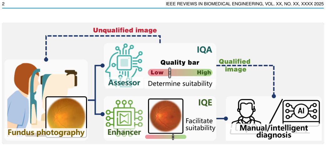
The central claim is that the field should be understood as a single pipeline with two cooperating functions. IQA determines whether a captured fundus image is usable; IQE restores images that are degraded but recoverable; together they decide the flow of accept, enhance, or recapture before diagnosis. The review systematises IQA by reference dependence (numerical and learning-based full-reference methods; numerical, end-to-end, and comprehensive no-reference methods) and IQE by technique origin (pixel processing, filtering, and statistical priors versus paired- and unpaired-data deep models), and it argues that the two literatures are converging: handcrafted priors such as the dark channel or green channel are being embedded into deep enhancers, quality sub-metrics are being predicted alongside overall grades for interpretability, and multi-task systems couple quality assessment with disease grading. On this view the main obstacles to clinical use are data availability, inconsistent quality labels across datasets, domain shift, and black-box behaviour, and the next advances will come from medical knowledge embedding, interference interpretation, information decoupling, personalisation, continual optimisation, and cross-modality generalisation.
Load-bearing premise
The review assumes that the papers it surveys, grouped under its full-reference versus no-reference and handcrafted versus learning-based taxonomy, are representative of the whole field, and that its qualitative claims about their strengths and weaknesses are accurate; because no reproducible search protocol is described, that coverage cannot be audited or independently reconstructed.
Editorial extensions
If this is right
- Treating IQA and IQE as one pipeline suggests that future systems should be built as closed loops, where an assessment module decides between accept, enhance, or recapture before diagnosis.
- Because both tasks share degradation models and datasets, progress in synthesizing realistic low-quality fundus images should improve both IQA training and IQE training at once.
- The review implies that handcrafted priors (dark channel, green channel, structure) will increasingly be embedded inside learning-based models to gain interpretability without giving up performance.
- Domain adaptation and domain generalization, not just benchmark accuracy, become the decisive criteria for whether an IQA and IQE method can be deployed in a new clinic.
- Multi-task models that couple quality assessment with disease grading or vessel segmentation are a natural end point of the integrated view, since they let quality features act as inductive bias for diagnosis.
Reading between the lines
- A testable extension the paper leaves implicit is an explicit ‘enhanceability’ label: instead of binary good or bad quality grades, datasets could carry a third label for images that are degraded but restorable, which would make the IQA–IQE loop directly trainable.
- The integrated perspective suggests that evaluation protocols for IQE should be standardised around downstream clinical tasks (segmentation, grading) rather than only around perceptual metrics, since the review repeatedly ties quality to diagnostic utility.
- One could check the review’s central claim empirically by measuring how often IQA and IQE methods are actually co-cited or jointly trained in the literature; if the two literatures barely overlap in practice, the ‘intimate relationship’ may be a design goal rather than a current fact.
- The emphasis on source-free domain adaptation and continual learning implies that post-deployment data, normally discarded, could become the primary resource for keeping quality models aligned with each clinic’s imaging conditions.
Signed reviews
Editorial analysis
A structured set of objections, weighed in public.
Referee Report
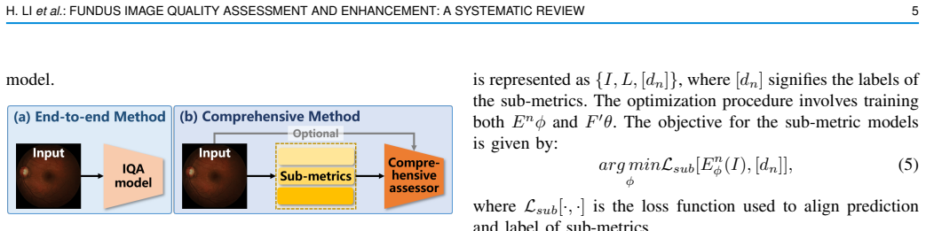
Summary. This manuscript is a review of fundus image quality assessment (IQA) and enhancement (IQE). It first describes the fundus camera imaging system and common sources of image degradation, then categorizes IQA methods into full-reference and no-reference approaches, subdividing NR-IQA into numerical metrics, end-to-end learning-based methods, and comprehensive methods that introduce intermediate sub-metrics. For IQE, the paper distinguishes handcrafted methods (pixel-based processing, image filtering, statistical priors) from learning-based methods (paired and unpaired data paradigms) and further separates real-world from synthesized paired data. The latter half of the paper discusses practical deployment challenges such as data availability, generalizability, and interpretability, reviews applications of IQA and IQE in diagnosis and segmentation, and closes with six future research directions. The paper is written as a tutorial-style survey with equations for standard metrics and enhancement operations, tables of representative models and datasets, and example figures illustrating degradations.
Significance. If the manuscript's scope claims are taken at face value, it fills a gap by co-reviewing fundus IQA and IQE, which earlier surveys treat separately. The tutorial material is solid: standard formulas for MSE, PSNR, SSIM, histogram equalization, CLAHE, gamma correction, the dark-channel model, and CycleGAN losses are presented in a self-contained way, and the tables of representative IQA/IQE models and datasets provide a useful quick reference. The discussion of synthesized versus real-world paired data, domain adaptation/generalization, and the use of IQA to guide enhancement is timely and practically relevant. However, the contribution is currently weakened by two structural issues: the 'systematic review' claim is not backed by a reproducible search protocol, and the promised integrated perspective is not fully realized because the IQA and IQE halves are largely separate surveys with only limited cross-referencing. These issues are fixable within the manuscript's scope and are the main reasons for requesting major revision.
major comments (2)
- [Title, Abstract, Section I] The manuscript is presented as a 'Systematic Review' in the title and abstract, but no systematic methodology is described anywhere. There is no statement of the databases queried, the search strings used, the inclusion/exclusion criteria, the screening procedure, or the time window of coverage, and no PRISMA-style flow diagram or study-selection protocol. The reference list also contains a substantial number of the authors' own prior publications (e.g., [14], [23], [24], [34], [68], [91], [101], [102]), which increases the risk that the corpus is a convenience sample rather than an auditable comprehensive survey. Because the paper's central value proposition is that it provides a thorough, comprehensive, integrated review, the absence of a reproducible corpus is a load-bearing omission. The authors should either add a complete search and selection protocol, or explicitly reframe the manuscript as a narrative review in the title and abstract.
- [Sections III-V] The Introduction and Abstract promise an integrated perspective grounded in the 'intimate relationship' between IQA and IQE, but the body of the paper does not deliver this synthesis. Sections III and IV are two largely independent surveys, and the connections between the two fields appear only in isolated remarks, such as Hou et al. [90] using IQA to guide enhancement and the quality-feature fusion examples in Section V.B. The paper would substantially strengthen its central claim if it made the interplay explicit, for example by mapping the sub-metrics used in comprehensive NR-IQA (Section III.B.3) to the degradation targets addressed by IQE methods, or by discussing how the same datasets and quality labels support both tasks. Without such a conceptual bridge, the integrative contribution asserted in the Introduction is not fully demonstrated.
minor comments (8)
- [Fig. 1 caption] The caption repeats '(b)' for the second and third images; the third image should be labeled '(c)'.
- [Section III.B.3] 'Biaswas et al. [41]' is a misspelling of Biswas; later in the same paragraph 'NF-IQA' should be 'NR-IQA'.
- [Section IV.A.1, Eq. (7) text] The prose definition 'CFD(l) = sum_{l=0}^{L-1} H(l)/N' is not the cumulative distribution function; it should be 'CDF(l) = sum_{i=0}^{l} H(i)/N'. The actual computation in Eq. (7) uses the correct lower limit, so only the defining sentence needs correction.
- [Fig. 6 caption] 'Scematics' should be 'Schematics'.
- [Section IV.B] There are two typos: 'funds image data' should be 'fundus image data', and 'paried' should be 'paired'.
- [Table IV] The column headings 'Instance #' and 'Quality #' are ambiguous; the table would be clearer if the headers were 'Images' and 'Quality levels'. The superscript footnote markers for ODIR and iSee are also placed awkwardly in the dataset-name column.
- [References] References [83] and [103] appear to be the same paper (Guo et al., 'Bridging synthetic and real images: a transferable and multiple consistency aided fundus image enhancement framework'); one should be removed and the in-text citations renumbered and checked.
- [Table I] The 'Aspect' entry for SSIM is listed as 'Pixel-level and structure fidelity', but Eq. (3) defines SSIM in terms of luminance, contrast, and structure; consider revising to 'Luminance, contrast, and structure fidelity' for consistency.
Circularity Check
No significant circularity: the review is descriptive and self-contained, with self-citations used as examples rather than as load-bearing justification.
full rationale
This manuscript is a literature review, not a derivation. It makes no empirical predictions, fits no parameters, and proves no theorems from assumptions. Its central claim, that an integrated treatment of fundus IQA and IQE is more logical than separate reviews, is an organizational judgment supported by the cited literature rather than by a chain of reasoning whose conclusion is equivalent to its premises. The paper borrows definitions (e.g., MSE, PSNR, SSIM, BRISQUE, NIQE, PIQE), standard imaging models (e.g., Eqs. 11-12), and prior algorithmic results from the cited works, but this borrowing is the normal substance of a review, not circularity. Self-citations are present (e.g., refs. [14], [23], [24], [34], [68], [91], [101], [102]) and are used to illustrate paradigms in IQA/IQE and deployment; the review does not invoke a self-cited theorem to forbid alternatives or to force a conclusion, and the cited works themselves contain independent algorithmic contributions. Even if the absence of a reproducible search protocol weakens the auditable 'systematic' claim, that is a methodological limitation about external validity, not a logical circularity. No step in the paper reduces by construction to its own inputs, no fitted quantity is renamed as a prediction, and no ansatz is smuggled in via self-citation. Accordingly, the circularity score is 0.
Assumptions & free parameters
assumptions (2)
- domain assumption Standard image quality metrics (MSE, PSNR, SSIM, NR-IQA metrics) are meaningful proxies for the clinical usefulness of fundus images.
- domain assumption The cited prior studies accurately describe their algorithms and reported performance.
Cite this review
Pith. "Pith review of Fundus Image Quality Assessment and Enhancement: a Systematic Review." pith.science (2026). https://pith.science/paper/S5VLLPKS
@misc{pith2026250111520,
  author       = {Pith},
  title        = {Pith review of: Fundus Image Quality Assessment and Enhancement: a Systematic Review},
  year         = {2026},
  howpublished = {\url{https://pith.science/paper/S5VLLPKS}},
  note         = {Machine review of arXiv:2501.11520}
}
read the original abstract
As an affordable and convenient eye scan, fundus photography holds the potential for preventing vision impairment, especially in resource-limited regions. However, fundus image degradation is common under intricate imaging environments, impacting following diagnosis and treatment. Consequently, image quality assessment (IQA) and enhancement (IQE) are essential for ensuring the clinical value and reliability of fundus images. While existing reviews offer some overview of this field, a comprehensive analysis of the interplay between IQA and IQE, along with their clinical deployment challenges, is lacking. This paper addresses this gap by providing a thorough review of fundus IQA and IQE algorithms, research advancements, and practical applications. We outline the fundamentals of the fundus photography imaging system and the associated interferences, and then systematically summarize the paradigms in fundus IQA and IQE. Furthermore, we discuss the practical challenges and solutions in deploying IQA and IQE, as well as offer insights into potential future research directions.
Figures


Figures from the paper (12 more)




Reference graph
Works this paper leans on
-
[14]
Deepfundus: a flow-cytometry-like image quality classi- fier for boosting the whole life cycle of medical artificial intelligence,
L. Liu, X. Wu, D. Lin, L. Zhao, M. Li, D. Yun, Z. Lin, J. Pang, L. Li, Y . Wuet al., “Deepfundus: a flow-cytometry-like image quality classi- fier for boosting the whole life cycle of medical artificial intelligence,” Cell Reports Medicine , vol. 4, no. 2, 2023
2023
-
[23]
An annotation-free restoration network for cataractous fundus images,
H. Li, H. Liu, Y . Hu, H. Fu, Y . Zhao, H. Miao, and J. Liu, “An annotation-free restoration network for cataractous fundus images,” IEEE Transactions on Medical Imaging , 2022
2022
-
[24]
A generic fundus image enhancement network boosted by frequency self- supervised representation learning,
H. Li, H. Liu, H. Fu, Y . Xu, H. Shu, K. Niu, Y . Hu, and J. Liu, “A generic fundus image enhancement network boosted by frequency self- supervised representation learning,” Medical Image Analysis , vol. 90, p. 102945, 2023
2023
-
[34]
Domain adapta- tive retinal image quality assessment with knowledge distillation using competitive teacher-student network,
Y . Lin, H. Li, H. Liu, H. Shu, Z. Li, Y . Hu, and J. Liu, “Domain adapta- tive retinal image quality assessment with knowledge distillation using competitive teacher-student network,” in 2023 IEEE 20th International Symposium on Biomedical Imaging (ISBI) . IEEE, 2023, pp. 1–5
2023
-
[68]
Structure-consistent restoration network for cataract fundus image enhancement,
H. Li, H. Liu, H. Fu, H. Shu, Y . Zhao, X. Luo, Y . Hu, and J. Liu, “Structure-consistent restoration network for cataract fundus image enhancement,” in Medical Image Computing and Computer Assisted Intervention–MICCAI 2022: 25th International Conference, Singapore, September 18–22, 2022, Proceedings, Part II . Springer, 2022, pp. 487–496
work page 2022
-
[91]
H. Li, Z. Lin, Z. Qiu, Z. Li, K. Niu, N. Guo, H. Fu, Y . Hu, and J. Liu, “Enhancing and adapting in the clinic: Source-free unsupervised do- main adaptation for medical image enhancement,” IEEE Transactions on Medical Imaging , vol. 43, no. 4, pp. 1323–1336, 2024
work page 2024
-
[101]
Degradation-invariant enhancement of fundus images via pyramid constraint network,
H. Liu, H. Li, H. Fu, R. Xiao, Y . Gao, Y . Hu, and J. Liu, “Degradation-invariant enhancement of fundus images via pyramid constraint network,” in Medical Image Computing and Computer Assisted Intervention–MICCAI 2022: 25th International Conference, Singapore, September 18–22, 2022, Proceedings, Part II . Springer, 2022, pp. 507–516
work page 2022
-
[102]
Restora- tion of cataract fundus images via unsupervised domain adaptation,
H. Li, H. Liu, Y . Hu, R. Higashita, Y . Zhao, H. Qi, and J. Liu, “Restora- tion of cataract fundus images via unsupervised domain adaptation,” in 2021 IEEE 18th International Symposium on Biomedical Imaging (ISBI). IEEE, 2021, pp. 516–520
work page 2021
-
[90]
A reference-free quality enhancement framework for low-quality fundus images,
Q. Hou, Y . Wang, L. Lan, P. Cao, J. Yang, X. Liu, M. Wang, Y . C. Tham, and O. R. Zaiane, “A reference-free quality enhancement framework for low-quality fundus images,” IEEE Transactions on Circuits and Systems for Video Technology , 2024
work page 2024
Show all 126 references
-
[1]
Blindness and vision impairment,
WHO, “Blindness and vision impairment,” https://www.who.int/ news-room/fact-sheets/detail/blindness-and-visual-impairment, 2023, 10 August 2023
2023
-
[2]
Artificial intelligence in ophthalmology: The path to the real-world clinic,
Z. Li, L. Wang, X. Wu, J. Jiang, W. Qiang, H. Xie, H. Zhou, S. Wu, Y . Shao, and W. Chen, “Artificial intelligence in ophthalmology: The path to the real-world clinic,” Cell Reports Medicine , vol. 4, no. 7, 2023
2023
-
[3]
Economic evaluation of combined population- based screening for multiple blindness-causing eye diseases in china: a cost-effectiveness analysis,
H. Liu, R. Li, Y . Zhang, K. Zhang, M. Yusufu, Y . Liu, D. Mou, X. Chen, J. Tian, H. Li et al. , “Economic evaluation of combined population- based screening for multiple blindness-causing eye diseases in china: a cost-effectiveness analysis,” The Lancet Global Health , vol. 1...
2023
-
[4]
Eye care in health systems: guide for action,
WHO, “Eye care in health systems: guide for action,” https://www. who.int/publications/i/item/9789240050068, 2022, 20 May 2022
2022
-
[5]
Image quality issues in teled- ermatology: a comparative analysis of artificial intelligence solutions,
K. Maier, L. Zaniolo, and O. Marques, “Image quality issues in teled- ermatology: a comparative analysis of artificial intelligence solutions,” Journal of the American Academy of Dermatology , vol. 87, no. 1, pp. 240–242, 2022
2022
-
[6]
Sensitivity and specificity of handheld fundus cameras for eye disease: a system- atic review and pooled analysis,
B. J. Palermo, S. L. D’Amico, B. Y . Kim, and C. J. Brady, “Sensitivity and specificity of handheld fundus cameras for eye disease: a system- atic review and pooled analysis,” Survey of Ophthalmology , vol. 67, no. 5, pp. 1531–1539, 2022
2022
-
[7]
Automated fundus image quality assessment in retinopathy of prematurity using deep convolutional neural networks,
A. S. Coyner, R. Swan, J. P. Campbell, S. Ostmo, J. M. Brown, J. Kalpathy-Cramer, S. J. Kim, K. E. Jonas, R. P. Chan, M. F. Chiang et al., “Automated fundus image quality assessment in retinopathy of prematurity using deep convolutional neural networks,” Ophthalmology retina, ...
2019
-
[8]
Quality assessment of non-mydriatic fundus photographs for glaucoma screening in primary healthcare centres: a real-world study,
Q. Chen, M. Zhou, Y . Cao, X. Zheng, H. Mao, C. Lei, W. Lin, J. Jiang, Y . Chen, D. Song et al., “Quality assessment of non-mydriatic fundus photographs for glaucoma screening in primary healthcare centres: a real-world study,” BMJ Open Ophthalmology, vol. 8, no. 1, p. e001493, 2023
2023
-
[9]
A human-centered evaluation of a deep learning system deployed in clinics for the detection of diabetic retinopathy,
E. Beede, E. Baylor, F. Hersch, A. Iurchenko, L. Wilcox, P. Ruamvi- boonsuk, and L. M. Vardoulakis, “A human-centered evaluation of a deep learning system deployed in clinics for the detection of diabetic retinopathy,” in Proceedings of the 2020 CHI conference on human factors...
2020
-
[10]
Understanding how fundus image quality degradation affects cnn- based diagnosis,
H. Liu, H. Li, X. Wang, H. Li, M. Ou, L. Hao, Y . Hu, and J. Liu, “Understanding how fundus image quality degradation affects cnn- based diagnosis,” in 2022 44th Annual International Conference of the IEEE Engineering in Medicine & Biology Society (EMBC) . IEEE, 2022, pp. 438–442
2022
-
[11]
Feasibility and clinical utility of handheld fundus cameras for retinal imaging,
S. Das, H. J. Kuht, I. De Silva, S. S. Deol, L. Osman, J. Burns, N. Sarvananthan, U. Sarodia, B. Kapoor, T. Islam et al. , “Feasibility and clinical utility of handheld fundus cameras for retinal imaging,” Eye, vol. 37, no. 2, pp. 274–279, 2023
2023
-
[12]
Fundus image quality as- sessment: survey, challenges, and future scope,
A. Raj, A. K. Tiwari, and M. G. Martini, “Fundus image quality as- sessment: survey, challenges, and future scope,” IET Image Processing, vol. 13, no. 8, pp. 1211–1224, 2019
2019
-
[13]
Computational single fundus image restoration techniques: a review,
S. Zhang, C. A. Webers, and T. T. Berendschot, “Computational single fundus image restoration techniques: a review,” Frontiers in Ophthalmology, vol. 4, p. 1332197, 2024
2024
-
[15]
Applications of deep learning in fundus images: A review,
T. Li, W. Bo, C. Hu, H. Kang, H. Liu, K. Wang, and H. Fu, “Applications of deep learning in fundus images: A review,” Medical Image Analysis, vol. 69, p. 101971, 2021
2021
-
[16]
Recent trends and advances in fundus image analysis: A review,
S. Iqbal, T. M. Khan, K. Naveed, S. S. Naqvi, and S. J. Nawaz, “Recent trends and advances in fundus image analysis: A review,” Computers in Biology and Medicine , vol. 151, p. 106277, 2022
2022
-
[17]
Assessment of image quality on color fundus retinal images using the automatic retinal image analysis,
C. Shi, J. Lee, G. Wang, X. Dou, F. Yuan, and B. Zee, “Assessment of image quality on color fundus retinal images using the automatic retinal image analysis,” Scientific Reports , vol. 12, no. 1, p. 10455, 2022
2022
-
[18]
Image quality assessment of retinal fundus photographs for diabetic retinopathy in the machine learning era: A review,
M. B. Gonc ¸alves, L. F. Nakayama, D. Ferraz, H. Faber, E. Korot, F. K. Malerbi, C. V . Regatieri, M. Maia, L. A. Celi, P. A. Keane et al., “Image quality assessment of retinal fundus photographs for diabetic retinopathy in the machine learning era: A review,” Eye, vol. 38, no...
2024
-
[19]
Fundus image quality assessment: a brief review of techniques,
E. V olkov and A. Averkin, “Fundus image quality assessment: a brief review of techniques,” in2024 International Conference on Information Processes and Systems Development and Quality Assurance (IPS) . IEEE, 2024, pp. 50–54
2024
-
[20]
A review on methods of enhancement and denoising in retinal fundus images,
P. Bindhya, C. Jegan, and V . Raj, “A review on methods of enhancement and denoising in retinal fundus images,” INTERNATIONAL JOURNAL OF COMPUTER SCIENCES AND ENGINEERING , vol. 8, pp. 1–9, 2020
2020
-
[21]
Image enhancement techniques for fundus images-a re- view,
J. R. Balashunmugam, M. M. R. Sindha, A. Makkie, and U. M. Pandiyan, “Image enhancement techniques for fundus images-a re- view,” in AIP Conference Proceedings , vol. 2857, no. 1. AIP Publishing, 2023
2023
-
[22]
Quality and perceived usefulness of patient-submitted store- and-forward teledermatology images,
S. W. Jiang, M. S. Flynn, J. T. Kwock, B. Liu, K. Quow, S. K. Blanchard, K. F. Breglio, A. Fresco, M. O. Jamison, E. Lesesky et al. , “Quality and perceived usefulness of patient-submitted store- and-forward teledermatology images,” JAMA dermatology , vol. 158, no. 10, pp. 118...
2022
-
[25]
Image quality assessment: from error visibility to structural similarity,
Z. Wang, A. C. Bovik, H. R. Sheikh, and E. P. Simoncelli, “Image quality assessment: from error visibility to structural similarity,” IEEE transactions on image processing , vol. 13, no. 4, pp. 600–612, 2004
2004
-
[26]
Multiscale structural sim- ilarity for image quality assessment,
Z. Wang, E. P. Simoncelli, and A. C. Bovik, “Multiscale structural sim- ilarity for image quality assessment,” in The Thrity-Seventh Asilomar Conference on Signals, Systems & Computers, 2003 , vol. 2. Ieee, 2003, pp. 1398–1402
2003
-
[27]
Information content weighting for perceptual image quality assessment,
Z. Wang and Q. Li, “Information content weighting for perceptual image quality assessment,” IEEE Transactions on image processing , vol. 20, no. 5, pp. 1185–1198, 2010
2010
-
[28]
Fsim: A feature similarity index for image quality assessment,
L. Zhang, L. Zhang, X. Mou, and D. Zhang, “Fsim: A feature similarity index for image quality assessment,” IEEE transactions on Image Processing, vol. 20, no. 8, pp. 2378–2386, 2011
2011
-
[29]
Deep neural networks for no-reference and full-reference image quality assessment,
S. Bosse, D. Maniry, K.-R. M ¨uller, T. Wiegand, and W. Samek, “Deep neural networks for no-reference and full-reference image quality assessment,” IEEE Transactions on image processing , vol. 27, no. 1, pp. 206–219, 2017
2017
-
[30]
Perceptual quality as- sessment of smartphone photography,
Y . Fang, H. Zhu, Y . Zeng, K. Ma, and Z. Wang, “Perceptual quality as- sessment of smartphone photography,” inProceedings of the IEEE/CVF Conference on Computer Vision and Pattern Recognition , 2020, pp. 3677–3686
2020
-
[31]
Retinal image quality assessment using deep learning,
G. T. Zago, R. V . Andreao, B. Dorizzi, and E. O. T. Salles, “Retinal image quality assessment using deep learning,” Computers in biology and medicine, vol. 103, pp. 64–70, 2018. H. LI et al.: FUNDUS IMAGE QUALITY ASSESSMENT AND ENHANCEMENT: A SYSTEMATIC REVIEW 17
2018
-
[32]
Metaiqa: Deep meta- learning for no-reference image quality assessment,
H. Zhu, L. Li, J. Wu, W. Dong, and G. Shi, “Metaiqa: Deep meta- learning for no-reference image quality assessment,” in Proceedings of the IEEE/CVF Conference on Computer Vision and Pattern Recogni- tion, 2020, pp. 14 143–14 152
2020
-
[33]
Small sample color fundus image quality assessment based on gcforest,
H. Liu, N. Zhang, S. Jin, D. Xu, and W. Gao, “Small sample color fundus image quality assessment based on gcforest,” Multimedia Tools and Applications, vol. 80, pp. 17 441–17 459, 2021
2021
-
[35]
Uncertainty-aware blind im- age quality assessment in the laboratory and wild,
W. Zhang, K. Ma, G. Zhai, and X. Yang, “Uncertainty-aware blind im- age quality assessment in the laboratory and wild,” IEEE Transactions on Image Processing , vol. 30, pp. 3474–3486, 2021
2021
-
[36]
Human visual system-based fundus image quality assessment of portable fundus camera photographs,
S. Wang, K. Jin, H. Lu, C. Cheng, J. Ye, and D. Qian, “Human visual system-based fundus image quality assessment of portable fundus camera photographs,” IEEE transactions on medical imaging , vol. 35, no. 4, pp. 1046–1055, 2015
2015
-
[37]
Eyequal: Accurate, explainable, retinal image quality assessment,
P. Costa, A. Campilho, B. Hooi, A. Smailagic, K. Kitani, S. Liu, C. Faloutsos, and A. Galdran, “Eyequal: Accurate, explainable, retinal image quality assessment,” in2017 16th IEEE International Conference on machine learning and applications (ICMLA). IEEE, 2017, pp. 323– 330
2017
-
[38]
Quality and content analysis of fundus images using deep learning,
R. J. Chalakkal, W. H. Abdulla, and S. S. Thulaseedharan, “Quality and content analysis of fundus images using deep learning,” Computers in biology and medicine , vol. 108, pp. 317–331, 2019
2019
-
[39]
Evalu- ation of retinal image quality assessment networks in different color- spaces,
H. Fu, B. Wang, J. Shen, S. Cui, Y . Xu, J. Liu, and L. Shao, “Evalu- ation of retinal image quality assessment networks in different color- spaces,” in International Conference on Medical Image Computing and Computer-Assisted Intervention. Springer, 2019, pp. 48–56
2019
-
[40]
Fundus image quality assessment through analysis of illumination, naturalness, and structure level,
J. Kanimozhi, P. Vasuki, and S. Mohamed Mansoor Roomi, “Fundus image quality assessment through analysis of illumination, naturalness, and structure level,” in Computer Vision, Pattern Recognition, Image Processing, and Graphics: 7th National Conference, NCVPRIPG 2019, Hubball...
2019
-
[41]
Grading quality of color retinal images to assist fundus camera operators,
S. Biswas, J. Rohdin, A. Kavetskyi, and M. Drahansky, “Grading quality of color retinal images to assist fundus camera operators,” in 2020 IEEE 33rd International Symposium on Computer-Based Medical Systems (CBMS). IEEE, 2020, pp. 77–82
2020
-
[42]
Domain-invariant interpretable fundus image quality assessment,
Y . Shen, B. Sheng, R. Fang, H. Li, L. Dai, S. Stolte, J. Qin, W. Jia, and D. Shen, “Domain-invariant interpretable fundus image quality assessment,” Medical image analysis , vol. 61, p. 101654, 2020
2020
-
[43]
A deep learning system for detecting diabetic retinopathy across the disease spectrum,
L. Dai, L. Wu, H. Li, C. Cai, Q. Wu, H. Kong, R. Liu, X. Wang, X. Hou, Y . Liuet al., “A deep learning system for detecting diabetic retinopathy across the disease spectrum,” Nature communications, vol. 12, no. 1, p. 3242, 2021
2021
-
[44]
A dark and bright channel prior guided deep network for retinal image quality assessment,
Z. Xu, B. Zou, and Q. Liu, “A dark and bright channel prior guided deep network for retinal image quality assessment,” Biocybernetics and Biomedical Engineering, vol. 42, no. 3, pp. 772–783, 2022
2022
-
[45]
A deep retinal image quality assessment network with salient structure priors,
——, “A deep retinal image quality assessment network with salient structure priors,” Multimedia Tools and Applications , vol. 82, no. 22, pp. 34 005–34 028, 2023
2023
-
[46]
Label-free medical image quality evaluation by semantics-aware contrastive learning in iomt,
D. Yi, Y . Hua, P. Murchie, and P. K. Sharma, “Label-free medical image quality evaluation by semantics-aware contrastive learning in iomt,” IEEE journal of biomedical and health informatics , 2023
2023
-
[47]
Blind image quality assessment: From natural scene statistics to perceptual quality,
A. K. Moorthy and A. C. Bovik, “Blind image quality assessment: From natural scene statistics to perceptual quality,” IEEE transactions on Image Processing , vol. 20, no. 12, pp. 3350–3364, 2011
2011
-
[48]
Blind predicting similar quality map for image quality assessment,
D. Pan, P. Shi, M. Hou, Z. Ying, S. Fu, and Y . Zhang, “Blind predicting similar quality map for image quality assessment,” in Proceedings of the IEEE conference on computer vision and pattern recognition, 2018, pp. 6373–6382
2018
-
[49]
Sdd-fiqa: unsupervised face image quality assessment with similarity distribution distance,
F.-Z. Ou, X. Chen, R. Zhang, Y . Huang, S. Li, J. Li, Y . Li, L. Cao, and Y .-G. Wang, “Sdd-fiqa: unsupervised face image quality assessment with similarity distribution distance,” in Proceedings of the IEEE/CVF conference on computer vision and pattern recognition , 2021, pp....
2021
-
[50]
Blind image quality assessment with active inference,
J. Ma, J. Wu, L. Li, W. Dong, X. Xie, G. Shi, and W. Lin, “Blind image quality assessment with active inference,” IEEE Transactions on Image Processing , vol. 30, pp. 3650–3663, 2021
2021
-
[51]
Which has better visual quality: The clear blue sky or a blurry animal?
D. Li, T. Jiang, W. Lin, and M. Jiang, “Which has better visual quality: The clear blue sky or a blurry animal?” IEEE Transactions on Multimedia, vol. 21, no. 5, pp. 1221–1234, 2018
2018
-
[52]
Learning transferable visual models from natural language supervision,
A. Radford, J. W. Kim, C. Hallacy, A. Ramesh, G. Goh, S. Agarwal, G. Sastry, A. Askell, P. Mishkin, J. Clark et al., “Learning transferable visual models from natural language supervision,” in International conference on machine learning . PMLR, 2021, pp. 8748–8763
2021
-
[53]
Enhancement and restoration of non-uniform illuminated fundus image of retina obtained through thin layer of cataract,
A. Mitra, S. Roy, S. Roy, and S. K. Setua, “Enhancement and restoration of non-uniform illuminated fundus image of retina obtained through thin layer of cataract,” Computer methods and programs in biomedicine, vol. 156, pp. 169–178, 2018
2018
-
[54]
Realization of the contrast limited adaptive histogram equalization (clahe) for real-time image enhancement,
A. M. Reza, “Realization of the contrast limited adaptive histogram equalization (clahe) for real-time image enhancement,” Journal of VLSI signal processing systems for signal, image and video technology , vol. 38, pp. 35–44, 2004
2004
-
[55]
Local gray level s-curve transformation–a generalized contrast en- hancement technique for medical images,
A. Gandhamal, S. Talbar, S. Gajre, A. F. M. Hani, and D. Kumar, “Local gray level s-curve transformation–a generalized contrast en- hancement technique for medical images,” Computers in biology and medicine, vol. 83, pp. 120–133, 2017
2017
-
[56]
Medical image enhancement based on histogram algorithms,
N. Salem, H. Malik, and A. Shams, “Medical image enhancement based on histogram algorithms,” Procedia Computer Science , vol. 163, pp. 300–311, 2019
2019
-
[57]
Retinal fundus image enhancement using adaptive clahe methods,
S. B. Patil and B. Patil, “Retinal fundus image enhancement using adaptive clahe methods,” Journal of Seybold Report ISSN NO , vol. 1533, p. 9211, 2020
2020
-
[58]
Fundus image enhancement through direct diffusion bridges,
S. Kim, H. Chung, S. H. Park, E.-S. Chung, K. Yi, and J. C. Ye, “Fundus image enhancement through direct diffusion bridges,” IEEE Journal of Biomedical and Health Informatics , 2024
2024
-
[59]
Blind inverse gamma correction,
H. Farid, “Blind inverse gamma correction,” IEEE transactions on image processing, vol. 10, no. 10, pp. 1428–1433, 2001
2001
-
[60]
Medical image contrast enhance- ment based on gamma correction,
K. Somasundaram and P. Kalavathi, “Medical image contrast enhance- ment based on gamma correction,” Int J Knowl Manag e-learning , vol. 3, no. 1, pp. 15–18, 2011
2011
-
[61]
Brightness preserving contrast enhancement of medical images using adaptive gamma correction and homomorphic filtering,
M. Tiwari and B. Gupta, “Brightness preserving contrast enhancement of medical images using adaptive gamma correction and homomorphic filtering,” in 2016 IEEE Students’ Conference on Electrical, Electronics and Computer Science (SCEECS) . IEEE, 2016, pp. 1–4
2016
-
[62]
Medical images contrast enhancement using quad weighted histogram equalization with adaptive gama cor- rection and homomorphic filtering,
M. Agarwal and R. Mahajan, “Medical images contrast enhancement using quad weighted histogram equalization with adaptive gama cor- rection and homomorphic filtering,” Procedia computer science , vol. 115, pp. 509–517, 2017
2017
-
[63]
Directed searching optimized texture based adaptive gamma correction (dsotagc) technique for medical image enhancement,
U. K. Acharya and S. Kumar, “Directed searching optimized texture based adaptive gamma correction (dsotagc) technique for medical image enhancement,” Multimedia Tools and Applications , vol. 83, no. 3, pp. 6943–6962, 2024
2024
-
[64]
Automatic 2-d/3-d vessel enhancement in multiple modality images using a weighted symmetry filter,
Y . Zhao, Y . Zheng, Y . Liu, Y . Zhao, L. Luo, S. Yang, T. Na, Y . Wang, and J. Liu, “Automatic 2-d/3-d vessel enhancement in multiple modality images using a weighted symmetry filter,” IEEE transactions on medical imaging, vol. 37, no. 2, pp. 438–450, 2017
2017
-
[65]
Retinal image enhancement using low- pass filtering and α-rooting,
L. Cao, H. Li, and Y . Zhang, “Retinal image enhancement using low- pass filtering and α-rooting,” Signal Processing, vol. 170, p. 107445, 2020
2020
-
[66]
Mr image enhancement using adaptive weighted mean filtering and homomorphic filtering,
P. Yugander, C. Tejaswini, J. Meenakshi, B. S. Varma, M. Jagannath et al., “Mr image enhancement using adaptive weighted mean filtering and homomorphic filtering,” Procedia Computer Science, vol. 167, pp. 677–685, 2020
2020
-
[67]
Medical image enhancement by a bilateral filter using optimization technique (retraction of vol 43, art no 240, 2019),
V . Anoop and P. Bipin, “Medical image enhancement by a bilateral filter using optimization technique (retraction of vol 43, art no 240, 2019),” 2022
2019
-
[69]
Joint denoising and demosaicking with green channel prior for real-world burst images,
S. Guo, Z. Liang, and L. Zhang, “Joint denoising and demosaicking with green channel prior for real-world burst images,” IEEE Transac- tions on Image Processing , vol. 30, pp. 6930–6942, 2021
2021
-
[70]
Color retinal image enhancement based on luminosity and contrast adjustment,
M. Zhou, K. Jin, S. Wang, J. Ye, and D. Qian, “Color retinal image enhancement based on luminosity and contrast adjustment,” IEEE Transactions on Biomedical engineering , vol. 65, no. 3, pp. 521–527, 2017
2017
-
[71]
Detail-richest-channel based enhancement for retinal image and beyond,
L. Cao and H. Li, “Detail-richest-channel based enhancement for retinal image and beyond,” Biomedical Signal Processing and Control, vol. 69, p. 102933, 2021
2021
-
[72]
A double-pass fundus reflection model for efficient single retinal image enhancement,
S. Zhang, C. A. Webers, and T. T. Berendschot, “A double-pass fundus reflection model for efficient single retinal image enhancement,” Signal Processing, vol. 192, p. 108400, 2022
2022
-
[73]
Retinal image enhancement based on color dominance of image,
C. Priyadharsini et al. , “Retinal image enhancement based on color dominance of image,” Scientific Reports, vol. 13, 2023. 18 IEEE REVIEWS IN BIOMEDICAL ENGINEERING, VOL. XX, NO. XX, XXXX 2025
2023
-
[74]
Single image haze removal using dark channel prior,
K. He, J. Sun, and X. Tang, “Single image haze removal using dark channel prior,” IEEE transactions on pattern analysis and machine intelligence, vol. 33, no. 12, pp. 2341–2353, 2010
2010
-
[75]
Restoration of retinal images obtained through cataracts,
E. Peli and T. Peli, “Restoration of retinal images obtained through cataracts,” IEEE transactions on medical imaging , vol. 8, no. 4, pp. 401–406, 1989
1989
-
[76]
Structure- preserving guided retinal image filtering and its application for optic disk analysis,
J. Cheng, Z. Li, Z. Gu, H. Fu, D. W. K. Wong, and J. Liu, “Structure- preserving guided retinal image filtering and its application for optic disk analysis,” IEEE Transactions on Medical Imaging, vol. 37, no. 11, pp. 2536–2546, 2018
2018
-
[77]
Dark channel processing for medical image enhancement,
A. Singh, A. Chandra, R. Kumar, K. Singh, and N. Dey, “Dark channel processing for medical image enhancement,” in 2019 IEEE International WIE Conference on Electrical and Computer Engineering (WIECON-ECE). IEEE, 2019, pp. 1–6
2019
-
[78]
Mute: A multilevel-stimulated denoising strategy for single cataractous retinal image dehazing,
S. Zhang, A. Mohan, C. A. Webers, and T. T. Berendschot, “Mute: A multilevel-stimulated denoising strategy for single cataractous retinal image dehazing,” Medical Image Analysis , vol. 88, p. 102848, 2023
2023
-
[79]
A mixed-supervision multilevel gan framework for image quality enhancement,
U. Upadhyay and S. P. Awate, “A mixed-supervision multilevel gan framework for image quality enhancement,” in International Confer- ence on Medical Image Computing and Computer-Assisted Interven- tion. Springer, 2019, pp. 556–564
2019
-
[80]
Rformer: Transformer-based generative adver- sarial network for real fundus image restoration on a new clinical benchmark,
Z. Deng, Y . Cai, L. Chen, Z. Gong, Q. Bao, X. Yao, D. Fang, W. Yang, S. Zhang, and L. Ma, “Rformer: Transformer-based generative adver- sarial network for real fundus image restoration on a new clinical benchmark,” IEEE Journal of Biomedical and Health Informatics , vol. 26, ...
2022
-
[81]
Modeling and enhancing low- quality retinal fundus images,
Z. Shen, H. Fu, J. Shen, and L. Shao, “Modeling and enhancing low- quality retinal fundus images,” IEEE transactions on medical imaging , vol. 40, no. 3, pp. 996–1006, 2020
2020
-
[82]
Dehaze of cataractous retinal images using an unpaired generative adversarial network,
Y . Luo, K. Chen, L. Liu, J. Liu, J. Mao, G. Ke, and M. Sun, “Dehaze of cataractous retinal images using an unpaired generative adversarial network,” IEEE Journal of Biomedical and Health Informatics, vol. 24, no. 12, pp. 3374–3383, 2020
2020
-
[84]
Unpaired image-to-image translation using cycle-consistent adversarial networks,
J.-Y . Zhu, T. Park, P. Isola, and A. A. Efros, “Unpaired image-to-image translation using cycle-consistent adversarial networks,” in Proceedings of the IEEE international conference on computer vision , 2017, pp. 2223–2232
2017
-
[85]
Structure and illumination constrained gan for medical image enhancement,
Y . Ma, J. Liu, Y . Liu, H. Fu, Y . Hu, J. Cheng, H. Qi, Y . Wu, J. Zhang, and Y . Zhao, “Structure and illumination constrained gan for medical image enhancement,” IEEE Transactions on Medical Imaging , vol. 40, no. 12, pp. 3955–3967, 2021
2021
-
[86]
Prior guided fundus image quality enhancement via contrastive learning,
P. Cheng, L. Lin, Y . Huang, J. Lyu, and X. Tang, “Prior guided fundus image quality enhancement via contrastive learning,” in 2021 IEEE 18th International Symposium on Biomedical Imaging (ISBI) . IEEE, 2021, pp. 521–525
2021
-
[87]
I-secret: Importance-guided fundus image enhancement via semi-supervised contrastive constraining,
——, “I-secret: Importance-guided fundus image enhancement via semi-supervised contrastive constraining,” in Medical Image Comput- ing and Computer Assisted Intervention–MICCAI 2021: 24th Interna- tional Conference, Strasbourg, France, September 27–October 1, 2021, Proceedings,...
2021
-
[88]
Retinal image enhancement with artifact reduction and structure retention,
B. Yang, H. Zhao, L. Cao, H. Liu, N. Wang, and H. Li, “Retinal image enhancement with artifact reduction and structure retention,” Pattern Recognition, vol. 133, p. 108968, 2023
2023
-
[89]
Hqg-net: Unpaired medical image enhancement with high-quality guidance,
C. He, K. Li, G. Xu, J. Yan, L. Tang, Y . Zhang, Y . Wang, and X. Li, “Hqg-net: Unpaired medical image enhancement with high-quality guidance,” IEEE Transactions on Neural Networks and Learning Systems, 2023
2023
-
[92]
Sample alignment for image-to-image translation based medical domain adap- tation,
H. Li, H. Liu, X. Wang, C. Yi, H. Chen, Y . Hu, and J. Liu, “Sample alignment for image-to-image translation based medical domain adap- tation,” in 2022 IEEE 19th International Symposium on Biomedical Imaging (ISBI). IEEE, 2022, pp. 1–5
2022
-
[93]
Data-driven enhancement of blurry retinal images via generative adversarial networks,
H. Zhao, B. Yang, L. Cao, and H. Li, “Data-driven enhancement of blurry retinal images via generative adversarial networks,” in Medical Image Computing and Computer Assisted Intervention–MICCAI 2019: 22nd International Conference, Shenzhen, China, October 13–17, 2019, Proceedi...
2019
-
[94]
Cycle structure and illumination constrained gan for medical image enhancement,
Y . Ma, Y . Liu, J. Cheng, Y . Zheng, M. Ghahremani, H. Chen, J. Liu, and Y . Zhao, “Cycle structure and illumination constrained gan for medical image enhancement,” in Medical Image Computing and Computer Assisted Intervention–MICCAI 2020: 23rd International Conference, Lima,...
2020
-
[95]
Contrastive learning for unpaired image-to-image translation,
T. Park, A. A. Efros, R. Zhang, and J.-Y . Zhu, “Contrastive learning for unpaired image-to-image translation,” in Computer Vision–ECCV 2020: 16th European Conference, Glasgow, UK, August 23–28, 2020, Proceedings, Part IX 16 . Springer, 2020, pp. 319–345
2020
-
[96]
Identification of suitable fundus images using automated quality assessment methods,
U. S ¸evik, C. K¨ose, T. Berber, and H. Erd ¨ol, “Identification of suitable fundus images using automated quality assessment methods,” Journal of biomedical optics , vol. 19, no. 4, pp. 046 006–046 006, 2014
2014
-
[97]
Fives: A fundus image dataset for artificial intelligence based vessel segmentation,
K. Jin, X. Huang, J. Zhou, Y . Li, Y . Yan, Y . Sun, Q. Zhang, Y . Wang, and J. Ye, “Fives: A fundus image dataset for artificial intelligence based vessel segmentation,” Scientific data, vol. 9, no. 1, p. 475, 2022
2022
-
[98]
Mshf: A multi-source heterogeneous fundus (mshf) dataset for image quality assessment,
K. Jin, Z. Gao, X. Jiang, Y . Wang, X. Ma, Y . Li, and J. Ye, “Mshf: A multi-source heterogeneous fundus (mshf) dataset for image quality assessment,” Scientific Data, vol. 10, no. 1, p. 286, 2023
2023
-
[99]
Deepdrid: Diabetic retinopathy—grading and image quality estimation challenge,
R. Liu, X. Wang, Q. Wu, L. Dai, X. Fang, T. Yan, J. Son, S. Tang, J. Li, Z. Gao, A. Galdran, J. Poorneshwaran, H. Liu, J. Wang, Y . Chen, P. Porwal, G. S. Wei Tan, X. Yang, C. Dai, H. Song, M. Chen, H. Li, W. Jia, D. Shen, B. Sheng, and P. Zhang, “Deepdrid: Diabetic retinopath...
2022
-
[100]
Automatic no-reference quality assessment for retinal fundus images using vessel segmentation,
T. K ¨ohler, A. Budai, M. F. Kraus, J. Odstr ˇcilik, G. Michelson, and J. Hornegger, “Automatic no-reference quality assessment for retinal fundus images using vessel segmentation,” in Proceedings of the 26th IEEE international symposium on computer-based medical systems . IEE...
2013
-
[103]
Bridging synthetic and real images: a transferable and multiple consistency aided fundus image enhancement framework,
E. Guo, H. Fu, L. Zhou, and D. Xu, “Bridging synthetic and real images: a transferable and multiple consistency aided fundus image enhancement framework,” IEEE Transactions on Medical Imaging , 2023
2023
-
[104]
Multi- degradation-adaptation network for fundus image enhancement with degradation representation learning,
R. Guo, Y . Xu, A. Tompkins, M. Pagnucco, and Y . Song, “Multi- degradation-adaptation network for fundus image enhancement with degradation representation learning,” Medical Image Analysis , vol. 97, p. 103273, 2024
2024
-
[105]
Domain generalization in restoration of cataract fundus images via high- frequency components,
H. Liu, H. Li, M. Ou, Y . Zhao, H. Qi, Y . Hu, and J. Liu, “Domain generalization in restoration of cataract fundus images via high- frequency components,” in 2022 IEEE 19th International Symposium on Biomedical Imaging (ISBI) . IEEE, 2022, pp. 1–5
2022
-
[106]
Deepquality improves infant retinopathy screening,
L. Li, D. Lin, Z. Lin, M. Li, Z. Lian, L. Zhao, X. Wu, L. Liu, J. Liu, X. Wei et al. , “Deepquality improves infant retinopathy screening,” NPJ Digital Medicine , vol. 6, no. 1, p. 192, 2023
2023
-
[107]
Fgr-net: interpretable fundus image gradeability classification based on deep reconstruction learning,
S. Khalid, H. A. Rashwan, S. Abdulwahab, M. Abdel-Nasser, F. M. Quiroga, and D. Puig, “Fgr-net: interpretable fundus image gradeability classification based on deep reconstruction learning,” Expert Systems With Applications, vol. 238, p. 121644, 2024
2024
-
[108]
Computer-aided diagnosis of cataract using deep transfer learning,
T. Pratap and P. Kokil, “Computer-aided diagnosis of cataract using deep transfer learning,” Biomedical Signal Processing and Control , vol. 53, p. 101533, 2019
2019
-
[109]
Computer-aided diagnosis of cataract severity using retinal fundus images and deep learning,
J. K. P. S. Yadav and S. Yadav, “Computer-aided diagnosis of cataract severity using retinal fundus images and deep learning,” Computational Intelligence, vol. 38, no. 4, pp. 1450–1473, 2022
2022
-
[110]
Diabetic retinopathy diagnosis from fundus images using stacked generalization of deep models,
H. Kaushik, D. Singh, M. Kaur, H. Alshazly, A. Zaguia, and H. Hamam, “Diabetic retinopathy diagnosis from fundus images using stacked generalization of deep models,” IEEE Access , vol. 9, pp. 108 276–108 292, 2021
2021
-
[111]
An automated system for the detection and classification of retinal changes due to red lesions in longitudinal fundus images,
K. M. Adal, P. G. Van Etten, J. P. Martinez, K. W. Rouwen, K. A. Vermeer, and L. J. van Vliet, “An automated system for the detection and classification of retinal changes due to red lesions in longitudinal fundus images,” IEEE transactions on biomedical engineering, vol. 65, ...
2017
-
[112]
Attennet: Deep attention based retinal disease classification in oct images,
J. Wu, Y . Zhang, J. Wang, J. Zhao, D. Ding, N. Chen, L. Wang, X. Chen, C. Jiang, X. Zou et al., “Attennet: Deep attention based retinal disease classification in oct images,” in MultiMedia Modeling: 26th International Conference, MMM 2020, Daejeon, South Korea, January 5–8, 2...
2020
-
[113]
Image quality assessment guided collaborative learning of image enhancement and classification for diabetic retinopathy grading,
Q. Hou, P. Cao, L. Jia, L. Chen, J. Yang, and O. R. Zaiane, “Image quality assessment guided collaborative learning of image enhancement and classification for diabetic retinopathy grading,” IEEE Journal of Biomedical and Health Informatics , vol. 27, no. 3, pp. 1455–1466, 2022
2022
-
[114]
Glaucoma detection with retinal fundus images using segmentation and classification,
T. Shyamalee and D. Meedeniya, “Glaucoma detection with retinal fundus images using segmentation and classification,” Machine Intel- ligence Research, vol. 19, no. 6, pp. 563–580, 2022
2022
-
[115]
Automatic blood vessel segmen- tation in retinal fundus images using image enhancement and dynamic gray-level thresholding,
J. J. Shanthamalar and R. G. Ramani, “Automatic blood vessel segmen- tation in retinal fundus images using image enhancement and dynamic gray-level thresholding,” in Proceedings of International Conference on Computational Intelligence and Data Engineering: ICCIDE 2021 . Sprin...
2021
-
[116]
Detection of retinal abnormalities in fundus image using transfer learning networks,
M. Kaur and A. Kamra, “Detection of retinal abnormalities in fundus image using transfer learning networks,” Soft Computing, vol. 27, no. 6, pp. 3411–3425, 2023
2023
-
[117]
Image quality-aware diagnosis via meta-knowledge co-embedding,
H. Che, S. Chen, and H. Chen, “Image quality-aware diagnosis via meta-knowledge co-embedding,” in Proceedings of the IEEE/CVF Conference on Computer Vision and Pattern Recognition , 2023, pp. 19 819–19 829
2023
-
[118]
Iterative prompt learning for unsupervised backlit image enhancement,
Z. Liang, C. Li, S. Zhou, R. Feng, and C. C. Loy, “Iterative prompt learning for unsupervised backlit image enhancement,” in Proceedings of the IEEE/CVF International Conference on Computer Vision , 2023, pp. 8094–8103
2023
-
[119]
Extracting noise and darkness: Low-light image enhancement via dual prior guidance,
H. Wang, X. Yan, X. Hou, K. Zhang, and Y . Dun, “Extracting noise and darkness: Low-light image enhancement via dual prior guidance,”IEEE Transactions on Circuits and Systems for Video Technology , 2024
2024
-
[120]
Towards a flexible semantic guided model for single image enhancement and restoration,
Y . Wu, G. Wang, S. Liu, Y . Yang, W. Li, X. Tang, S. Gu, C. Li, and H. T. Shen, “Towards a flexible semantic guided model for single image enhancement and restoration,” IEEE Transactions on Pattern Analysis and Machine Intelligence , 2024
2024
-
[121]
Seesr: Towards semantics-aware real-world image super-resolution,
R. Wu, T. Yang, L. Sun, Z. Zhang, S. Li, and L. Zhang, “Seesr: Towards semantics-aware real-world image super-resolution,” in Proceedings of the IEEE/CVF conference on computer vision and pattern recognition , 2024, pp. 25 456–25 467
2024
-
[122]
Prompt-guided image-adaptive neural implicit lookup ta- bles for interpretable image enhancement,
S. Kosugi, “Prompt-guided image-adaptive neural implicit lookup ta- bles for interpretable image enhancement,” in Proceedings of the 32nd ACM International Conference on Multimedia , 2024, pp. 6463–6471
2024
-
[123]
Diffusion-based image-to-image trans- lation by noise correction via prompt interpolation,
J. Lee, M. Kang, and B. Han, “Diffusion-based image-to-image trans- lation by noise correction via prompt interpolation,” in European Conference on Computer Vision . Springer, 2025, pp. 289–304
2025
-
[124]
Continuous learning ai in radiology: implementation principles and early applications,
O. S. Pianykh, G. Langs, M. Dewey, D. R. Enzmann, C. J. Herold, S. O. Schoenberg, and J. A. Brink, “Continuous learning ai in radiology: implementation principles and early applications,” Radiology, vol. 297, no. 1, pp. 6–14, 2020
2020
-
[125]
Clode: Continuous exposure learning for low-light image enhancement using neural odes,
D. Jung, D. Kim, and T. H. Kim, “Clode: Continuous exposure learning for low-light image enhancement using neural odes,” OpenReview, 2024
2024
-
[126]
Aif-sfda: Autonomous information filter-driven source-free domain adaptation for medical image segmentation,
H. Li, H. Li, J. Chen, R. Zhong, K. Niu, H. Fu, and J. Liu, “Aif-sfda: Autonomous information filter-driven source-free domain adaptation for medical image segmentation,” in Proceedings of the AAAI Confer- ence on Artificial Intelligence , 2025
2025
-
[127]
Source- free active domain adaptation for diabetic retinopathy grading based on ultra-wide-field fundus images,
J. Ran, G. Zhang, F. Xia, X. Zhang, J. Xie, and H. Zhang, “Source- free active domain adaptation for diabetic retinopathy grading based on ultra-wide-field fundus images,” Computers in Biology and Medicine , vol. 174, p. 108418, 2024
2024
Reviewed August 10, 2026 · model on record in the stance chip above.
Discussion (0). Continue with ORCID to comment.