REVIEW 5 major objections 5 minor 1 cited by
An efficient end-to-end computational framework for the generation of ECG calibrated volumetric models of human atrial electrophysiology
T0 review · 5 major / 5 minor · reviewed 2026-08-09 · deepseek-v4-flash
Pith's one-line read The paper claims an automated workflow can generate ECG-calibrated volumetric atrial models from CT scans in under ten minutes, with fast-model P-waves matching full biophysical simulation to about 3.7% RMSE.
desk verdict A genuinely useful automation pipeline for biatrial modeling, but the ECG-calibration claim outruns the evidence and one mesh-quality statement in the paper contradicts another. read the letter →
The pith
A machine-rendered reading of the paper's core claim, the machinery that carries it, and where it could break.
The reading
What carries the argument
The load-bearing object is the end-to-end processing chain built on a volumetric image-stack representation. Instead of extruding a surface manifold, atrial walls are grown by rule-based erosion and dilation directly on the segmented image stack, which avoids topological errors and yields mesh quality above the 0.99 threshold used for bidomain solvers. Multiple Laplace-Dirichlet problems with Dirichlet boundary conditions on automatically detected orifices generate both the anatomical labels (sino-atrial node, crista terminalis, pectinate muscles, Bachmann's bundle, fossa ovalis) and the universal atrial coordinates that make parameter fields addressable. Inter-atrial conduction is parameterized by auto-generated conducting cables anchored at coordinate-defined sites, so insertion points can be swept without remeshing. The forward ECG uses a simplified reaction-eikonal model, which tracks activation times rather than full wavefront dynamics, combined with lead fields; reaction-diffusion conductivities are calibrated so both models propagate at matching velocities, reducing a full P-wave computation from roughly nineteen minutes to about one second.
What would settle it
Run the calibrated reaction-diffusion model on a cohort of patients and compare its simulated activation times and P-waves against clinically recorded electro-anatomical maps; if the model systematically misses measured local activation times by more than a small threshold, then the 3.71% agreement between the fast and full models does not establish clinical accuracy.
Extended reading notes
Core claim
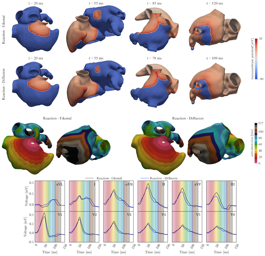
The central discovery is that the bottleneck to scalable atrial modelling is not any single component but the integration: a volumetric, image-stack-based wall extrusion that avoids topological errors, a set of Laplace-Dirichlet solutions that turn automatically detected orifices into anatomical labels and universal atrial coordinates, and a cable-based parameterization of inter-atrial conduction that allows insertion sites and velocities to be swept without remeshing. On 50 atrial fibrillation patients, the workflow produced meshes with element quality above 0.99, required no manual correction in 38 of 50 cases at the main processing stage, and generated ready-to-simulate models in an average of about 9 minutes at 0.90 mm resolution. The fast reaction-eikonal model, calibrated to the reaction-diffusion model's conduction velocities, produced activation maps and P-waves closely matching the gold standard, with an average P-wave RMSE of 3.71% across the 12 leads.
Load-bearing premise
The claim that the fast reaction-eikonal ECG matches the clinical gold standard rests on the assumption that the reaction-diffusion model with its calibrated conductivities is itself a faithful representation of human atrial electrophysiology; if that gold standard is wrong, the 3.71% error only measures agreement between two models.
Editorial extensions
If this is right
- Each patient CT can yield a simulation-ready biatrial-torso model in under ten minutes at reaction-eikonal resolution, making large virtual cohorts feasible.
- The fast reaction-eikonal forward ECG can replace reaction-diffusion simulations for calibration sweeps, since the average P-wave RMSE between the two is 3.71%.
- Inter-atrial conduction pathways can be parameterized as automatically generated cables with tunable anchoring sites and conduction velocities, enabling sweeps over Bachmann's bundle insertion geometry without remeshing.
- Parameters governing the P-wave, such as the sino-atrial node exit location and right-atrial conduction velocity, can be sampled to produce envelopes that cover clinical P-waves in most leads.
- Generated meshes have element quality above the threshold considered critical for bidomain simulations, so the same anatomy supports both fast calibration and high-fidelity mechanistic studies.
Reading between the lines
- The 3.71% RMSE is a model-to-model consistency check, not validation against measured human atrial electrophysiology; clinical validity would require comparing against invasive activation maps or ECG data in a cohort.
- The cable-based inter-atrial parameterization could be reused to model reentrant circuits such as atrial flutter once a forward model capable of reentry is used, since the framework exposes inter-atrial anatomy as a tunable parameter.
- The assumption of uniform atrial wall thickness likely limits patient specificity in regions where wall thickness varies, and a testable extension is to integrate patient-specific thickness maps once imaging resolution permits.
- Because the P-wave is a global observation, the workflow does not by itself resolve the identifiability problem: many parameter sets may reproduce the same P-wave, so a dedicated optimization with uniqueness analysis is needed.
Editorial analysis
A structured set of objections, weighed in public.
Referee Report
Summary. The manuscript describes an end-to-end computational workflow for generating volumetric biatrial models from patient CT images, annotating anatomical structures and fiber architecture, computing volumetric universal atrial coordinates, modeling inter-atrial conduction with auto-generated cables, and computing 12-lead atrial P-waves using a reaction-eikonal lead-field forward model. The workflow was evaluated on 50 atrial fibrillation patients, with reported processing times for coarse (0.90 mm, reaction-eikonal) and fine (0.25 mm, reaction-diffusion) meshes, an R-E versus R-D comparison of activation and P-waves, parametric studies of Bachmann bundle insertion and SAN location, and a one-patient pseudo-calibration of the simulated P-wave to clinical ECG data.
Significance. The paper's concrete strengths are the large cohort of 50 real CT datasets, the reported per-stage processing times, the automated anatomical labeling pipeline, the flexible cable-based representation of inter-atrial connections, and the very fast R-E forward model for P-wave computation. If the mesh-quality and automation claims are made internally consistent, and if the ECG-calibration claim is appropriately scoped, the framework would be a useful contribution to scalable atrial digital twin generation. The R-E/R-D comparison, while currently only a model-to-model consistency check, is valuable as a numerical verification, but it is not a clinical validation of the relationship between simulated and measured P-waves.
major comments (5)
- [§3.1 and §4.3] The mesh-quality claims are contradictory. Section 3.1 states "Worst element quality was always below 0.99, according to the quality metric [55], which is considered a critical threshold in simulations using R-D solvers such as openCARP", while Section 4.3 states "achieving element quality exceeding a threshold of 0.99, which has been empirically identified as critical for cardiac bidomain simulations." These statements cannot both describe the same 50-model cohort. If the worst element quality is below 0.99, then the R-D meshes do not meet the stated critical threshold, which undermines both the "suitable for R-D" claim and the reliability of the R-D "gold standard" simulations used in Table 7. Please report the actual element-quality distribution, the minimum value, and the fraction of elements below the threshold, and correct the contradictory wording.
- [§3.7 and Table 11] The ECG-calibration demonstration is limited to a single patient and is essentially a fit to the data used for evaluation. Section 3.7 states "One anatomical model of a patient was selected", and the parameters (SAN location, RA conduction velocities, BB insertion sites) are chosen by minimizing RMSE against the measured P-wave, with no independent test set, cross-validation, or uncertainty quantification. Moreover, the simulated P-wave envelope does not cover the clinical signal in leads aVL, -aVR, and V1, and the Discussion attributes the V1 discrepancy to fibrotic tissue that is not modeled. The title's "ECG calibrated" claim is therefore stronger than what is demonstrated; the paper should reframe this as a feasibility demonstration of a pseudo-calibration workflow and explicitly state the identifiability and generalization limitations.
- [§3.3, Table 7, §2.2.2 and Table 4] The R-E/R-D comparison is presented as the validation of the fast forward model, but Section 3.3 says the results are "illustrated for a representative test case", and Table 7 reports a single row of RMSE values without indicating whether the 3.71% average is over the 50-patient cohort or one geometry. Please state the sample size and report per-case variability. In addition, because the R-D conductivities in Table 4 are calibrated with the ForCEPSS framework to reproduce the R-E conduction velocities, the RMSE in Table 7 measures model-to-model consistency under matched parameters, not accuracy against independent human electrophysiology; the "gold standard" wording in Section 3.3 should be qualified accordingly.
- [§3.1, Table 5 and §3.1.1] The automation statistics are internally inconsistent. Section 3.1 states that "all 50 cases in both resolutions were processed automatically in the majority of cases (38), with only minimal user intervention required in 22 cases", which does not add to 50. Table 5 lists 19 and 22 cases requiring manual correction at the first two stages, while Section 3.1.1 reports 21 cases for the CS/IVC landmark switch and 22 cases for IVC discard-tissue adjustment. Since "highly automated" is a headline claim, please provide a coherent per-stage and per-case accounting (for example, a table of correction types and a clear statement of how many of the 50 cases required any manual intervention).
- [§3.2, Table 6 and §4.6] The reported computational performance of the R-E model is inconsistent. Section 3.2 states that computation of a full biatrial activation sequence with a high-fidelity ECG takes about 1.00 s, while Table 6 reports "EP simulation ≈ 27 s" for the 0.90 mm/R-E case, and Section 4.6 states "A full single forward simulation lasted ≈ 27.00 s only where the actual evaluation of the EP model amounted only to ≈ 3.00 s." These numbers need to be reconciled with a precise definition of what is included in each timing (setup, lead-field computation, eikonal solve, ECG time-series generation) and on which hardware and mesh size they were measured.
minor comments (5)
- [§2.2.2] The global ECG scaling factor of 0.2 is introduced without justification; since the RMSE metric is amplitude-sensitive, the scaling should be reported as part of the model definition or the calibration procedure, not as an ad hoc post-processing step.
- [§3.7] There is a typo in "electrophysiologicalglsep excitability"; it should presumably be "electrophysiological excitability".
- [Table 4] The table heading contains a typo: "surface-to-volume ration" should be "surface-to-volume ratio".
- [§2.2.1] The sentence "The origin of the cable in the RA was chosen superior to the CS, at a distance between 3 and 8 .00 mm [21] from the CS ostium" could be clarified, since a range without a prescription for how it was chosen in the implemented models makes the parameter not fully reproducible.
- [§5] The Limitations section is candid about the uniform wall-thickness assumption and the cable approximation for inter-atrial connections, but the Discussion in Section 4.6 still uses the P-wave fit results as evidence of clinical compatibility; the wording should be adjusted to match the caveats stated in Section 5.
Circularity Check
Partial circularity: the R-D 'gold standard' is calibrated to match R-E conduction velocities, and the P-wave calibration is fit to the same clinical signal; a separate mesh-quality contradiction is a correctness risk, not circularity.
-
fitted input called prediction
[Section 2.2.2, Table 4; Section 3.3, Table 7]
"To identify the electrical conductivities for the R-D model at the targeted mesh resolution that match the R-E conduction velocities, the ForCEPSS framework [47] was employed, using reported values [92] for initialization."
The R-D model is designated the 'gold standard' in Section 3.3, but its conductivities are fitted by the authors' ForCEPSS framework to reproduce the R-E model's prescribed conduction velocities (Table 4). The subsequent comparison reports an average RMSE of 3.71% between R-E and R-D P-waves (Table 7). This is not an independent test of R-E accuracy: the target quantity, conduction velocity, is an input to the R-D fit, so the agreement largely reflects the success of that fit. It measures model-to-model consistency under matched parameters, not validation against an external, clinically calibrated gold standard.
-
fitted input called prediction
[Section 3.7, Table 11 and Figure 13]
"In a first pseudo-calibration step for a fixed SAN exit site and baseline velocities in the RA, the location of BB was varied in interactive simulation runs to obtain a close approximation of the terminal half of the P-wave."
The clinical P-wave used as the evaluation target is the same signal used to steer the interactive BB placement and to sample the SAN exit site and RA conduction velocities. The reported RMSE range [1.72%, 2.79%] (Table 11) is therefore a fit residual to the calibration data, not a prediction on held-out data. Since the paper frames this as a demonstration of calibration feasibility rather than a blinded validation, this is a partial circularity; the conclusion that 'the P-wave can be closely captured' is the success criterion of the fitting procedure itself.
full rationale
The anatomical model generation (Section 2.1), UAC computation (Section 2.1.5), inter-atrial cable formulation (Section 2.2.1), and rule-based fiber/label annotation are self-contained engineering contributions benchmarked on 50 CT datasets; these are not circular. The main circularity is in the R-E/R-D 'gold standard' comparison: Table 4's R-D conductivities are explicitly fitted with ForCEPSS to match the R-E conduction velocities, so the 3.71% average RMSE in Table 7 is a consistency measure between two models sharing the same prescribed CVs rather than an independent accuracy assessment of R-E against a clinically validated gold standard. This is partial, not total, because matching local CVs does not by itself force identical wavefront dynamics or ECGs. The Section 3.7 calibration demonstrates fitting rather than prediction: the BB anchor was chosen interactively to approximate the same clinical P-wave, and the SAN/velocity sample RMSE is evaluated on the same signal, so the reported [1.72%, 2.79%] RMSE is a fit residual. The paper is candid about the latter ('pseudo-calibration') and about identifiability limitations in Section 5, which lowers the severity. Self-citations to [42, 74, 77] support the RELF fidelity claim, but the paper also provides its own R-E/R-D comparison, so the claim does not reduce solely to a self-citation chain. Separately flagged as a correctness risk, not circularity: Section 3.1 states 'Worst element quality was always below 0.99' while Section 4.3 claims 'element quality exceeding a threshold of 0.99'; this internal contradiction should be resolved but does not by itself constitute a circular derivation.
Assumptions & free parameters
free parameters (8)
- UAC boundary constants R and r =
R=0.1, r=0.04
- UAC orifice target positions alpha_cs, beta_cs, alpha_ipv, beta_ipv =
0.2, 0.8, 0.25, 0.25
- Regional wall thickness values =
Prescribed per region (Section 2.1.2), not patient-specific
- Per-region conduction velocities (vl, vt) =
RA 0.97/0.74, LA 0.98/0.76, CT 1.21/0.92, PM 1.30/0.99, BB 1.40/1.08, FO 0.33/0.24 m/s
- R-D tissue conductivities (gi,l, ge,l, gi,t, ge,t) =
Table 4 values
- SAN radius and length =
2.5 mm radius (Section 2.1.4), 2.2 cm band (Section 3.7)
- IC cable conduction velocities =
Tuned to physiological inter-atrial delays (Section 2.2.2)
- ECG scaling factor =
0.2
assumptions (6)
- standard math Laplace-Dirichlet solutions provide valid distance-like coordinates for atrial parametrization.
- domain assumption The Courtemanche et al. cell model with region-specific parameterization adequately represents human atrial cellular dynamics.
- domain assumption The five inter-atrial pathways (BB, CS, FO rim, superior-posterior, middle-posterior) are the only electrical connections between the atria.
- domain assumption The template torso from a different subject is a valid volume conductor for each patient.
- ad hoc to paper Uniform regional wall thickness is an acceptable anatomical approximation.
- domain assumption Rule-based fiber orientations approximate patient-specific fiber architecture.
invented entities (2)
-
Auto-generated inter-atrial conduction cables
-
Cigarette-shaped SAN focal activation site with insulating boundary
Cite this review
Pith. "Pith review of An efficient end-to-end computational framework for the generation of ECG calibrated volumetric models of human atrial electrophysiology." pith.science (2026). https://pith.science/paper/JBOBRGLL
@misc{pith2026250203322,
  author       = {Pith},
  title        = {Pith review of: An efficient end-to-end computational framework for the generation of ECG calibrated volumetric models of human atrial electrophysiology},
  year         = {2026},
  howpublished = {\url{https://pith.science/paper/JBOBRGLL}},
  note         = {Machine review of arXiv:2502.03322}
}
read the original abstract
Computational models of atrial electrophysiology (EP) are increasingly utilized for applications such as the development of advanced mapping systems, personalized clinical therapy planning, and the generation of virtual cohorts and digital twins. These models have the potential to establish robust causal links between simulated in silico behaviors and observed human atrial EP, enabling safer, cost-effective, and comprehensive exploration of atrial dynamics. However, current state-of-the-art approaches lack the fidelity and scalability required for regulatory-grade applications, particularly in creating high-quality virtual cohorts or patient-specific digital twins. Challenges include anatomically accurate model generation, calibration to sparse and uncertain clinical data, and computational efficiency within a streamlined workflow. This study addresses these limitations by introducing novel methodologies integrated into an automated end-to-end workflow for generating high-fidelity digital twin snapshots and virtual cohorts of atrial EP. These innovations include: (i) automated multi-scale generation of volumetric biatrial models with detailed anatomical structures and fiber architecture; (ii) a robust method for defining space-varying atrial parameter fields; (iii) a parametric approach for modeling inter-atrial conduction pathways; and (iv) an efficient forward EP model for high-fidelity electrocardiogram computation. We evaluated this workflow on a cohort of 50 atrial fibrillation patients, producing high-quality meshes suitable for reaction-eikonal and reaction-diffusion models and demonstrating the ability to simulate atrial ECGs under parametrically controlled conditions. These advancements represent a critical step toward scalable, precise, and clinically applicable digital twin models and virtual cohorts, enabling enhanced patient-specific predictions and therapeutic planning.
Figures


Figures from the paper (10 more)




Forward citations
Cited by 1 Pith paper
-
Integrating anatomy and electrophysiology in the healthy human heart: Insights from biventricular statistical shape analysis using universal coordinates
A biventricular statistical shape model from 271 healthy CT scans shows heart size and elongation are the main shape variations and correlate with ECG intervals and amplitudes, and provides 100 synthetic heart meshes ...
Reference graph
Works this paper leans on
-
[55]
Karabelas, E., Gsell, M.A.F., Augustin, C.M., Marx, L., Neic, A., Prassl, A.J., Goubergrits, L., Kuehne, T., Plank, G.,
-
[1]
A method of bivariate interpolation and smooth surface fitting for irregularly distributed data points
Akima, H., 1978. A method of bivariate interpolation and smooth surface fitting for irregularly distributed data points. ACM Transactions on Mathematical Software (TOMS) 4, 148–159
1978
-
[2]
The surgical anatomy of the conduction tissues
Anderson, R.H., Ho, S.Y., Becker, A.E., 1983. The surgical anatomy of the conduction tissues. Thorax 38, 408–420
1983
-
[3]
The development of the sinoatrial node, in: The Sinus Node: Structure, Function, and Clinical Relevance
Anderson, R.H., Siew Yen, H., Becker, A.E., Gosling, J.A., 1978. The development of the sinoatrial node, in: The Sinus Node: Structure, Function, and Clinical Relevance. Springer, pp. 166–182
1978
-
[4]
Electrical conduction between the right atrium and the left atrium via the musculature of the coronary sinus
Antz, M., Otomo, K., Arruda, M., Scherlag, B.J., Pitha, J., Tondo, C., Lazzara, R., Jackman, W.M., 1998a. Electrical conduction between the right atrium and the left atrium via the musculature of the coronary sinus. Circulation 98, 1790–1795
-
[5]
Antz, M., Otomo, K., Arruda, M., Scherlag, B.J., Pitha, J., Tondo, C., Lazzara, R., Jackman, W.M., 1998b. Electrical Conduction Between the Right Atrium and the Left Atrium via the Musculature of the Coronary Sinus. Circulation 98, 1790–1795. URL: https://www.ahajournals.org/doi/10.1161/01.CIR.98.17.1790, doi:10.1161/01.CIR.98.17.1790
-
[6]
Anatomically accurate high resolution modeling of human whole heart electromechanics: a strongly scalable algebraic multigrid solver method for nonlinear deformation
Augustin, C.M., Neic, A., Liebmann, M., Prassl, A.J., Niederer, S.A., Haase, G., Plank, G., 2016. Anatomically accurate high resolution modeling of human whole heart electromechanics: a strongly scalable algebraic multigrid solver method for nonlinear deformation. Journal of computational physics 305, 622–646
2016
-
[7]
AugmentA: Patient-specific augmented atrial model generation tool
Azzolin, L., Eichenlaub, M., Nagel, C., Nairn, D., S´ anchez, J., Unger, L., Arentz, T., Westermann, D., D¨ ossel, O., Jadidi, A., Loewe, A., 2023. AugmentA: Patient-specific augmented atrial model generation tool. Computerized Medical Imaging and Graphics 108, 102265. URL: https://linkinghub.elsevier.com/retrieve/pii/S0895611123000836, doi: 10.1016/ j.co...
Show all 120 references
-
[8]
Influence of gradient and smoothness of atrial wall thickness on initiation and maintenance of atrial fibrillation, in: 2020 Computing in Cardiology, IEEE
Azzolin, L., Luongo, G., Ventura, S.R., Saiz, J., D¨ osse, O., Loewe, A., 2020. Influence of gradient and smoothness of atrial wall thickness on initiation and maintenance of atrial fibrillation, in: 2020 Computing in Cardiology, IEEE. pp. 1–4
2020
-
[9]
The inter-auricular time interval
Bachmann, G., 1916. The inter-auricular time interval. American Journal of Physiology-Legacy Content 41, 309–320. doi:10.1152/ajplegacy.1916.41.3.309
1916 doi
-
[10]
Computational study of the effect of unstructured mesh quality on solution efficiency, in: 13th Computational Fluid Dynamics Conference, p
Batdorf, M., Freitag, L., Ollivier-Gooch, C., Batdorf, M., Freitag, L., Ollivier-Gooch, C., 1997. Computational study of the effect of unstructured mesh quality on solution efficiency, in: 13th Computational Fluid Dynamics Conference, p. 1888
1997
-
[11]
Universal ventricular coordinates: A generic framework for describing position within the heart and transferring data
Bayer, J., Prassl, A.J., Pashaei, A., Gomez, J.F., Frontera, A., Neic, A., Plank, G., Vigmond, E.J., 2018. Universal ventricular coordinates: A generic framework for describing position within the heart and transferring data. Medical Image Analysis 45, 83–93
2018
-
[12]
Left atrial wall thickness vari- ability measured by ct scans in patients undergoing pulmonary vein isolation
Beinart, R., Abbara, S., Blum, A., Ferencik, M., Heist, K., Ruskin, J., Mansour, M., 2011. Left atrial wall thickness vari- ability measured by ct scans in patients undergoing pulmonary vein isolation. Journal of cardiovascular electrophysiology 22, 1232–1236
2011
-
[13]
From bits to bedside: Entering the age of digital twins in cardiac electrophysiology
Bhagirath, P., Strocchi, M., Bishop, M.J., Boyle, P., Plank, G., 2024. From bits to bedside: Entering the age of digital twins in cardiac electrophysiology. Europace
2024
-
[14]
Stochastic behaviour of arrhythmia induction in virtual heart models suggests caution in offering mechanistic insights
Bishop, M., Plank, G., 2025. Stochastic behaviour of arrhythmia induction in virtual heart models suggests caution in offering mechanistic insights. Nature Cardiovascular Research In press
2025
-
[15]
Three- dimensional atrial wall thickness maps to inform catheter ablation procedures for atrial fibrillation
Bishop, M., Rajani, R., Plank, G., Gaddum, N., Carr-White, G., Wright, M., O’Neill, M., Niederer, S., 2016. Three- dimensional atrial wall thickness maps to inform catheter ablation procedures for atrial fibrillation. Europace 18, 376–383
2016
-
[16]
Bidomain ECG simulations using an augmented monodomain model for the cardiac source
Bishop, M.J., Plank, G., 2011a. Bidomain ECG simulations using an augmented monodomain model for the cardiac source. IEEE transactions on biomedical engineering 58, 2297–2307
-
[17]
Representing cardiac bidomain bath-loading effects by an augmented monodomain approach: application to complex ventricular models
Bishop, M.J., Plank, G., 2011b. Representing cardiac bidomain bath-loading effects by an augmented monodomain approach: application to complex ventricular models. IEEE transactions on bio-medical engineering 58, 1066–1075. doi:10.1109/TBME.2010.2096425
-
[18]
The sinoatrial node, a heterogeneous pacemaker structure
Boyett, M.R., Honjo, H., Kodama, I., 2000. The sinoatrial node, a heterogeneous pacemaker structure. Cardiovascular research 47, 658–687
2000
-
[19]
Computationally guided personalized targeted ablation of persistent atrial fibrillation
Boyle, P.M., Zghaib, T., Zahid, S., Ali, R.L., Deng, D., Franceschi, W.H., Hakim, J.B., Murphy, M.J., Prakosa, A., Zimmerman, S.L., et al., 2019. Computationally guided personalized targeted ablation of persistent atrial fibrillation. Nature biomedical engineering 3, 870–879
2019
-
[20]
Bachmann’s bundle
van Campenhout, M.J., Yaksh, A., Kik, C., de Jaegere, P.P., Ho, S.Y., Allessie, M.A., de Groot, N.M., 2013. Bachmann’s bundle. Circulation: Arrhythmia and Electrophysiology 6, 1041–1046. doi: 10.1161/CIRCEP.113.000758
2013 doi
-
[21]
The anatomic basis of connections between the coronary sinus musculature and the left atrium in humans
Chauvin, M., Shah, D.C., Ha ¨ ıssaguerre, M., Marcellin, L., Brechenmacher, C., 2000. The anatomic basis of connections between the coronary sinus musculature and the left atrium in humans. Circulation 101, 647–652
2000
-
[22]
Overcoming element quality dependence of finite elements with adaptive extended stencil fem (aes-fem)
Conley, R., Delaney, T.J., Jiao, X., 2016. Overcoming element quality dependence of finite elements with adaptive extended stencil fem (aes-fem). International Journal for Numerical Methods in Engineering 108, 1054–1085
2016
-
[23]
The atria: from morphology to function
Corradi, D., Maestri, R., Macchi, E., Callegari, S., 2011. The atria: from morphology to function. Journal of Cardiovas- cular Electrophysiology 22, 223–235
2011
-
[24]
The ’Digital Twin’ to enable the vision of precision cardiology
Corral-Acero, J., Margara, F., Marciniak, M., Rodero, C., Loncaric, F., Feng, Y., Gilbert, A., Fernandes, J.F., Bukhari, H.A., Wajdan, A., Martinez, M.V., Santos, M.S., Shamohammdi, M., Luo, H., Westphal, P., Leeson, P., DiAchille, P., Gurev, V., Mayr, M., Geris, L., Pathmanat...
2020
-
[25]
Costa, C.M., Campos, F.O., Prassl, A.J., Dos Santos, R.W., Sanchez-Quintana, D., Ahammer, H., Hofer, E., Plank, G., 35
-
[26]
Ionic mechanisms underlying human atrial action potential properties: insights from a mathematical model
Courtemanche, M., Ramirez, R.J., Nattel, S., 1998. Ionic mechanisms underlying human atrial action potential properties: insights from a mathematical model. American Journal of Physiology-Heart and Circulatory Physiology 275, H301–H321
1998
-
[27]
Calibrating cardiac electrophysiology models using latent gaussian processes on atrial manifolds
Coveney, S., Roney, C.H., Corrado, C., Wilkinson, R.D., Oakley, J.E., Niederer, S.A., Clayton, R.H., 2022. Calibrating cardiac electrophysiology models using latent gaussian processes on atrial manifolds. Scientific Reports 12, 16572
2022
-
[28]
Image-based personalization of cardiac anatomy for coupled electromechanical modeling
Crozier, A., Augustin, C., Neic, A., Prassl, A., Holler, M., Fastl, T., Hennemuth, A., Bredies, K., Kuehne, T., Bishop, M., et al., 2016. Image-based personalization of cardiac anatomy for coupled electromechanical modeling. Annals of biomedical engineering 44, 58–70
2016
-
[29]
Human sinoatrial node structure: 3D microanatomy of sinoatrial conduction pathways
Csepe, T.A., Zhao, J., Hansen, B.J., Li, N., Sul, L.V., Lim, P., Wang, Y., Simonetti, O.P., Kilic, A., Mohler, P.J., Janssen, P.M., Fedorov, V.V., 2016. Human sinoatrial node structure: 3D microanatomy of sinoatrial conduction pathways. Progress in Biophysics and Molecular Bio...
2016 doi
-
[30]
Harrild, C.S.H.., 2000
David M. Harrild, C.S.H.., 2000. A Computer Model of Normal Conduction in the Human Atria. Circulation Research 87, e25–e36. URL: https://www.ahajournals.org/doi/10.1161/01.res.87.7.e25, doi:10.1161/01.RES.87.7.e25. pub- lisher: American Heart Association
2000 doi
-
[31]
Simulation of biatrial conduction via different pathways during sinus rhythm with a detailed human atrial model
Deng, D.d., Gong, Y.l., Shou, G.f., Jiao, P.f., Zhang, H.g., Ye, X.s., Xia, L., 2012. Simulation of biatrial conduction via different pathways during sinus rhythm with a detailed human atrial model. Journal of Zhejiang University Science B 13, 676–694
2012
-
[32]
Use of computed tomography to identify atrial fibrillation associated differences in left atrial wall thickness and density
Dewland, T.A., Wintermark, M., Vaysman, A., Smith, L.M., Tong, E., Vittinghoff, E., Marcus, G.M., 2013. Use of computed tomography to identify atrial fibrillation associated differences in left atrial wall thickness and density. Pacing and Clinical Electrophysiology 36, 55–62
2013
-
[33]
Computer three-dimensional reconstruction of the sinoatrial node
Dobrzynski, H., Li, J., Tellez, J., Greener, I., Nikolski, V., Wright, S., Parson, S., Jones, S., Lancaster, M., Yamamoto, M., et al., 2005. Computer three-dimensional reconstruction of the sinoatrial node. Circulation 111, 846–854
2005
-
[34]
Computational modeling of the human atrial anatomy and electrophysiology
D¨ ossel, O., Krueger, M.W., Weber, F.M., Wilhelms, M., Seemann, G., 2012. Computational modeling of the human atrial anatomy and electrophysiology. Medical & biological engineering & computing 50, 773–799
2012
-
[35]
A comprehensive and biophysically detailed computational model of the whole human heart electromechanics
Fedele, M., Piersanti, R., Regazzoni, F., Salvador, M., Africa, P.C., Bucelli, M., Zingaro, A., Quarteroni, A., et al., 2023. A comprehensive and biophysically detailed computational model of the whole human heart electromechanics. Computer Methods in Applied Mechanics and Eng...
2023
-
[36]
Conduction barriers and pathways of the sinoatrial pacemaker com- plex: their role in normal rhythm and atrial arrhythmias
Fedorov, V.V., Glukhov, A.V., Chang, R., 2012. Conduction barriers and pathways of the sinoatrial pacemaker com- plex: their role in normal rhythm and atrial arrhythmias. American Journal of Physiology-Heart and Circulatory Physiology 302, H1773–H1783. URL: https://www.physiol...
2012
-
[37]
Optical Mapping of the Isolated Coronary-Perfused Human Sinus Node
Fedorov, V.V., Glukhov, A.V., Chang, R., Kostecki, G., Aferol, H., Hucker, W.J., Wuskell, J.P., Loew, L.M., Schuessler, R.B., Moazami, N., Efimov, I.R., 2010. Optical Mapping of the Isolated Coronary-Perfused Human Sinus Node. Jour- nal of the American College of Cardiology 56...
2010 doi
-
[38]
Detailed anatomical and electrophysiological models of human atria and torso for the simulation of atrial activation
Ferrer, A., Sebasti´ an, R., S´ anchez-Quintana, D., Rodriguez, J.F., Godoy, E.J., Martinez, L., Saiz, J., 2015. Detailed anatomical and electrophysiological models of human atria and torso for the simulation of atrial activation. PloS one 10, e0141573
2015
-
[39]
On the theory of the electrocardiogram
Geselowitz, D.B., 1989. On the theory of the electrocardiogram. Proceedings of the IEEE 77, 857–876
1989
-
[40]
Automated framework for the inclusion of a his–purkinje system in cardiac digital twins of ventricular electrophysiology
Gillette, K., Gsell, M.A., Bouyssier, J., Prassl, A.J., Neic, A., Vigmond, E.J., Plank, G., 2021a. Automated framework for the inclusion of a his–purkinje system in cardiac digital twins of ventricular electrophysiology. Annals of biomedical engineering 49, 3143–3153
-
[41]
A personalized real-time virtual model of whole heart electrophysiology
Gillette, K., Gsell, M.A., Strocchi, M., Grandits, T., Neic, A., Manninger, M., Scherr, D., Roney, C.H., Prassl, A.J., Augustin, C.M., et al., 2022. A personalized real-time virtual model of whole heart electrophysiology. Frontiers in Physiology 13, 907190
2022
-
[42]
A Framework for the generation of digital twins of cardiac electrophysiology from clinical 12-leads ECGs
Gillette, K., Gsell, M.A.F., Prassl, A.J., Karabelas, E., Reiter, U., Reiter, G., Grandits, T., Payer, C., ˇStern, D., Urschler, M., Bayer, J.D., Augustin, C.M., Neic, A., Pock, T., Vigmond, E.J., Plank, G., 2021b. A Framework for the generation of digital twins of cardiac ele...
-
[43]
GEASI: Geodesic-based earliest activation sites identification in cardiac models
Grandits, T., Effland, A., Pock, T., Krause, R., Plank, G., Pezzuto, S., 2021. GEASI: Geodesic-based earliest activation sites identification in cardiac models. International journal for numerical methods in biomedical engineering 37, e3505. URL: http://arxiv.org/abs/2102.0996...
2021 arXiv
-
[44]
Accurate and Efficient Cardiac Digital Twin from surface ECGs: Insights into Identifiability of Ventricular Conduction System
Grandits, T., Gillette, K., Plank, G., Pezzuto, S., 2024a. Accurate and Efficient Cardiac Digital Twin from surface ECGs: Insights into Identifiability of Ventricular Conduction System. URL: http://arxiv.org/abs/2411.00165, doi:10.48550/ arXiv.2411.00165. arXiv:2411.00165
-
[45]
Digital Twinning of Cardiac Electrophysiology Models From the Surface ECG: A Geodesic Backpropagation Approach
Grandits, T., Verh¨ ulsdonk, J., Haase, G., Effland, A., Pezzuto, S., 2024b. Digital Twinning of Cardiac Electrophysiology Models From the Surface ECG: A Geodesic Backpropagation Approach. IEEE Transactions on Biomedical Engineering 71, 1281–1288. URL: https://ieeexplore.ieee....
-
[46]
Incomplete reentry and epicardial breakthrough patterns during atrial fibrillation in the sheep heart
Gray, R.A., Pertsov, A.M., Jalife, J., 1996. Incomplete reentry and epicardial breakthrough patterns during atrial fibrillation in the sheep heart. Circulation 94, 2649–2661
1996
-
[47]
For- CEPSS—A framework for cardiac electrophysiology simulations standardization
Gsell, M.A., Neic, A., Bishop, M.J., Gillette, K., Prassl, A.J., Augustin, C.M., Vigmond, E.J., Plank, G., 2024. For- CEPSS—A framework for cardiac electrophysiology simulations standardization. Computer Methods and Programs in 36 Biomedicine 251, 108189. URL: https://linkingh...
2024
-
[48]
Right atrial free wall conduction velocity and degree of anisotropy in patients with stable sinus rhythm studied during open heart surgery
Hansson, A., Holm, M., Blomstr¨ om, P., Johansson, R., L¨ uhrs, C., Brandt, J., Olsson, S., 1998. Right atrial free wall conduction velocity and degree of anisotropy in patients with stable sinus rhythm studied during open heart surgery. European heart journal 19, 293–300
1998
-
[49]
A computer model of normal conduction in the human atria
Harrild, D.M., Henriquez, C.S., 2000. A computer model of normal conduction in the human atria. Circulation research 87, e25–e36
2000
-
[50]
The importance of atrial structure and fibers
Ho, S., S´ anchez-Quintana, D., 2009. The importance of atrial structure and fibers. Clinical Anatomy: The Official Journal of the American Association of Clinical Anatomists and the British Association of Clinical Anatomists 22, 52–63
2009
-
[51]
Correlation between electrical activity and the size of rabbit sino-atrial node cells
Honjo, H., Boyett, M., Kodama, I., Toyama, J., 1996. Correlation between electrical activity and the size of rabbit sino-atrial node cells. The Journal of physiology 496, 795–808
1996
-
[52]
Right atrial function and fibrosis in relation to successful atrial fibrillation ablation
Hopman, L.H., Visch, J.E., Bhagirath, P., van der Laan, A.M., Mulder, M.J., Razeghi, O., Kemme, M.J., Niederer, S.A., Allaart, C.P., G¨ otte, M.J., 2023. Right atrial function and fibrosis in relation to successful atrial fibrillation ablation. European Heart Journal-Cardiovas...
2023
-
[53]
Propagation through electrically coupled cells
Joyner, R., Van Capelle, F., 1986. Propagation through electrically coupled cells. How a small SA node drives a large atrium. Biophysical Journal 50, 1157–1164. URL: https://linkinghub.elsevier.com/retrieve/pii/ S0006349586835597, doi:10.1016/S0006-3495(86)83559-7
1986 doi
-
[54]
A 3d adaptive mesh moving scheme
Kanchi, H., Masud, A., 2007. A 3d adaptive mesh moving scheme. International Journal for Numerical Methods in Fluids 54, 923–944
2007
-
[56]
The form and nature of the muscular connections between the primary divisions of the vertebrate heart
Keith, A., Flack, M., 1907. The form and nature of the muscular connections between the primary divisions of the vertebrate heart. Journal of anatomy and physiology 41, 172
1907
-
[57]
Ranking the influence of tissue conductivities on forward- calculated ecgs
Keller, D.U., Weber, F.M., Seemann, G., Dossel, O., 2010. Ranking the influence of tissue conductivities on forward- calculated ecgs. IEEE Transactions on Biomedical Engineering 57, 1568–1576
2010
-
[58]
Current concepts of anatomy, electrophysiology, and therapeutic implications of the interatrial septum
Kharbanda, R.K., ¨Ozdemir, E.H., Taverne, Y.J., Kik, C., Bogers, A.J., de Groot, N.M., 2019. Current concepts of anatomy, electrophysiology, and therapeutic implications of the interatrial septum. JACC: Clinical Electrophysiology 5, 647–656
2019
-
[59]
The bachmann bundle and interatrial conduction: comparing atrial morphology to electrical activity
Knol, W.G., Teuwen, C.P., Kleinrensink, G.J., Bogers, A.J., de Groot, N.M., Taverne, Y.J., 2019. The bachmann bundle and interatrial conduction: comparing atrial morphology to electrical activity. Heart Rhythm 16, 606–614
2019
-
[60]
Worst case mesh quality in the target matrix optimization paradigm
Knupp, P., 2022. Worst case mesh quality in the target matrix optimization paradigm. Engineering with Computers 38, 5695–5711
2022
-
[61]
Personalization of atrial anatomy and electrophysiology as a basis for clinical modeling of radio-frequency ablation of atrial fibrillation
Krueger, M.W., Seemann, G., Rhode, K., Keller, D.U., Schilling, C., Arujuna, A., Gill, J., O’Neill, M.D., Razavi, R., Dossel, O., 2012. Personalization of atrial anatomy and electrophysiology as a basis for clinical modeling of radio-frequency ablation of atrial fibrillation. ...
2012
-
[62]
A bilayer model of human atria: mathematical background, construction, and assessment
Labarthe, S., Bayer, J., Coudiere, Y., Henry, J., Cochet, H., Jais, P., Vigmond, E., 2014. A bilayer model of human atria: mathematical background, construction, and assessment. Europace 16, iv21–iv29. URL: https://academic.oup. com/europace/article-lookup/doi/10.1093/europace...
2014 doi
-
[63]
Imaging assessment of the right atrium: anatomy and function
Lang, R.M., Cameli, M., Sade, L.E., Faletra, F.F., Fortuni, F., Rossi, A., Soulat-Dufour, L., 2022. Imaging assessment of the right atrium: anatomy and function. European Heart Journal-Cardiovascular Imaging 23, 867–884
2022
-
[64]
Normal atrial activation and voltage during sinus rhythm in the human heart: an endocardial and epicardial mapping study in patients with a history of atrial fibrillation
Lemery, R., Birnie, D., Tang, A.S., Green, M., Gollob, M., Hendry, M., Lau, E., 2007. Normal atrial activation and voltage during sinus rhythm in the human heart: an endocardial and epicardial mapping study in patients with a history of atrial fibrillation. Journal of cardiova...
2007
-
[65]
Anatomic description of bachmann’s bundle and its relation to the atrial septum
Lemery, R., Guiraudon, G., Veinot, J.P., 2003. Anatomic description of bachmann’s bundle and its relation to the atrial septum. The American journal of cardiology 91, 1482–1482
2003
-
[66]
Re- dundant and diverse intranodal pacemakers and conduction pathways protect the human sinoatrial node from fail- ure
Li, N., Hansen, B.J., Csepe, T.A., Zhao, J., Ignozzi, A.J., Sul, L.V., Zakharkin, S.O., Kalyanasundaram, A., Davis, J.P., Biesiadecki, B.J., Kilic, A., Janssen, P.M.L., Mohler, P.J., Weiss, R., Hummel, J.D., Fedorov, V.V., 2017. Re- dundant and diverse intranodal pacemakers an...
2017 doi
-
[67]
Influence of the earliest right atrial activation site and its proximity to interatrial connections on p-wave morphology
Loewe, A., Krueger, M.W., Holmqvist, F., D¨ ossel, O., Seemann, G., Platonov, P.G., 2016. Influence of the earliest right atrial activation site and its proximity to interatrial connections on p-wave morphology. EP Europace 18, iv35–iv43
2016
-
[68]
Loewe, A., Krueger, M.W., Platonov, P.G., Holmqvist, F., D¨ ossel, O., Seemann, G., 2015. Left and right atrial contribu- tion to the p-wave in realistic computational models, in: Functional Imaging and Modeling of the Heart: 8th International Conference, FIMH 2015, Maastricht...
2015
-
[69]
Lubrecht, J.M., Grandits, T., Gharaviri, A., Schotten, U., Pock, T., Plank, G., Krause, R., Auricchio, A., Conte, G., Pezzuto, S., 2021. Automatic reconstruction of the left atrium activation from sparse intracardiac contact recordings by inverse estimate of fibre structure an...
2021 doi
-
[70]
Anatomical diversity and age-related histological changes in the human right atrial posterolateral wall
Matsuyama, T.a., Inoue, S., Kobayashi, Y., Sakai, T., Saito, T., Katagiri, T., Ota, H., 2004. Anatomical diversity and age-related histological changes in the human right atrial posterolateral wall. EP Europace 6, 307–315
2004
-
[71]
The anatomy and physiology of the sinoatrial node—a contemporary review
Monfredi, O., Dobrzynski, H., Mondal, T., Boyett, M.R., Morris, G.M., 2010. The anatomy and physiology of the sinoatrial node—a contemporary review. Pacing and clinical electrophysiology 33, 1392–1406
2010
-
[72]
Uncertainty quantification for the 12-lead ecg: a lead field approach
Multerer, M., Pezzuto, S., 2021. Uncertainty quantification for the 12-lead ecg: a lead field approach. arXiv preprint 37 arXiv:2102.09960
2021 arXiv
-
[73]
Onset of atrial arrhythmias elicited by autonomic modulation of rabbit sinoatrial node activity: a modeling study
Mu˜ noz, M.A., Kaur, J., Vigmond, E.J., 2011. Onset of atrial arrhythmias elicited by autonomic modulation of rabbit sinoatrial node activity: a modeling study. American Journal of Physiology-Heart and Circulatory Physiology 301, H1974–H1983. doi: 10.1152/ajpheart.00059.2011
2011
-
[74]
Comparison of propagation models and forward calculation methods on cellular, tissue and organ scale atrial electrophysiology
Nagel, C., Espinosa, C.B., Gillette, K., Gsell, M.A., S´ anchez, J., Plank, G., D¨ ossel, O., Loewe, A., 2022. Comparison of propagation models and forward calculation methods on cellular, tissue and organ scale atrial electrophysiology. IEEE Transactions on Biomedical Enginee...
2022
-
[75]
A bi-atrial statistical shape model for large-scale in silico studies of human atria: Model development and application to ECG simulations
Nagel, C., Schuler, S., D¨ ossel, O., Loewe, A., 2021. A bi-atrial statistical shape model for large-scale in silico studies of human atria: Model development and application to ECG simulations. Medical Image Analysis 74, 102210. URL: https://linkinghub.elsevier.com/retrieve/p...
2021
-
[76]
Efficient computa- tion of electrograms and ecgs in human whole heart simulations using a reaction-eikonal model
Neic, A., Campos, F.O., Prassl, A.J., Neiderer, S.A., Bishop, M.J., Vigmond, E.J., Plank, G., 2017a. Efficient computa- tion of electrograms and ecgs in human whole heart simulations using a reaction-eikonal model
-
[77]
Efficient computa- tion of electrograms and ecgs in human whole heart simulations using a reaction-eikonal model
Neic, A., Campos, F.O., Prassl, A.J., Niederer, S.A., Bishop, M.J., Vigmond, E.J., Plank, G., 2017b. Efficient computa- tion of electrograms and ecgs in human whole heart simulations using a reaction-eikonal model. Journal of computational physics 346, 191–211. doi: 10.1016/j....
-
[78]
Automating image-based mesh generation and manipulation tasks in cardiac modeling workflows using meshtool
Neic, A., Gsell, M., Karabelas, E., Prassl, A., Plank, G., under review. Automating image-based mesh generation and manipulation tasks in cardiac modeling workflows using meshtool. Software X
-
[79]
Automating image-based mesh generation and manipulation tasks in cardiac modeling workflows using meshtool
Neic, A., Gsell, M.A., Karabelas, E., Prassl, A.J., Plank, G., 2020. Automating image-based mesh generation and manipulation tasks in cardiac modeling workflows using meshtool. SoftwareX 11, 100454
2020
-
[80]
Creation and application of virtual patient cohorts of heart models
Niederer, S.A., Aboelkassem, Y., Cantwell, C.D., Corrado, C., Coveney, S., Cherry, E.M., Delhaas, T., Fenton, F.H., Panfilov, A.V., Pathmanathan, P., Plank, G., Riabiz, M., Roney, C.H., Dos Santos, R.W., Wang, L., 2020. Creation and application of virtual patient cohorts of he...
2020
-
[81]
Anatomy of the cardiac conduction system
Padala, S.K., Cabrera, J.A., Ellenbogen, K.A., 2021. Anatomy of the cardiac conduction system. Pacing and Clinical Electrophysiology 44, 15–25
2021
-
[82]
Payer, C., ˇStern, D., Bischof, H., Urschler, M., 2017. Multi-label whole heart segmentation using cnns and anatomical label configurations, in: International Workshop on Statistical Atlases and Computational Models of the Heart, Springer. pp. 190–198
2017
-
[83]
Evaluation of a rapid anisotropic model for ECG simulation
Pezzuto, S., Kal’avsk´ y, P., Potse, M., Prinzen, F.W., Auricchio, A., Krause, R., 2017. Evaluation of a rapid anisotropic model for ECG simulation. Front. Physiol. 8. doi: 10.3389/fphys.2017.00265
2017
-
[84]
Modeling cardiac muscle fibers in ventricular and atrial electrophysiology simulations
Piersanti, R., Africa, P.C., Fedele, M., Vergara, C., Ded` e, L., Corno, A.F., Quarteroni, A., 2021. Modeling cardiac muscle fibers in ventricular and atrial electrophysiology simulations. Computer Methods in Applied Mechanics and Engineering 373, 113468. URL: https://doi.org/...
2021
-
[85]
The opencarp simulation environment for cardiac electrophysiology
Plank, G., Loewe, A., Neic, A., Augustin, C., Huang, Y.L., Gsell, M.A., Karabelas, E., Nothstein, M., Prassl, A.J., S´ anchez, J., et al., 2021. The opencarp simulation environment for cardiac electrophysiology. Computer methods and Programs in Biomedicine 208, 106223
2021
-
[86]
Morphology of inter-atrial conduction routes in patients with atrial fibrillation
Platonov, P., Mitrofanova, L., Chireikin, L., Olsson, S., 2002. Morphology of inter-atrial conduction routes in patients with atrial fibrillation. Europace 4, 183–192
2002
-
[87]
Interatrial conduction in the mechanisms of atrial fibrillation: from anatomy to cardiac signals and new treatment modalities
Platonov, P.G., 2007. Interatrial conduction in the mechanisms of atrial fibrillation: from anatomy to cardiac signals and new treatment modalities. Europace 9, vi10–vi16
2007
-
[88]
Scalable and accurate ECG simulation for reaction-diffusion models of the human heart
Potse, M., 2018. Scalable and accurate ECG simulation for reaction-diffusion models of the human heart. Frontiers in physiology 9, 370
2018
-
[89]
A comparison of monodomain and bidomain reaction- diffusion models for action potential propagation in the human heart
Potse, M., Dub´ e, B., Richer, J., Vinet, A., Gulrajani, R.M., 2006. A comparison of monodomain and bidomain reaction- diffusion models for action potential propagation in the human heart. IEEE Transactions on Biomedical Engineering 53, 2425–2435
2006
-
[90]
Automatically generated, anatomically accurate meshes for cardiac electrophysiology problems
Prassl, A.J., Kickinger, F., Ahammer, H., Grau, V., Schneider, J.E., Hofer, E., Vigmond, E.J., Trayanova, N.A., Plank, G., 2009. Automatically generated, anatomically accurate meshes for cardiac electrophysiology problems. IEEE trans- actions on bio-medical engineering 56, 131...
2009
-
[91]
Electrographic flow mapping of persistent atrial fibrillation: intra-and inter-procedure reproducibility in the absence of ‘ground truth’
Reddy, V.Y., Kong, M.H., Petru, J., Maan, A., Funasako, M., Minami, K., Ruppersberg, P., Dukkipati, S., Neuzil, P., 2023. Electrographic flow mapping of persistent atrial fibrillation: intra-and inter-procedure reproducibility in the absence of ‘ground truth’. Europace 25, euad308
2023
-
[92]
Influence of cardiac fiber orientation on wavefront voltage, conduction velocity, and tissue resistivity in the dog
Roberts, D.E., Hersh, L.T., Scher, A.M., 1979. Influence of cardiac fiber orientation on wavefront voltage, conduction velocity, and tissue resistivity in the dog. Circulation research 44, 701–712
1979
-
[93]
Linking statistical shape models and simulated function in the healthy adult human heart
Rodero, C., Strocchi, M., Marciniak, M., Longobardi, S., Whitaker, J., O’Neill, M.D., Gillette, K., Augustin, C., Plank, G., Vigmond, E.J., Lamata, P., Niederer, S.A., 2021. Linking statistical shape models and simulated function in the healthy adult human heart. PLOS Computat...
2021 doi
-
[94]
Constructing bilayer and volumetric atrial models at scale
Roney, C., Solis Lemus, J.A., et al, 2023. Constructing bilayer and volumetric atrial models at scale. Interface Focus , 20230038
2023
-
[95]
Constructing a human atrial fibre atlas
Roney, C.H., Bendikas, R., Pashakhanloo, F., Corrado, C., Vigmond, E.J., McVeigh, E.R., Trayanova, N.A., Niederer, S.A., 2021. Constructing a human atrial fibre atlas. Annals of biomedical engineering 49, 233–250
2021
-
[96]
Universal atrial coordinates applied to visualisation, registration and construction of patient specific meshes
Roney, C.H., Pashaei, A., Meo, M., Dubois, R., Boyle, P.M., Trayanova, N.A., Cochet, H., Niederer, S.A., Vigmond, E.J., 2019a. Universal atrial coordinates applied to visualisation, registration and construction of patient specific meshes. Med. Image Anal. 55, 65–75. doi: 10.1...
-
[97]
Universal atrial coordinates applied to visualisation, registration and construction of patient specific meshes
Roney, C.H., Pashaei, A., Meo, M., Dubois, R., Boyle, P.M., Trayanova, N.A., Cochet, H., Niederer, S.A., Vigmond, 38 E.J., 2019b. Universal atrial coordinates applied to visualisation, registration and construction of patient specific meshes. Medical Image Analysis 55, 65–75. ...
-
[98]
Interatrial electrical connections: the precise location and preferential conduction
Sakamoto, S.I., Nitta, T., Ishii, Y., Miyagi, Y., Ohmori, H., Shimizu, K., 2005. Interatrial electrical connections: the precise location and preferential conduction. Journal of cardiovascular electrophysiology 16, 1077–1086
2005
-
[99]
Assessing the arrhyth- mogenic propensity of fibrotic substrate using digital twins to inform a mechanisms-based atrial fibrillation ablation strategy
Sakata, K., Bradley, R.P., Prakosa, A., Yamamoto, C.A.P., Ali, S.Y., Loeffler, S., Tice, B.M., Boyle, P.M., Kholmovski, E.G., Yadav, R., Sinha, S.K., Marine, J.E., Calkins, H., Spragg, D.D., Trayanova, N.A., 2024. Assessing the arrhyth- mogenic propensity of fibrotic substrate...
2024 doi
-
[100]
Decoupling simulation accuracy from mesh quality
Schneider, T., Hu, Y., Dumas, J., Gao, X., Panozzo, D., Zorin, D., 2018. Decoupling simulation accuracy from mesh quality. ACM transactions on graphics
2018
-
[101]
Pathophysiological mechanisms of atrial fibrillation: a translational appraisal
Schotten, U., Verheule, S., Kirchhof, P., Goette, A., 2011. Pathophysiological mechanisms of atrial fibrillation: a translational appraisal. Physiological reviews 91, 265–325
2011
-
[102]
Cobiveco: Consistent biventricular coordinates for precise and intuitive description of position in the heart–with matlab implementation
Schuler, S., Pilia, N., Potyagaylo, D., Loewe, A., 2021. Cobiveco: Consistent biventricular coordinates for precise and intuitive description of position in the heart–with matlab implementation. Medical Image Analysis 74, 102247
2021
-
[103]
Spatial Downsampling of Surface Sources in the Forward Problem of Electrocardiography, in: Coudi` ere, Y., Ozenne, V., Vigmond, E., Zemzemi, N
Schuler, S., Tate, J.D., Oostendorp, T.F., MacLeod, R.S., D¨ ossel, O., 2019. Spatial Downsampling of Surface Sources in the Forward Problem of Electrocardiography, in: Coudi` ere, Y., Ozenne, V., Vigmond, E., Zemzemi, N. (Eds.), Functional Imaging and Modeling of the Heart, S...
2019
-
[104]
Heterogeneous three-dimensional anatomical and electrophysiological model of human atria
Seemann, G., H¨ oper, C., Sachse, F.B., D¨ ossel, O., Holden, A.V., Zhang, H., 2006. Heterogeneous three-dimensional anatomical and electrophysiological model of human atria. Philosophical transactions. Series A, Mathematical, physical, and engineering sciences 364, 1465–81. U...
2006
-
[105]
Electrographic flow–guided ablation in redo patients with persistent atrial fibrillation (flow-af): design and rationale
Szili-Torok, T., Neuzil, P., Langbein, A., Petru, J., Funasako, M., Dinshaw, L., Wijchers, S., Bhagwandien, R., Rillig, A., Spitzer, S.G., et al., 2023. Electrographic flow–guided ablation in redo patients with persistent atrial fibrillation (flow-af): design and rationale. He...
2023
-
[106]
Detection of the qrs complex, p wave and t wave in electrocardiogram, in: 2000 First International Conference Advances in Medical Signal and Information Processing (IEE Conf
Tan, K., Chan, K., Choi, K., 2000. Detection of the qrs complex, p wave and t wave in electrocardiogram, in: 2000 First International Conference Advances in Medical Signal and Information Processing (IEE Conf. Publ. No. 476), IET. pp. 41–47
2000
-
[107]
Efficient multi-organ segmentation using spatial configuration-net with low gpu memory requirements
Thaler, F., Payer, C., Bischof, H., Stern, D., 2021. Efficient multi-organ segmentation using spatial configuration-net with low gpu memory requirements. arXiv preprint arXiv:2111.13630
2021 arXiv
-
[108]
Novel mri technique enables non-invasive measurement of atrial wall thickness
Varela, M., Morgan, R., Theron, A., Dillon-Murphy, D., Chubb, H., Whitaker, J., Henningsson, M., Aljabar, P., Scha- effter, T., Kolbitsch, C., et al., 2017. Novel mri technique enables non-invasive measurement of atrial wall thickness. IEEE transactions on medical imaging 36, ...
2017
-
[109]
In silico trials: Verification, validation and uncertainty quantification of predictive models used in the regulatory evaluation of biomedical products
Viceconti, M., Pappalardo, F., Rodriguez, B., Horner, M., Bischoff, J., Musuamba Tshinanu, F., 2020. In silico trials: Verification, validation and uncertainty quantification of predictive models used in the regulatory evaluation of biomedical products. Methods doi: 10.1016/j....
2020 doi
-
[110]
Construction of a computer model to investigate sawtooth effects in the Purkinje system
Vigmond, E.J., Clements, C., 2007. Construction of a computer model to investigate sawtooth effects in the Purkinje system. IEEE Transactions on Biomedical Engineering 54, 389–399. doi: 10.1109/TBME.2006.888817. iSBN: 0018-9294 (Print)\r0018-9294 (Linking)
2007
-
[111]
Mesh structure-independent modeling of patient-specific atrial fiber orientation
Wachter, A., Loewe, A., Krueger, M.W., D¨ ossel, O., Seemann, G., 2015. Mesh structure-independent modeling of patient-specific atrial fiber orientation. Current Directions in Biomedical Engineering 1, 409–412
2015
-
[112]
The role of myocardial wall thickness in atrial arrhythmogenesis
Whitaker, J., Rajani, R., Chubb, H., Gabrawi, M., Varela, M., Wright, M., Niederer, S., O’Neill, M.D., 2016. The role of myocardial wall thickness in atrial arrhythmogenesis. Ep Europace 18, 1758–1772
2016
-
[113]
Calibration of ionic and cellular cardiac electrophysiology models
Whittaker, D.G., Clerx, M., Lei, C.L., Christini, D.J., Mirams, G.R., 2020. Calibration of ionic and cellular cardiac electrophysiology models. Wiley Interdisciplinary Reviews: Systems Biology and Medicine 12, e1482
2020
-
[114]
Go-icp: A globally optimal solution to 3d icp point-set registration
Yang, J., Li, H., Campbell, D., Jia, Y., 2015. Go-icp: A globally optimal solution to 3d icp point-set registration. IEEE transactions on pattern analysis and machine intelligence 38, 2241–2254
2015
-
[115]
Go-icp: Solving 3d registration efficiently and globally optimally, in: Proceedings of the IEEE International Conference on Computer Vision, pp
Yang, J., Li, H., Jia, Y., 2013. Go-icp: Solving 3d registration efficiently and globally optimally, in: Proceedings of the IEEE International Conference on Computer Vision, pp. 1457–1464
2013
-
[116]
User-guided 3D active contour segmentation of anatomical structures: Significantly improved efficiency and reliability
Yushkevich, P.A., Piven, J., Cody Hazlett, H., Gimpel Smith, R., Ho, S., Gee, J.C., Gerig, G., 2006. User-guided 3D active contour segmentation of anatomical structures: Significantly improved efficiency and reliability. Neuroimage 31, 1116–1128
2006
-
[117]
A non-conforming-in-space numerical framework for realistic cardiac electrophysiological outputs
Zappon, E., Manzoni, A., Quarteroni, A., 2024. A non-conforming-in-space numerical framework for realistic cardiac electrophysiological outputs. Journal of Computational Physics 502, 112815. URL: https://www.sciencedirect.com/ science/article/pii/S0021999124000640, doi:https:/...
2024
-
[118]
An automate pipeline for generating fiber orientation and region annotation in patient specific atrial models
Zheng, T., Azzolin, L., S´ anchez, J., D¨ ossel, O., Loewe, A., 2021. An automate pipeline for generating fiber orientation and region annotation in patient specific atrial models. Current Directions in Biomedical Engineering 7, 136–139. URL: https://doi.org/10.1515/cdbme-2021...
2021 doi
-
[2014]
IEEE Transactions on Biomedical Engineering 61, 900–910
An efficient finite element approach for modeling fibrotic clefts in the heart. IEEE Transactions on Biomedical Engineering 61, 900–910. doi: 10.1109/TBME.2013.2292320
2013
-
[2018]
Frontiers in physiology 9, 538
Towards a Computational Framework for Modeling the Impact of Aortic Coarctations Upon Left Ventricular Load. Frontiers in physiology 9, 538. URL: http://www.ncbi.nlm.nih.gov/pubmed/29892227, doi:10.3389/fphys.2018.00538
2018
Reviewed August 9, 2026 · model on record in the stance chip above.
Discussion (0). Continue with ORCID to comment.