REVIEW 4 major objections 6 minor 1 cited by
Fully Automated SAM for Single-source Domain Generalization in Medical Image Segmentation
T0 review · 4 major / 6 minor · reviewed 2026-08-06 · deepseek-v4-flash
Pith's one-line read This paper claims that SAM can segment medical images from unseen clinical sites automatically, by generating its own bounding-box prompts from uncertainty-modeled features and by making the SAM mask decoder tolerant of imperfect prompts.
desk verdict A practical auto-prompting SAM pipeline, but target-domain leakage in model selection inflates the headline Dice gain; the fix is straightforward and the idea is worth refereeing. read the letter →
The pith
A machine-rendered reading of the paper's core claim, the machinery that carries it, and where it could break.
The reading
What carries the argument
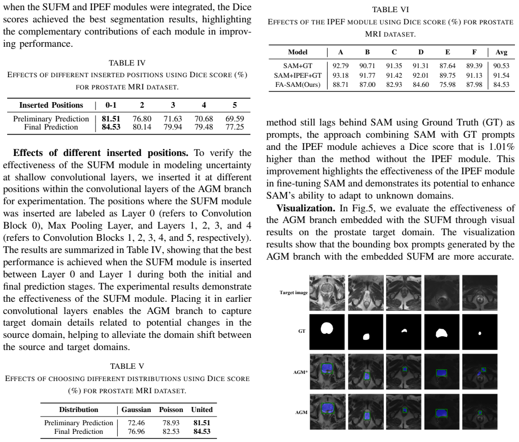
The load-bearing object is the SUFM module inside the AGM branch. SUFM takes the channel-wise mean $\mu$ and standard deviation $\sigma$ of shallow encoder features, estimates their batch-level uncertainty, samples perturbed statistics from $\mathcal{N}(\mu, \Sigma_\mu^2)$ and $\mathcal{N}(\sigma, \Sigma_\sigma^2)$, adds Poisson-distributed noise, and renormalizes the features via $\hat{f} = \gamma(x)\cdot (x-\mu(x))\cdot\sigma(x)^{-1} + \beta(x)$. This simulates target-domain feature statistics from source data alone, so the AGM's predicted mask leads to a better bounding-box prompt for SAM. The second mechanism is IPEF, which concatenates multi-scale SAM image embeddings with prompt embeddings, passes them through SEResBlocks, and upsamples with skip connections before the mask decoder, letting SAM recover local detail even when the box is too small or too large. Together these two modules carry the claim that SAM can be fully automated and made robust to poor prompts.
What would settle it
For each target site, compute the actual channel-wise mean and variance of the AGM's shallow features and check whether they are drawn from the distribution SUFM samples; a site whose statistics fall far outside that envelope, or whose generated boxes consistently cut off the target structure, would disconfirm the paper's core claim. A direct test is to report box-IoU between AGM-generated boxes and ground-truth masks per site: box-IoU near chance would show the automated prompting does not work even if final Dice is aided by IPEF.
Extended reading notes
Core claim
FA-SAM claims fully automated SAM-based segmentation under single-source domain generalization. The method treats SAM's prompt dependency as the bottleneck: instead of asking an expert for bounding boxes, a separate segmentation network (AGM) predicts a preliminary mask on the target image, keeps the largest connected component via breadth-first search, and converts it into a box prompt. Domain shift is handled by SUFM, which models the channel-wise mean and variance of the AGM encoder's shallow features as uncertain, samples perturbed statistics from a multivariate Gaussian, and adds Poisson-distributed noise to simulate real medical noise. The IPEF module then concatenates multi-scale SAM image embeddings with prompt embeddings, enhances them with SEResBlocks, and upsamples with skip connections so the mask decoder recovers local detail that a too-small or too-large box would otherwise erase. The paper reports the best average Dice on both benchmark suites and a 4.99% average gain over SAMMed on prostate.
Load-bearing premise
Everything rests on the assumption that unseen target domains can be simulated by perturbing the channel-wise mean and variance of shallow features with Gaussian samples and Poisson noise; if actual target shifts involve new anatomy, field-of-view changes, or artifacts these perturbations do not capture, the AGM will emit poor boxes and the automated pipeline loses its advantage.
Editorial extensions
If this is right
- SAM can be prompted without expert annotation: at test time the AGM branch generates the boxes, so FA-SAM runs end-to-end from raw target image to mask.
- On the prostate cross-site benchmark, average Dice over six sites improves to 84.53%, a 4.99% gain over the best SAM-based comparison, with the largest per-site gains on sites B and C.
- Fusing image and prompt embeddings with IPEF also helps when prompts are ground-truth boxes: SAM with IPEF and ground-truth prompts averages 91.54% versus 90.53% without IPEF.
- SUFM works best when inserted between the first and second convolutional blocks and when Gaussian and Poisson disturbances are combined, according to the paper's ablations.
Reading between the lines
- A straightforward extension would evaluate the generated bounding boxes themselves, measuring box IoU and size ratio relative to the mask on each target site; the paper reports final Dice but not box quality, so box quality is the unstated intermediate claim.
- If the SUFM assumption holds, the same auto-prompting recipe could apply to other prompt-based vision foundation models, with uncertainty modeled on whatever encoder features they expose.
- The reported gains leave open whether the method generalizes to 3D volumes, CT or ultrasound, or small lesions; testing those modalities would separate the benefit of automated prompting from the benefit of the two specific datasets used here.
Editorial analysis
A structured set of objections, weighed in public.
Referee Report
Summary. The paper proposes FA-SAM, a framework for fully automated SAM-based medical image segmentation under single-source domain generalization. It has two main components: an Auto-prompted Generation Model (AGM) branch with a Shallow Feature Uncertainty Modeling (SUFM) module that generates bounding-box prompts for unseen target domains, and an Image-Prompt Embedding Fusion (IPEF) module inserted in the SAM mask decoder to reduce sensitivity to poor prompts. The method is evaluated on a six-site prostate MRI dataset and a three-site fundus vessel dataset, reporting average Dice improvements over prior SAM-based and data-augmentation SDG baselines, including a claimed 4.99% average Dice improvement over SAMMed on prostate.
Significance. If the quantitative claims held, FA-SAM would address a genuine clinical bottleneck: removing the need for expert prompts while improving SAM's robustness under domain shift. The paper attacks a worthwhile problem, uses publicly available datasets, and includes ablations of the two proposed modules. The idea of coupling feature-statistics uncertainty with prompt generation is plausible and worth investigating. However, the current manuscript's evaluation protocol undermines the central generalization claim, because key design choices are selected using target-domain performance. The paper also lacks error bars and statistical tests, and some textual claims are contradicted by the tables. These issues must be resolved before the results can be interpreted as evidence of source-only domain generalization.
major comments (4)
- [Section IV-D (Tables IV and V)] The model-selection protocol leaks target-domain information into the reported generalization results. Table IV selects the SUFM insertion position '0-1' because it yields the best Final Prediction Dice on sites B–F, and Table V selects the combined Gaussian+Poisson distribution for the same reason. These choices are made using the very unseen domains on which the method is later evaluated in Table I, so the reported 84.53% average is not the performance of a fixed source-only model. The comparison with SAMMed and other baselines in Table I is therefore not a clean single-source generalization comparison. Please fix all hyperparameters and architectural choices using only the source domain (e.g., a source-domain validation split) and re-report all main results, or present the current numbers as an oracle-model-selection upper bound.
- [Tables I and II] All reported Dice scores are single runs with no standard deviations, confidence intervals, or significance tests. On the fundus dataset the target domains contain only 30 and 20 images, and on prostate many per-site differences between FA-SAM and SAMMed are small (e.g., site D 84.60 vs 82.40), while site E actually favors SAMMed. Without repeated-seed variability or a statistical test, the headline 4.99% average improvement over SAMMed cannot be distinguished from noise. Please provide at least three seeds and report mean ± std, and perform a paired significance test where the same test images are used.
- [Section IV-C and Table I] The text states that 'FA-SAM achieved the best overall performance in all unseen domains' and 'Our method outperformed all others,' but Table I shows that on site E, SAMMed scores 80.47% while FA-SAM scores 75.98%, and in Table II on site C, RAS4DG scores 72.88% while FA-SAM scores 71.67%. The claims should be restricted to average Dice or to the specific domains where the improvement holds. This is not a purely cosmetic issue: the paper's argument for robustness across unseen sites rests on the per-domain consistency that the data do not support.
- [Section III-B] The core assumption that source-to-target domain shift is representable by Gaussian/Poisson perturbations of channel-wise feature statistics is not validated, and the evidence offered in Table V is affected by the target-domain selection problem described above. Please provide a direct test of this assumption, for example by measuring the feature-statistic discrepancy between source and target encoders, or by showing that SUFM improves performance on a target domain that was not used in selecting the noise model. Absent such a validation, the mechanism behind the proposed uncertainty modeling remains unsupported.
minor comments (6)
- [Section III-B, Eqs. (1)–(5)] The notation is inconsistent: in Eq. (1) the feature f is written without indices, and the same symbol x is used both for the input image and for the argument of µ(x) and σ(x); Eq. (5) appears to normalize the feature map x using the original statistics but the notation is ambiguous. Please rewrite with consistent subscripts, e.g., f_bchw and normalized features, and define all variables.
- [Section III-D and Fig. 2] The IPEF module is repeatedly described as being inserted into the 'SAM mask encoder' (e.g., 'we introduced an Image-Prompt Embedding Fusion (IPEF) module into the SAM mask encoder' in the first paragraph and the caption of Fig. 2(B)), while the architecture and context indicate the mask decoder. Please correct the terminology throughout.
- [Section IV-B] The phrase 'the Adam decay optimizer' is not a standard optimizer name; please specify whether AdamW or Adam with weight decay is used, and clarify whether '200 iterations' and '100 iterations' refer to epochs or to gradient-descent iterations.
- [Table III] The checkmark pattern under the header (SUFM, AGM∗, IPEF, SAM) is ambiguous; the first row appears to combine SUFM with the base AGM and SAM, but it is marked only with two checkmarks. Please label each configuration explicitly.
- [Tables IV and V] The terms 'Preliminary Prediction' and 'Final Prediction' are used without a definition in the main text. Please define these as the AGM-only output and the final SAM output, respectively, and state what is averaged for the reported numbers.
- [Section III-C] The pipeline relies on taking the largest connected component of the AGM mask; please state explicitly that this post-processing is applied automatically, and discuss the case of disconnected or multiple target structures where the largest-component heuristic may fail.
Circularity Check
No significant circularity: FA-SAM's derivation chain is not self-referential, and its components are tested against external benchmarks; the target-domain leakage in hyperparameter selection is an evaluation-protocol concern, not a circular derivation.
full rationale
The paper's claimed derivation chain is not circular. The AGM branch with SUFM is trained on the source domain with a supervised loss and generates bounding-box prompts; the IPEF module fuses SAM image and prompt embeddings in the mask decoder; the final Dice scores are measured against external baselines on unseen target sites. No prediction is constructed by definition from a fitted quantity: SUFM perturbs source feature statistics with uncertainty sampling and Poisson noise, and the AGM is a separate trained network, not an inverse of the reported metric. The comparison to SAMMed, DeSAM, MedSAM, and other published methods provides independent external grounding, and the only author self-citations are routine context references, not load-bearing premises. The main validity concern is that Tables IV and V select the SUFM insertion position ('0-1') and the combined Gaussian+Poisson distribution using Dice on the target domains B-F, so the headline 84.53% average is partly a selection artifact and may overstate clean source-only generalization. That is a serious fairness and leakage issue, and a source-domain validation split would strengthen the evaluation, but it is not a mathematical circularity: the reported performance is not equal by construction to an input, and no fitted parameter is renamed as an independent prediction. The paper also honestly acknowledges in Table VI that FA-SAM trails SAM with ground-truth prompts, which further indicates the evaluation is not being presented as a tautological win.
Assumptions & free parameters
free parameters (4)
- SUFM insertion position =
between Layer 0 and Layer 1 (Convolution Blocks 0 and 1)
- Number of AGM and SAM training iterations =
200 and 100 iterations respectively
- Noise model for SUFM =
Gaussian plus Poisson
- Multi-scale encoder channels for IPEF =
256, 128, 64
assumptions (4)
- domain assumption Domain shift in medical images is primarily captured by shallow feature statistics such as color, texture, and contrast, so perturbing those statistics approximates unseen target distributions.
- ad hoc to paper The largest connected component of the AGM's preliminary mask contains the target structure, so all other components can be discarded before box-prompt generation.
- domain assumption Poisson plus Gaussian noise is an appropriate model for real medical imaging noise in feature-statistics space.
- domain assumption A Transformer-based SAM mask decoder neglects local and high-frequency details and therefore benefits from skip connections and multi-scale fusion.
Cite this review
Pith. "Pith review of Fully Automated SAM for Single-source Domain Generalization in Medical Image Segmentation." pith.science (2026). https://pith.science/paper/ZK37E72Q
@misc{pith2026250717281,
  author       = {Pith},
  title        = {Pith review of: Fully Automated SAM for Single-source Domain Generalization in Medical Image Segmentation},
  year         = {2026},
  howpublished = {\url{https://pith.science/paper/ZK37E72Q}},
  note         = {Machine review of arXiv:2507.17281}
}
read the original abstract
Although SAM-based single-source domain generalization models for medical image segmentation can mitigate the impact of domain shift on the model in cross-domain scenarios, these models still face two major challenges. First, the segmentation of SAM is highly dependent on domain-specific expert-annotated prompts, which prevents SAM from achieving fully automated medical image segmentation and therefore limits its application in clinical settings. Second, providing poor prompts (such as bounding boxes that are too small or too large) to the SAM prompt encoder can mislead SAM into generating incorrect mask results. Therefore, we propose the FA-SAM, a single-source domain generalization framework for medical image segmentation that achieves fully automated SAM. FA-SAM introduces two key innovations: an Auto-prompted Generation Model (AGM) branch equipped with a Shallow Feature Uncertainty Modeling (SUFM) module, and an Image-Prompt Embedding Fusion (IPEF) module integrated into the SAM mask decoder. Specifically, AGM models the uncertainty distribution of shallow features through the SUFM module to generate bounding box prompts for the target domain, enabling fully automated segmentation with SAM. The IPEF module integrates multiscale information from SAM image embeddings and prompt embeddings to capture global and local details of the target object, enabling SAM to mitigate the impact of poor prompts. Extensive experiments on publicly available prostate and fundus vessel datasets validate the effectiveness of FA-SAM and highlight its potential to address the above challenges.
Figures


Figures from the paper (2 more)

Forward citations
Cited by 1 Pith paper
-
Enhancing MedSAM with a Lightweight Box Predictor for Medical Image Segmentation
Enhances MedSAM with a 1.6M-parameter Box Predictor trained in two stages to convert single clicks to bounding boxes, reporting Dice scores of 0.89-0.98 on four medical datasets across CT, MRI, and ultrasound.
Reference graph
Works this paper leans on
-
[1]
L. Tang, Z. Yin, H. Su, W. Lyu, and B. Luo, “Wfss: weighted fusion of spectral transformer and spatial self-attention for robust hyperspectral image classification against adversarial attacks,” Visual Intelligence , vol. 2, no. 1, p. 5, 2024
work page 2024
-
[2]
Correl- ative and discriminative label grouping for multi-label visual prompt tuning,
L. Ma, S. Xu, M.-K. Xie, L. Wang, D. Sun, and H. Zhao, “Correl- ative and discriminative label grouping for multi-label visual prompt tuning,” in CVPR, 2025, pp. 25 434–25 443
work page 2025
-
[3]
Text-region matching for multi-label image recognition with missing labels,
L. Ma, H. Xie, L. Wang, Y . Fu, D. Sun, and H. Zhao, “Text-region matching for multi-label image recognition with missing labels,” in ACM MM, 2024, pp. 6133–6142
work page 2024
-
[4]
Uav-ground visual tracking: A unified dataset and collaborative learning approach,
D. Sun, L. Cheng, S. Chen, C. Li, Y . Xiao, and B. Luo, “Uav-ground visual tracking: A unified dataset and collaborative learning approach,” IEEE TCSVT , vol. 34, no. 5, pp. 3619–3632, 2023
work page 2023
-
[5]
Semantic-aware dual contrastive learning for multi-label image classification,
L. Ma, D. Sun, L. Wang, H. Zhao, and B. Luo, “Semantic-aware dual contrastive learning for multi-label image classification,” in ECAI, 2023, pp. 1656–1663
work page 2023
-
[6]
Domain adaptive lung nodule detection in x-ray image,
H. Zhao, L. Jiang, L. Ma, D. Sun, and Y . Fu, “Domain adaptive lung nodule detection in x-ray image,” in SMC. IEEE, 2024, pp. 1684– 1690
work page 2024
-
[7]
Semantic knowledge transfer for semi-supervised medical image segmentation,
S. Zhou, H. Zhao, L. Ma, and D. Sun, “Semantic knowledge transfer for semi-supervised medical image segmentation,” EAAI, vol. 158, p. 111235, 2025
work page 2025
-
[8]
High-order paired- aspp for deep semantic segmentation networks,
X. Sun, Y . Zhang, C. Chen, S. Xie, and J. Dong, “High-order paired- aspp for deep semantic segmentation networks,” Information Sciences, vol. 646, p. 119364, 2023
work page 2023
Show all 45 references
-
[9]
Domain generalization: A survey,
K. Zhou, Z. Liu, Y . Qiao, T. Xiang, and C. C. Loy, “Domain generalization: A survey,” IEEE TPAMI , vol. 45, no. 4, pp. 4396– 4415, 2022
2022
-
[10]
Rethinking data augmentation for single-source domain generalization in medical image segmentation,
Z. Su, K. Yao, X. Yang, K. Huang, Q. Wang, and J. Sun, “Rethinking data augmentation for single-source domain generalization in medical image segmentation,” in AAAI, vol. 37, 2023
2023
-
[11]
Exact feature distribu- tion matching for arbitrary style transfer and domain generalization,
Y . Zhang, M. Li, R. Li, K. Jia, and L. Zhang, “Exact feature distribu- tion matching for arbitrary style transfer and domain generalization,” in WACV, 2022, pp. 8035–8045
2022
-
[12]
Segment anything,
A. Kirillov, E. Mintun, N. Ravi, H. Mao, C. Rolland, L. Gustafson, T. Xiao, S. Whitehead, A. C. Berg, W.-Y . Lo et al. , “Segment anything,” in ICCV, 2023, pp. 4015–4026
2023
-
[13]
A comprehensive survey on segment anything model for vision and beyond,
C. Zhang, L. Liu, Y . Cui, G. Huang, W. Lin, Y . Yang, and Y . Hu, “A comprehensive survey on segment anything model for vision and beyond,” arXiv:2305.08196, 2023
2023 arXiv
-
[14]
Weaksam: Segment anything meets weakly-supervised instance-level recogni- tion,
L. Zhu, J. Zhou, Y . Liu, X. Hao, W. Liu, and X. Wang, “Weaksam: Segment anything meets weakly-supervised instance-level recogni- tion,” in ACM MM, 2024, pp. 7947–7956
2024
-
[15]
Segment anything in medical images,
J. Ma, Y . He, F. Li, L. Han, C. You, and B. Wang, “Segment anything in medical images,” Nature Communications , vol. 15, no. 1, p. 654, 2024
2024
-
[16]
Sam-u: Multi-box prompts triggered uncertainty estimation for reli- able sam in medical image,
G. Deng, K. Zou, K. Ren, M. Wang, X. Yuan, S. Ying, and H. Fu, “Sam-u: Multi-box prompts triggered uncertainty estimation for reli- able sam in medical image,” in MICCAI. Springer, 2023, pp. 368–377
2023
-
[17]
Ur- sam: Enhancing the reliability of segment anything model for auto- prompting medical image segmentation with uncertainty rectification,
Y . Zhang, S. Hu, S. Ren, T. Pan, C. Jiang, Y . Cheng, and Y . Qi, “Ur- sam: Enhancing the reliability of segment anything model for auto- prompting medical image segmentation with uncertainty rectification,” Available at SSRN 4878606 , 2023
2023
-
[18]
Leveraging sam for single-source domain generalization in medical image segmentation,
H. Wang, H. Ye, Y . Xia, and X. Zhang, “Leveraging sam for single-source domain generalization in medical image segmentation,” arXiv:2401.02076, 2024
2024 arXiv
-
[19]
Desam: Decoupled segment anything model for generalizable medical image segmenta- tion,
Y . Gao, W. Xia, D. Hu, W. Wang, and X. Gao, “Desam: Decoupled segment anything model for generalizable medical image segmenta- tion,” in MICCAI. Springer, 2024, pp. 509–519
2024
-
[20]
False negative/positive control for sam on noisy medical images,
X. Yao, H. Liu, D. Hu, D. Lu, A. Lou, H. Li, R. Deng, G. Arenas, B. Oguz, N. Schwartz et al. , “False negative/positive control for sam on noisy medical images,” arXiv:2308.10382, 2023
2023 arXiv
-
[21]
Samus: Adapting segment anything model for clinically-friendly and general- izable ultrasound image segmentation,
X. Lin, Y . Xiang, L. Zhang, X. Yang, Z. Yan, and L. Yu, “Samus: Adapting segment anything model for clinically-friendly and general- izable ultrasound image segmentation,” arXiv:2309.06824, 2023
2023 arXiv
-
[22]
Skinsam: Empowering skin can- cer segmentation with segment anything model. arxiv 2023,
M. Hu, Y . Li, and X. Yang, “Skinsam: Empowering skin can- cer segmentation with segment anything model. arxiv 2023,” arXiv:2304.13973, 2023
2023 arXiv
-
[23]
Segment anything model for med- ical image segmentation: Current applications and future directions,
Y . Zhang, Z. Shen, and R. Jiao, “Segment anything model for med- ical image segmentation: Current applications and future directions,” Computers in Biology and Medicine , p. 108238, 2024
2024
-
[24]
Causality-inspired single-source domain generalization for medical image segmentation,
C. Ouyang, C. Chen, S. Li, Z. Li, C. Qin, W. Bai, and D. Rueckert, “Causality-inspired single-source domain generalization for medical image segmentation,” IEEE TMI, vol. 42, no. 4, pp. 1095–1106, 2022
2022
-
[25]
Maxstyle: Adversarial style composition for robust medical image segmentation,
C. Chen, Z. Li, C. Ouyang, M. Sinclair, W. Bai, and D. Rueckert, “Maxstyle: Adversarial style composition for robust medical image segmentation,” in MICCAI. Springer, 2022, pp. 151–161
2022
-
[26]
Robust and generalizable visual representation learning via random convolutions,
Z. Xu, D. Liu, J. Yang, C. Raffel, and M. Niethammer, “Robust and generalizable visual representation learning via random convolutions,” arXiv:2007.13003, 2020
2007 arXiv
-
[27]
Domain generalization with mixstyle,
K. Zhou, Y . Yang, Y . Qiao, and T. Xiang, “Domain generalization with mixstyle,” arXiv:2104.02008, 2021
2021 arXiv
-
[28]
Uncertainty modeling for out-of-distribution generalization,
X. Li, Y . Dai, Y . Ge, J. Liu, Y . Shan, and L. Y . Duan, “Uncertainty modeling for out-of-distribution generalization,” arXiv e-prints , 2022
2022
-
[29]
From shallow feature learning to deep learning: Benefits from the width and depth of deep architec- tures,
G. Zhong, X. Ling, and L.-N. Wang, “From shallow feature learning to deep learning: Benefits from the width and depth of deep architec- tures,” Wiley Interdisciplinary Reviews: Data Mining and Knowledge Discovery, vol. 9, no. 1, p. e1255, 2019
2019
-
[30]
Meganet: Multi-scale edge-guided attention network for weak bound- ary polyp segmentation,
N.-T. Bui, D.-H. Hoang, Q.-T. Nguyen, M.-T. Tran, and N. Le, “Meganet: Multi-scale edge-guided attention network for weak bound- ary polyp segmentation,” in WACV, 2024, pp. 7985–7994
2024
-
[31]
Positron emission tomography,
G. Muehllehner and J. S. Karp, “Positron emission tomography,” Physics in Medicine & Biology , vol. 51, no. 13, p. R117, 2006
2006
-
[32]
Asps: Aug- mented segment anything model for polyp segmentation,
H. Li, D. Zhang, J. Yao, L. Han, Z. Li, and J. Han, “Asps: Aug- mented segment anything model for polyp segmentation,” in MICCAI. Springer, 2024, pp. 118–128
2024
-
[33]
Deep residual learning for image recognition,
K. He, X. Zhang, S. Ren, and J. Sun, “Deep residual learning for image recognition,” in CVPR, 2016, pp. 770–778
2016
-
[34]
Shape-aware meta-learning for gen- eralizing prostate mri segmentation to unseen domains,
Q. Liu, Q. Dou, and P. A. Heng, “Shape-aware meta-learning for gen- eralizing prostate mri segmentation to unseen domains,” in MICCAI. Springer, 2020
2020
-
[35]
Ridge-based vessel segmentation in color images of the retina
J. Staal, M. Abramoff, M. Niemeijer, M. A. Viergever, and B. Van Gin- neken, “Ridge-based vessel segmentation in color images of the retina.” IEEE TMI , vol. 23, no. 4, pp. 501–509, 2004
2004
-
[36]
Automatic detection of vascular bifurcations and crossings in retinal images using orientation scores,
S. Abbasi-Sureshjani, I. Smit-Ockeloen, E. Bekkers, B. Dashtbozorg, and B. ter Haar Romeny, “Automatic detection of vascular bifurcations and crossings in retinal images using orientation scores,” in ISBI, 2016, pp. 189–192
2016
-
[37]
Towards a glaucoma risk index based on simulated hemodynamics from fundus images,
J. I. Orlando, J. Barbosa Breda, K. Van Keer, M. B. Blaschko, P. J. Blanco, and C. A. Bulant, “Towards a glaucoma risk index based on simulated hemodynamics from fundus images,” in MICCAI. Springer, 2018, pp. 65–73
2018
-
[38]
Self-challenging improves cross-domain generalization,
Z. Huang, H. Wang, E. P. Xing, and D. Huang, “Self-challenging improves cross-domain generalization,” in ECCV. Springer, 2020, pp. 124–140
2020
-
[39]
Realistic adversarial data augmentation for mr image segmentation,
C. Chen, C. Qin, H. Qiu, C. Ouyang, S. Wang, L. Chen, G. Tarroni, W. Bai, and D. Rueckert, “Realistic adversarial data augmentation for mr image segmentation,” in MICCAI. Springer, 2020, pp. 667–677
2020
-
[40]
Cutmix: Regularization strategy to train strong classifiers with localizable features,
S. Yun, D. Han, S. J. Oh, S. Chun, J. Choe, and Y . Yoo, “Cutmix: Regularization strategy to train strong classifiers with localizable features,” in WACV, 2019, pp. 6023–6032
2019
-
[41]
Deep symmetric adaptation network for cross-modality medical image seg- mentation,
X. Han, L. Qi, Q. Yu, Z. Zhou, Y . Zheng, Y . Shi, and Y . Gao, “Deep symmetric adaptation network for cross-modality medical image seg- mentation,” IEEE TMI , vol. 41, no. 1, pp. 121–132, 2021
2021
-
[42]
Generalizable cross- modality medical image segmentation via style augmentation and dual normalization,
Z. Zhou, L. Qi, X. Yang, D. Ni, and Y . Shi, “Generalizable cross- modality medical image segmentation via style augmentation and dual normalization,” CVPR, 2021
2021
-
[43]
Semantic-aware domain generalized segmentation,
D. Peng, Y . Lei, M. Hayat, Y . Guo, and W. Li, “Semantic-aware domain generalized segmentation,” in CVPR, 2022
2022
-
[44]
Frequency- mixed single-source domain generalization for medical image segmen- tation,
H. Li, H. Li, W. Zhao, H. Fu, X. Su, Y . Hu, and J. Liu, “Frequency- mixed single-source domain generalization for medical image segmen- tation,” MICCAI, 2023
2023
-
[45]
Medical image segmentation via single-source domain generalization with random amplitude spectrum synthesis,
Q. Qiao, W. Wang, Q. Meixia, S. Kun, J. Bin, and G. Qiang, “Medical image segmentation via single-source domain generalization with random amplitude spectrum synthesis,” in MICCAI, 2024
2024
Reviewed August 6, 2026 · model on record in the stance chip above.
Discussion (0). Sign in to comment.