REVIEW 4 major objections 4 minor 1 cited by
Estimation of Hemodynamic Parameters via Physics Informed Neural Networks including Hematocrit Dependent Rheology
T0 review · 4 major / 4 minor · reviewed 2026-08-06 · deepseek-v4-flash
Pith's one-line read The paper claims that a physics-informed neural network can reconstruct aortic velocity, pressure, and hematocrit-dependent viscosity from low-resolution 4D-flow MRI-like data, and that combining it with the vWERP estimator yields the…
desk verdict Solid synthetic benchmark for PINN-based hemodynamic state estimation, but the abstract's pressure-drop accuracy claim is contradicted by the paper's own Table 5. read the letter →
The pith
A machine-rendered reading of the paper's core claim, the machinery that carries it, and where it could break.
The reading
What carries the argument
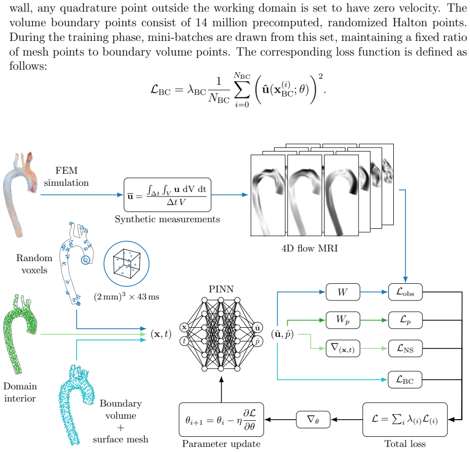
The load-bearing object is the neural field together with a measurement operator that mimics 4D-flow MRI: instead of point samples, the observation loss compares the network to local spatiotemporal averages over 2-mm voxels and 42.6-ms cardiac phases, computed by quasi-Monte Carlo quadrature with scrambled low-discrepancy points. This operator connects the low-resolution data to the high-resolution field, and a boundary-volume loss makes the network's local averages see zero velocity outside the lumen. The physics are enforced pointwise by automatic differentiation of the power-law Navier-Stokes residual at roughly 20 million collocation points, with adaptive inverse-Dirichlet loss weighting and a two-stage curriculum that first fits the data alone and then activates the physics. For pressure drops, the vWERP estimator, a virtual work-energy identity that uses solenoidal Stokes test functions to convert the weak Navier-Stokes equations into outlet pressure differences, is fed either the raw images or the PINN's smooth velocity; the second route is the paper's winning strategy.
What would settle it
A direct test would run the same pipeline on real 4D-flow MRI acquisitions of an aorta with a known pressure-drop reference (for example, from catheterization or a phantom with a known stenosis). If, under real noise and artifacts, the combined PINN+vWERP estimate's peak pressure-drop error exceeds the roughly 8-11% range seen in the synthetic experiments, the claimed clinically admissible accuracy would be falsified for clinical data.
Extended reading notes
Core claim
On its own terms, the work establishes a reconstruction pipeline: a fully connected neural field maps space-time points to velocity and pressure, and is trained by minimizing a sum of four losses: voxel-averaged velocity observations through a realistic observer operator, one scalar mean-pressure observation, Navier-Stokes residuals with a power-law viscosity whose consistency index and power-law index depend on hematocrit, and a no-slip wall condition enforced in a boundary volume. The paper's central quantitative claims are that this yields pressure fields with relative error between 0.93% and 1.66% over the whole space-time domain across all five hematocrit levels, that the velocity reconstructions reproduce the bifurcation of flow through the aortic branches with errors of a few percent, and that the best pressure-drop estimates come from post-processing the PINN velocity with vWERP, outperforming both direct PINN pressure reads and raw-image vWERP. In the descending aorta, the best peak pressure-drop error drops to about 8% with the combined method, while direct PINN errors range roughly from 27% to 70% depending on outlet and hematocrit. The authors conclude that the best strategy is to reconstruct first a high-fidelity, Navier-Stokes-consistent velocity field and then apply a work-energy estimator to recover the pressure drop.
Load-bearing premise
The load-bearing premise is that the synthetic 4D-flow MRI measurements are representative of real clinical data, meaning they include no noise, aliasing, flow-displacement artifacts, or other image degradation beyond local spatiotemporal averaging.
Editorial extensions
If this is right
- Pressure-drop biomarkers for aortic coarctation, valve stenosis, and congenital heart disease could be computed non-invasively from low-quality 4D-flow acquisitions, avoiding risky catheterization.
- The PINN+vWERP pipeline recovers systolic pressure-drop peaks that raw low-time-resolution images miss, because the network supplies about 1000 samples per cardiac cycle instead of the 22 image phases.
- Estimates remain accurate across a wide hematocrit range, suggesting the method applies to anemic, normal, and polycythemic patients without switching rheology models.
- The same reconstructed fields locate high wall-shear-stress regions near the inflow jet impact, a region relevant for identifying disease-prone areas of the aortic wall.
Reading between the lines
- This is an editorial inference, not the paper's claim: because the same power-law model generates the synthetic data and supplies the physics loss, the reported accuracy is a self-consistency check, and real MRI noise, aliasing, and flow-displacement artifacts would likely degrade it in ways the paper does not quantify.
- The hybrid strategy suggests a general principle: for derivative-based or integral biomarkers, smooth physics-constrained velocity reconstruction can act as a denoiser and time-supersampler upstream of classical estimators, and testing this on wall shear stress and oscillatory shear index is a natural next step.
- Replacing the power-law model with a different constitutive law, or adding vessel-wall compliance, would test whether the gains persist when the data-generation model and the network's physics model are not identical.
- Because the measurement operator already handles spatial and temporal voxel averaging, adding realistic noise models directly into the synthetic data generation would give a low-cost read on how much of the claimed accuracy survives clinical conditions.
Editorial analysis
A structured set of objections, weighed in public.
Referee Report
Summary. The paper presents a physics-informed neural network (PINN) framework for estimating velocity, pressure, and apparent viscosity fields from synthetic 4D-flow MRI data in a realistic aortic geometry, using a hematocrit-dependent power-law rheology model. Methodological contributions include a voxel-averaging observation operator, curriculum training, adaptive loss balancing, and residual-based collocation refinement. The authors evaluate five hematocrit levels, report pressure-field reconstruction errors below 1.66%, and compare pressure-drop estimates from direct PINN evaluation, the vWERP estimator alone, and a hybrid PINN+vWERP strategy that super-samples the PINN velocity field. The central quantitative claims are that pressure fields are accurate, that pressure-drop estimates are clinically admissible, and that PINN-based approaches outperform vWERP.
Significance. If the claims are appropriately scoped, the paper contributes a useful demonstration of PINN state estimation with a realistic 4D-flow measurement operator and systematic variation of hematocrit-driven rheology. The pressure-field errors (0.93-1.66%, Fig. 10) are well quantified and support the core methodological claim of accurate whole-field pressure reconstruction. The hybrid PINN+vWERP idea is sensible and the qualitative improvement in time resolution is plausible. However, the paper's headline claims about pressure-drop accuracy and superiority over vWERP are not supported by the tabulated numbers, and the noiseless synthetic-data setup limits the clinical interpretation. These issues are fixable by reframing claims and adding missing quantitative comparisons.
major comments (4)
- [Abstract; Section 5.3; Table 5] The abstract's phrase "pressure drops with relative errors below the 5% in the whole pressure field" is ambiguous, and the Introduction states "particularly accurate pressure drop estimations, relative errors as low as 1%" (end of Section 1). Table 5 reports e_dp values between 27.30% and 69.68% for the direct PINN and between 7.97% and 36.61% for PINN+vWERP across outlets and hematocrit levels. Unless the intended claim concerns only the whole pressure field (Fig. 10, errors 0.93-1.66%), the abstract and Introduction overstate the accuracy of pressure-drop estimates. Please rewrite these claims to distinguish whole-field pressure errors from the pressure-drop biomarker, and provide an explicit clinical threshold if "clinically admissible accuracy" is retained.
- [Abstract; Section 5.3; Table 5; Figure 12] The claim that PINN-based methods "outperform vWERP in terms of both accuracy and time resolution" is not quantitatively verifiable from the paper. Table 5 reports errors only for the three PINN-based strategies, and no numerical e_dp values are given for vWERP alone. Figure 12 shows only one hematocrit level (32.5%) and no error metric. Please add vWERP1st and vWERP2nd columns to Table 5 (or a companion table) and specify the comparison metric. In addition, e_dp is defined on the maximum absolute pressure drop over the cycle; a time-resolved error measure (e.g., L2 relative error over the cardiac phase per outlet) would strengthen the claim that the super-sampled methods are more accurate throughout the cycle, not only at the peak.
- [Section 3.3; Abstract] The synthetic 4D-flow data are generated without noise or artifacts (Section 3.3: "without taking into account noise or the more complex artifacts typically associated with MR imaging"), and the same power-law Navier-Stokes model generates the data and acts as the physics prior in the PINN loss. The experiments are therefore a consistency check rather than a clinical validation. The abstract's "clinically admissible accuracy" overreaches this evidence. Please add a limitations paragraph and reframe the clinical claims as proof-of-concept, or include experiments with realistic noise, aliasing, and displacement artifacts.
- [Eq. (3.1); Table 3; Section 5.2; Figure 8] The pressure inference relies on the ground-truth global mean pressure pmean as a data term (Eq. 3.1, Table 3), and all pressure-error metrics are computed after shifting the estimated pressure to match the reference mean at each time instant (Section 5.2, Fig. 8 caption). This should be stated explicitly as a limitation: the PINN does not estimate the pressure constant from 4D-flow velocity data alone. The paper should discuss how pmean would be obtained in a clinical setting and assess the sensitivity of the reported pressure-field errors to uncertainty in pmean. Relative pressure drops are less affected, but the presentation of pressure-field accuracy needs this caveat.
minor comments (4)
- [Section 5.1, Figure 6 discussion] The text says "For the 35% hematocrit, the difference was 4.4%" but the simulated hematocrit levels are 20%, 32.5%, 45%, 57.5%, and 70%; this should read 32.5%.
- [Table 5 caption and Eq. (5.1)] The caption of Table 5 says "Errors in absolute maxima" while the text defines e_dp using max|δp|; please clarify that the maximum is taken over time and that the error is relative to the reference maximum, and state whether all outlets follow the same convention.
- [Figure 8 caption] The figure caption mentions that pressure is corrected to the same average as the reference at each timestep, but the main text also applies this correction for the error metrics in Section 5.2; making this explicit in the text near Eq. (5.1) would avoid confusion about what the reported pressure errors represent.
- [Section 3.2, implementation details] The statement that all code is written in PyTorch could be complemented by a data/code availability statement; this is not required for acceptance but would improve reproducibility.
Circularity Check
No significant circularity: the pressure-drop estimates are genuinely inferred from velocity observations and the Navier-Stokes constraint, not defined in terms of the target; the main issues are in-sample validation and overstated accuracy, not circular derivation.
full rationale
Tracing the derivation chain, the PINN reconstruction is produced from sparse voxel-averaged velocity observations, the global mean pressure pmean, the Navier-Stokes residual with a power-law viscosity, and no-slip boundary enforcement. The pressure drop of Eq. (4.1) is a spatial average difference, so the pmean anchor in Eq. (3.1) fixes only the arbitrary pressure constant and does not determine the drops by construction. The vWERP benchmark is an independently published estimator ([6], with overlapping authorship) and is used as a comparison, not as a premise from which the PINN result is derived. The apparent viscosity field is indeed computed from the assumed constitutive law with tabulated m and n, so the 'viscosity estimation' claim is a derived-field claim rather than a parameter identification; this limits novelty but is not circular because the velocity field itself is inferred from data. The substantive limitations are clearly located in the paper: Section 3.3 states that the synthetic data are noiseless and artifact-free, Section 2.4 and Section 3.1 use the same forward model both to generate the ground truth and to build the physics loss, making the evaluation a consistency check rather than an independent clinical validation, and the pressure-field errors are reported after shifting the estimated pressure to the reference mean (Section 5.1 and Figure 8 caption). Additionally, Table 5 reports pressure-drop errors of 7.97% to 69.68%, which contradict the abstract's 'below the 5%' phrasing and weaken the 'clinically admissible accuracy' claim. These are correctness and generalization concerns, not reductions of a predicted quantity to its inputs. No equation in the paper is defined in terms of the quantity it purports to predict, and no fitted parameter is renamed as a prediction. Therefore no significant circularity is present in the claimed derivation chain.
Assumptions & free parameters
free parameters (1)
- pmean (global mean pressure) =
136381 to 136530 Ba depending on hematocrit
assumptions (5)
- domain assumption Incompressible Navier-Stokes equations with rigid walls describe thoracic aortic blood flow.
- domain assumption Power-law constitutive model with parameters from Table 2 accurately represents blood rheology at the five hematocrit levels.
- domain assumption Synthetic MRI voxel values are noise-free local spatiotemporal averages of velocity.
- ad hoc to paper A fully connected neural network with 6 hidden layers of 256 neurons can represent the velocity and pressure fields.
- domain assumption Windkessel outlet parameters from the Vascular Model Repository are known inputs.
Cite this review
Pith. "Pith review of Estimation of Hemodynamic Parameters via Physics Informed Neural Networks including Hematocrit Dependent Rheology." pith.science (2026). https://pith.science/paper/3U5PTWJ6
@misc{pith2026250803326,
  author       = {Pith},
  title        = {Pith review of: Estimation of Hemodynamic Parameters via Physics Informed Neural Networks including Hematocrit Dependent Rheology},
  year         = {2026},
  howpublished = {\url{https://pith.science/paper/3U5PTWJ6}},
  note         = {Machine review of arXiv:2508.03326}
}
read the original abstract
Physics-Informed Neural Networks (PINNs) show significant potential for solving inverse problems, especially when observations are limited and sparse, provided that the relevant physical equations are known. We use PINNs to estimate smooth velocity and pressure fields from synthetic 4D flow Magnetic Resonance Imaging (MRI) data. We analyze five non-Newtonian dynamic 3D blood flow cases within a realistic aortic model, covering a range of hematocrit values from anemic to polycythemic conditions. To enhance state estimation results, we consider various design and training techniques for PINNs, including adaptive loss balancing, curriculum training, and a realistic measurement operator. Regarding blood rheology, the PINN approach accurately estimates viscosity globally and locally under peak systolic conditions. It also provides a clear pattern recognition for diastolic stages. Regarding mass conservation, PINN estimations effectively reproduce the bifurcation of flow through the different branches of the aorta, demonstrate an excellent representation of the non-slip conditions at the walls, and accurately estimate pressure drops with relative errors below the 5% in the whole pressure field. We test our pressure drop estimations against the state of the art Virtual Work Energy Relative Pressure (vWERP) estimator, and we observe how our results outperform vWERP in terms of both accuracy and time resolution. Additionally, we find that the best results are achieved by computing the velocity field using the PINN, which is then integrated into the vWERP framework, leading to time super-sampled and high-order approximations, with a clinically admissible accuracy.
Figures


Figures from the paper (9 more)


Forward citations
Cited by 1 Pith paper
-
Wall Shear Stress Reconstruction from Concentration: Differentiable Physics and Physics-Informed Neural Networks
Differentiable physics recovers accurate wall shear stress from concentration observations across measurement scenarios where PINNs fail.
Reference graph
Works this paper leans on
-
[1]
A. Azarine, P. Gar¸ con, A. Stansal, N. Canepa, G. Angelopoulos, S. Silvera, D. Sidi, V. Marteau, M. Zins, Four-dimensional Flow MRI: Principles and Cardiovascular Ap- plications, RadioGraphics 39 (3) (2019) 632–648. doi:https://doi.org/10.1148/rg. 2019180091
work page doi:10.1148/rg 2019
-
[2]
G. Soulat, P. McCarthy, M. Markl, 4D Flow with MRI, Annual review of biomedical engineering 22 (1) (2020) 103–126, publisher: Annual Reviews. doi:https://doi.org/ 10.1146/annurev-bioeng-100219-110055
-
[3]
M. Terada, Y. Takehara, H. Isoda, T. Wakayama, A. Nozaki, Technical Background for 4D Flow MR Imaging, Magnetic Resonance in Medical Sciences 21 (2) (2022) 267–277. doi:https://doi.org/10.2463/mrms.rev.2021-0104
-
[4]
Fumagalli, S
I. Fumagalli, S. Pagani, C. Vergara, L. Dede, A. D.A., M. Del Greco, A. Frontera, G. Luciani, G. Pontone, R. Scrofani, A. Quarteroni, The role of computational methods in cardiovascular medicine: a narrative review, Transl Pediatr. 13 (2024). doi:10. 21037/tp-23-184
2024
-
[5]
D. Nolte, C. Bertoglio, Inverse problems in blood flow modeling: A review, International Journal for Numerical Methods in Biomedical Engineering 38 (8) (2022) e3613. doi: https://doi.org/10.1002/cnm.3613
doi:10.1002/cnm.3613 2022
-
[6]
C. Bertoglio, R. Nu˜ nez, F. Galarce, D. Nordsletten, A. Osses, Relative pressure estima- tion from velocity measurements in blood flows: State-of-the-art and new approaches, International Journal for Numerical Methods in Biomedical Engineering 34 (2), cited by: 24; All Open Access, Green Open Access (2018). doi:10.1002/cnm.2925. URL https://www.scopus.co...
-
[7]
F. M. Rijnberg, H. C. van Assen, J. F. Juffermans, L. J. M. Kroft, P. J. van den Boogaard, P. J. H. de Koning, M. G. Hazekamp, S. F. S. van der Woude, E. G. Warmer- dam, T. Leiner, H. B. Grotenhuis, J. J. Goeman, H. J. Lamb, A. A. W. Roest, J. J. M. Westenberg, Reduced scan time and superior image quality with 3D flow MRI compared to 4D flow MRI for hemod...
-
[8]
M. J. F. G. Ramaekers, J. J. M. Westenberg, B. P. Adriaans, E. C. Nijssen, J. E. Wildberger, H. J. Lamb, S. Schalla, A clinician’s guide to understanding aortic 4D flow MRI, Insights into Imaging 14 (1) (2023) 114. doi:10.1186/s13244-023-01458-x
Show all 87 references
-
[9]
Montalba, J
C. Montalba, J. Urbina, J. Sotelo, M. E. Andia, C. Tejos, P. Irarrazaval, D. E. Hurtado, I. Valverde, S. Uribe, Variability of 4D flow parameters when subjected to changes in MRI acquisition parameters using a realistic thoracic aortic phantom, Magnetic Res- onance in Medicine...
2018 doi
-
[10]
Demirkiran, P
A. Demirkiran, P. van Ooij, J. J. M. Westenberg, M. B. M. Hofman, H. C. van Assen, L. J. Schoonmade, U. Asim, C. P. S. Blanken, A. J. Nederveen, A. C. van Rossum, M. J. W. G¨ otte, Clinical intra-cardiac 4D flow CMR: Acquisition, analysis, and clinical applications, European H...
2022 doi
-
[11]
Berhane, M
H. Berhane, M. B. Scott, A. J. Barker, P. McCarthy, R. Avery, B. Allen, C. Malaisrie, J. D. Robinson, C. K. Rigsby, M. Markl, Deep learning–based velocity antialiasing of 4D-flow MRI, Magnetic Resonance in Medicine 88 (1) (2022) 449–463. doi:10.1002/ mrm.29205
2022
-
[12]
Schmidt, S
S. Schmidt, S. Flassbeck, S. Schmelter, E. Schmeyer, M. E. Ladd, S. Schmitter, The impact of 4D flow displacement artifacts on wall shear stress estimation, Magnetic Resonance in Medicine 85 (6) (2021) 3154–3168. doi:10.1002/mrm.28641
2021 doi
-
[13]
S. You, E. M. Masutani, M. T. Alley, S. S. Vasanawala, P. R. Taub, J. Liau, A. C. Roberts, A. Hsiao, Deep Learning Automated Background Phase Error Cor- rection for Abdominopelvic 4D Flow MRI, Radiology 302 (3) (2022) 584–592. doi: 10.1148/radiol.2021211270
2022 doi
-
[14]
Mart ´ ınez, M
A. Mart ´ ınez, M. Hoeijmakers, L. Geronzi, V. Morgenthaler, J. Tomasi, M. Rochette, M. E. Biancolini, Effect of turbulence and viscosity models on wall shear stress derived biomarkers for aorta simulations, Computers in Biology and Medicine 167 (2023) 107603. doi:https://doi....
2023
-
[15]
Z. Yin, C. Armour, H. Kandail, D. P. O’Regan, T. Bahrami, S. Mirsadraee, S. Pirola, X. Y. Xu, The impact of coronary outflow and non-Newtonian fluid property on aor- 33 tic valve haemodynamics, Biomechanics and Modeling in Mechanobiology (Jun. 2025). doi:10.1007/s10237-025-01975-2
2025 doi
-
[16]
Mella, F
H. Mella, F. Galarce, T. Sekine, J. Sotelo, E. Castillo, Evaluating the impact of blood rheology in hemodynamic parameters by 4D Flow MRI in large vessels considering the hematocrit effect, Biomedical Signal Processing and Control 111 (2026) 108145. doi:10.1016/j.bspc.2025.108145
2026
-
[17]
Carvalho, D
V. Carvalho, D. Pinho, R. A. Lima, J. C. Teixeira, S. Teixeira, Blood Flow Modeling in Coronary Arteries: A Review, Fluids 6 (2) (2021). doi:https://doi.org/10.3390/ fluids6020053
2021
-
[18]
K. Zhao, P. Malhotra, D. Rosen, P. Dalton, E. A. Pribitkin, Computational Fluid Dynamics as Surgical Planning Tool: A Pilot Study on Middle Turbinate Resection, The Anatomical Record 297 (11) (2014) 2187–2195. doi:https://doi.org/10.1002/ ar.23033
2014
-
[19]
J. Li, X. Cao, L. Xu, L. Qi, Finite element analysis for the effects of the descending aorta tortuosity on aortic hemodynamics, Procedia Computer Science 209 (2022) 148– 156, proceedings of the 2022 International Symposium on Biomimetic Intelligence and Robotics (ISBIR). doi:h...
2022 doi
-
[20]
X. Liu, H. Zhang, L. Ren, H. Xiong, Z. Gao, P. Xu, W. Huang, W. Wu, Functional assessment of the stenotic carotid artery by CFD-based pressure gradient evaluation, American Journal of Physiology-Heart and Circulatory Physiology 311 (3) (2016) H645– H653. doi:https://doi.org/10...
2016
-
[21]
Galarce, D
F. Galarce, D. Lombardi, O. Mula, Reconstructing haemodynamics quantities of inter- est from Doppler ultrasound imaging, International Journal for Numerical Methods in Biomedical Engineering 37 (2) (2021) e3416. doi:10.1002/cnm.3416. URL https://onlinelibrary.wiley.com/doi/10....
2021 doi
-
[22]
Lombardi, Chapter 12 - reduced order modelling for direct and in- verse problems in haemodynamics, in: F
D. Lombardi, Chapter 12 - reduced order modelling for direct and in- verse problems in haemodynamics, in: F. Chinesta, E. Cueto, Y. Payan, J. Ohayon (Eds.), Reduced Order Models for the Biomechanics of Living Or- gans, Biomechanics of Living Organs, Academic Press, 2023, pp. 2...
2023 doi
-
[23]
P. D. Morris, A. Narracott, H. von Tengg-Kobligk, D. A. Silva Soto, S. Hsiao, A. Lungu, P. Evans, N. W. Bressloff, P. V. Lawford, D. R. Hose, J. P. Gunn, Computational fluid dynamics modelling in cardiovascular medicine, Heart 102 (1) (2016) 18–28, publisher: BMJ Publishing Gr...
2016 doi
-
[24]
Abugattas, A
C. Abugattas, A. Aguirre, E. Castillo, M. Cruchaga, Numerical study of bifurcation blood flows using three different non-newtonian constitutive models, Applied Mathe- matical Modelling 88 (2020) 529–549. doi:https://doi.org/10.1016/j.apm.2020. 06.066. URL https://www.sciencedi...
2020 doi
-
[25]
Lynch, N
S. Lynch, N. Nama, C. A. Figueroa, Effects of non-newtonian viscosity on arterial and venous flow and transport, Scientific Reports 12 (1) (2022) 20568
2022
-
[26]
Far ´ ıas, C
C. Far ´ ıas, C. Bayona-Roa, E. Castillo, R. C. Cabrales, R. Reyes, Reduced order modeling of parametrized pulsatile blood flows: Hematocrit percentage and heart rate, International Journal of Engineering Science 193 (2023) 103943. doi:https: //doi.org/10.1016/j.ijengsci.2023....
2023
-
[27]
Chauhan, C
A. Chauhan, C. Sasmal, Effect of real and whole blood rheology on flow through an axisymmetric stenosed artery, International Journal of Engineering Science 169 (2021) 103565. doi:https://doi.org/10.1016/j.ijengsci.2021.103565. URL https://www.sciencedirect.com/science/article...
2021
-
[28]
Elhanafy, Y
A. Elhanafy, Y. Abuouf, S. Elsagheer, S. Ookawara, S. Nada, M. Ahmed, Effect of micro-vessel stenosis severity and hematocrit level on red blood cell dynamics and platelet margination: A numerical study, International Journal of Engineering Science 205 (2024) 104155. doi:https...
2024
-
[29]
H. J. Carpenter, A. Gholipour, M. H. Ghayesh, A. C. Zander, P. J. Psaltis, A review on the biomechanics of coronary arteries, International Journal of Engineering Science 147 (2020) 103201. doi:https://doi.org/10.1016/j.ijengsci.2019.103201. URL https://www.sciencedirect.com/s...
2020
-
[30]
Y. Qiao, J. Fan, K. Luo, Mechanism of blood flow energy loss in real healthy aorta using computational fluid-structure interaction framework, International Journal of En- gineering Science 192 (2023) 103939. doi:https://doi.org/10.1016/j.ijengsci. 2023.103939. URL https://www....
2023
-
[31]
Huang, M
H. Huang, M. Yang, W. Zang, S. Wu, Y. Pang, In Vitro Identification of Four-Element Windkessel Models Based on Iterated Unscented Kalman Filter, IEEE Transactions on Biomedical Engineering 58 (9) (2011) 2672–2680. doi:https://doi.org/10.1109/ TBME.2011.2161477
2011
-
[32]
Fevola, F
E. Fevola, F. Ballarin, L. Jim´ enez-Juan, S. Fremes, S. Grivet-Talocia, G. Rozza, P. Trive- rio, An optimal control approach to determine resistance-type boundary conditions from 35 in-vivo data for cardiovascular simulations, International Journal for Numerical Meth- ods in ...
2021
-
[33]
C. A. Warnes, R. G. Williams, T. M. Bashore, J. S. Child, H. M. Connolly, J. A. Dearani, P. del Nido, J. W. Fasules, T. P. Graham, Z. M. Hijazi, S. A. Hunt, M. E. King, M. J. Landzberg, P. D. Miner, M. J. Radford, E. P. Walsh, G. D. Webb, Acc/aha 2008 guidelines for the manage...
2008
-
[34]
S. Zhou, K. Xu, Y. Fang, J. Alastruey, S. Vennin, J. Yang, J. Wang, L. Xu, X. Wang, S. E. Greenwald, Patient-specific non-invasive estimation of the aortic blood pressure waveform by ultrasound and tonometry, Computer Methods and Programs in Biomedicine 247 (2024) 108082. doi:...
2024 doi
-
[35]
Ebbers, L
T. Ebbers, L. Wigstr¨ om, A. Bolger, J. Engvall, M. Karlsson, Estimation of relative car- diovascular pressures using time-resolved three-dimensional phase contrast MRI, Mag- netic Resonance in Medicine 45 (5) (2001) 872–879. doi:https://doi.org/10.1002/ mrm.1116
2001
-
[36]
Donati, C
F. Donati, C. A. Figueroa, N. P. Smith, P. Lamata, D. A. Nordsletten, Non-invasive pressure difference estimation from pc-mri using the work-energy equation, Medical Im- age Analysis 26 (1) (2015) 159–172. doi:https://doi.org/10.1016/j.media.2015. 08.012. URL https://www.scien...
2015 doi
-
[37]
ˇSvihlov´ a, J
H. ˇSvihlov´ a, J. Hron, J. M´ alek, K. Rajagopal, K. Rajagopal, Determination of pressure data from velocity data with a view toward its application in cardiovascular mechanics. Part 1: Theoretical considerations, International Journal of Engineering Science 105 (2016) 108–12...
2016 doi
-
[38]
Galarce, J.-F
F. Galarce, J.-F. Gerbeau, D. Lombardi, O. Mula, Fast reconstruction of 3D blood flows from Doppler ultrasound images and reduced models, Computer Methods in Applied Mechanics and Engineering 375 (2021) 113559. doi:10.1016/j.cma.2020.113559. URL https://linkinghub.elsevier.com...
2021
-
[39]
Alzubaidi, J
L. Alzubaidi, J. Bai, A. Al-Sabaawi, J. Santamar ´ ıa, A. S. Albahri, B. S. N. Al-Dabbagh, M. A. Fadhel, M. Manoufali, J. Zhang, A. H. Al-Timemy, et al., A survey on deep learning tools dealing with data scarcity: definitions, challenges, solutions, tips, and applications, Jou...
2023
-
[40]
Cuomo, V
S. Cuomo, V. Di Cola, F. Giampaolo, G. Rozza, M. Raissi, F. Piccialli, Scientific ma- chine learning through physics–informed neural networks: Where we are and what’s next, Journal of Scientific Computing 92 (2022). doi:10.1007/s10915-022-01939-z
2022 doi
-
[41]
Raissi, P
M. Raissi, P. Perdikaris, G. E. Karniadakis, Physics-informed neural networks: A deep learning framework for solving forward and inverse problems involving nonlinear partial differential equations, Journal of Computational Physics 378 (2019) 686–707. doi: https://doi.org/10.10...
2019 doi
-
[42]
S. Wang, Y. Teng, P. Perdikaris, Understanding and mitigating gradient pathologies in physics-informed neural networks, SIAM Journal on Scientific Computing 43 (5) (2021) A3055–A3081. doi:https://doi.org/10.1137/20M1318043
2021 doi
- [43]
-
[44]
Fabra, O
A. Fabra, O. Guasch, J. Baiges, R. Codina, Approximation of acoustic black holes with finite element mixed formulations and artificial neural network correction terms, Finite Elements in Analysis and Design 241 (2024) 104236. doi:https://doi.org/10.1016/ j.finel.2024.104236. U...
2024
-
[45]
G. E. Karniadakis, I. G. Kevrekidis, L. Lu, P. Perdikaris, S. Wang, L. Yang, Physics- informed machine learning, Nature Reviews Physics 3 (6) (2021) 422–440, publisher: Nature Publishing Group. doi:https://doi.org/10.1038/s42254-021-00314-5
2021 doi
-
[46]
J. D. Toscano, V. Oommen, A. J. Varghese, Z. Zou, N. Ahmadi Daryakenari, C. Wu, G. E. Karniadakis, From PINNs to PIKANs: recent advances in physics-informed ma- chine learning, Machine Learning for Computational Science and Engineering 1 (1) (2025) 15. doi:10.1007/s44379-025-0...
2025 doi
-
[47]
Kissas, Y
G. Kissas, Y. Yang, E. Hwuang, W. R. Witschey, J. A. Detre, P. Perdikaris, Machine learning in cardiovascular flows modeling: Predicting arterial blood pressure from non- invasive 4D flow MRI data using physics-informed neural networks, Computer Methods in Applied Mechanics an...
2020
-
[48]
Sarabian, H
M. Sarabian, H. Babaee, K. Laksari, Physics-Informed Neural Networks for Brain Hemo- dynamic Predictions Using Medical Imaging, IEEE Transactions on Medical Imaging 41 (9) (2022) 2285–2303. doi:10.1109/TMI.2022.3161653. URL https://ieeexplore.ieee.org/document/9740143/
2022
-
[49]
Changdar, B
S. Changdar, B. Bhaumik, N. Sadhukhan, S. Pandey, S. Mukhopadhyay, S. De, S. Bakalis, Integrating symbolic regression with physics-informed neural net- works for simulating nonlinear wave dynamics in arterial blood flow, Physics of Fluids 36 (12) (2024) 121924, tex.eprint: htt...
2024 doi
-
[50]
M. F. Fathi, I. Perez-Raya, A. Baghaie, P. Berg, G. Janiga, A. Arzani, R. M. D’Souza, Super-resolution and denoising of 4D-Flow MRI using physics-Informed deep neural nets, Computer Methods and Programs in Biomedicine 197 (2020) 105729. doi:https: //doi.org/10.1016/j.cmpb.2020.105729
2020
-
[51]
Arzani, J.-X
A. Arzani, J.-X. Wang, R. M. D’Souza, Uncovering near-wall blood flow from sparse data with physics-informed neural networks, Physics of Fluids 33 (7) (2021) 071905. doi:10.1063/5.0055600. URL https://pubs.aip.org/pof/article/33/7/071905/1058848/ Uncovering-near-wall-blood-flo...
2021 doi
-
[52]
J. F. Du Toit, R. Laubscher, Evaluation of Physics-Informed Neural Network Solution Accuracy and Efficiency for Modeling Aortic Transvalvular Blood Flow, Mathematical and Computational Applications 28 (2) (2023) 62. doi:10.3390/mca28020062. URL https://www.mdpi.com/2297-8747/28/2/62
2023 doi
-
[53]
Sengupta, A
U. Sengupta, A. Kontogiannis, M. P. Juniper, Simultaneous boundary shape estimation and velocity field de-noising in Magnetic Resonance Velocimetry using Physics-informed Neural Networks, version Number: 1 (2021). doi:10.48550/ARXIV.2107.07863. URL https://arxiv.org/abs/2107.07863
- [54]
-
[55]
Zhang, R
H. Zhang, R. H. Chan, X.-C. Tai, A Meshless Solver for Blood Flow Simulations in Elastic Vessels Using a Physics-Informed Neural Network, SIAM Journal on Scientific Computing 46 (4) (2024) C479–C507. doi:10.1137/23M1622696. URL https://epubs.siam.org/doi/10.1137/23M1622696
2024 doi
-
[56]
Aghaee, M
A. Aghaee, M. O. Khan, Pinning down the accuracy of physics-informed neural networks under laminar and turbulent-like aortic blood flow conditions, Computers in Biology and 38 Medicine 185 (2025) 109528. doi:10.1016/j.compbiomed.2024.109528. URL https://linkinghub.elsevier.com...
2025
-
[57]
Garay, J
J. Garay, J. Dunstan, S. Uribe, F. S. Costabal, Physics-informed neural networks for parameter estimation in blood flow models, Computers in Biology and Medicine 178 (2024) 108706. doi:https://doi.org/10.1016/j.compbiomed.2024.108706
2024
-
[58]
Zhang, B
X. Zhang, B. Mao, Y. Che, J. Kang, M. Luo, A. Qiao, Y. Liu, H. Anzai, M. Ohta, Y. Guo, G. Li, Physics-informed neural networks (PINNs) for 4D hemodynamics pre- diction: An investigation of optimal framework based on vascular morphology, Com- puters in Biology and Medicine 164 ...
2023 doi
-
[59]
Mahmoudabadbozchelou, G
M. Mahmoudabadbozchelou, G. E. Karniadakis, S. Jamali, nn-PINNs: Non-Newtonian physics-informed neural networks for complex fluid modeling, Soft Matter 18 (1) (2022) 172–185. doi:10.1039/D1SM01298C. URL https://xlink.rsc.org/?DOI=D1SM01298C
2022 doi
-
[60]
L. Lu, X. Meng, Z. Mao, G. E. Karniadakis, DeepXDE: A deep learning library for solving differential equations, SIAM Review 63 (1) (2021) 208–228, arXiv:1907.04502 [cs]. doi:10.1137/19M1274067. URL http://arxiv.org/abs/1907.04502
2021 arXiv
-
[61]
Maddu, D
S. Maddu, D. Sturm, C. L. M¨ uller, I. F. Sbalzarini, Inverse Dirichlet weighting enables reliable training of physics informed neural networks, Machine Learning: Science and Technology 3 (1) (2022) 015026, publisher: IOP Publishing. doi:https://doi.org/ 10.1088/2632-2153/ac3712
2022 doi
-
[62]
M. M. Bissell, F. Raimondi, L. Ait Ali, B. D. Allen, A. J. Barker, A. Bolger, N. Burris, C.-J. Carh¨ all, J. D. Collins, T. Ebbers, C. J. Francois, A. Frydrychowicz, P. Garg, J. Geiger, H. Ha, A. Hennemuth, M. D. Hope, A. Hsiao, K. Johnson, S. Kozerke, L. E. Ma, M. Markl, D. M...
2023
-
[63]
Mehri, C
R. Mehri, C. Mavriplis, M. Fenech, Red blood cell aggregates and their effect on non- Newtonian blood viscosity at low hematocrit in a two-fluid low shear rate microfluidic system, Plos One 13 (7) (2018) e0199911. doi:10.1371/journal.pone.0199911
2018 doi
-
[64]
Montecinos, L
G. Montecinos, L. M¨ uller, E. Toro, Hyperbolic reformulation of a 1D viscoelastic blood flow model and ader finite volume schemes, Journal of Computational Physics 266 (2014) 39 101–123. doi:https://doi.org/10.1016/j.jcp.2014.02.013. URL https://www.sciencedirect.com/science/...
2014 doi
-
[66]
Deparis, G
S. Deparis, G. Grandperrin, A. Quarteroni, Parallel preconditioners for the unsteady Navier-Stokes equations and applications to hemodynamics simulations, Computers & Fluids 92 (2014) 253–273. doi:10.1016/j.compfluid.2013.10.034
2014 doi
-
[67]
Bertoglio, D
C. Bertoglio, D. Chapelle, M. Fern´ andez, J.-F. Gerbeau, P. Moireau, State observers of a vascular fluid–structure interaction model through measurements in the solid, Computer Methods in Applied Mechanics and Engineering 256 (2013) 149–168. doi:10.1016/j. cma.2012.12.010. UR...
2013 doi
-
[68]
Zingaro, M
A. Zingaro, M. Bucelli, R. Piersanti, F. Regazzoni, L. Dede’, A. Quarteroni, An electromechanics-driven fluid dynamics model for the simulation of the whole human heart, Journal of Computational Physics 504 (2024) 112885. doi:10.1016/j.jcp. 2024.112885. URL https://linkinghub....
2024
-
[69]
O. Ruz, J. Diaz, M. Vidrascu, P. Moireau, D. Chapelle, M. A. Fern´ andez, Left heart hemodynamics simulations with fluid-structure interaction and reduced valve modeling, working paper or preprint (Oct 2024). URL https://hal.science/hal-04733426
2024
-
[70]
Walburn, D
F. Walburn, D. Schneck, A constitutive equation for whole human blood, Biorheology 13 (3) (1976) 201–210. doi:10.3233/BIR-1976-13307
1976 doi
-
[71]
Keramat, J
A. Keramat, J. Flores-Geronimo, J. Alastruey, Y. Zhang, Uncertainty quantification of the pressure waveform using a windkessel model, IJNMBE 40 (12) (DEC 2024). doi:10.1002/cnm.3867
2024 doi
-
[72]
Formaggia, A
L. Formaggia, A. Quarteroni, A. Veneziani (Eds.), Cardiovascular Mathematics, Springer Milan, Milano, 2009. doi:10.1007/978-88-470-1152-6 . URL http://link.springer.com/10.1007/978-88-470-1152-6
2009 doi
-
[73]
Updegrove, N
A. Updegrove, N. Wilson, J. Merkow, H. Lan, A. Marsden, S. Shadden, Simvascular: An open source pipeline for cardiovascular simulation, Ann Biomed Eng 45 (2017). doi:10.1007/s10439-016-1762-8 . 40
2017 doi
-
[74]
J. F. J. LaDisa, R. J. Dholakia, C. A. Figueroa, I. E. Vignon-Clementel, F. P. Chan, M. M. Samyn, J. R. Cava, C. A. Taylor, J. A. Feinstein, Computational Simulations Demonstrate Altered Wall Shear Stress in Aortic Coarctation Patients Treated by Resec- tion with End-to-end An...
2011 arXiv
-
[75]
John, Finite Element Methods for Incompressible Flow Problems, Vol
V. John, Finite Element Methods for Incompressible Flow Problems, Vol. 51 of Springer Series in Computational Mathematics, Springer International Publishing, Cham, 2016. doi:10.1007/978-3-319-45750-5 . URL http://link.springer.com/10.1007/978-3-319-45750-5
2016 doi
-
[76]
Galarce, Inverse problems in hemodynamics
F. Galarce, Inverse problems in hemodynamics. Fast estimation of blood flows from medical data, Theses, Inria Paris. Sorbonne Universite. Laboratoire Jacques-Louis Li- ons. (Apr. 2021). URL https://hal.science/tel-03221563
2021
-
[77]
Galarce, Mad: Multi-physics simulations for engineering and data assimilation, https://gitlab.com/felipe.galarce.m/mad (2024)
F. Galarce, Mad: Multi-physics simulations for engineering and data assimilation, https://gitlab.com/felipe.galarce.m/mad (2024). URL https://gitlab.com/felipe.galarce.m/mad
2024
-
[78]
Balay, S
S. Balay, S. Abhyankar, A. M.D., J. Brown, P. Brune, K. Buschelman, L. Dalcin, V. Eijkhout, W. Gropp, D. Kaushik, M. Knepley, L. McInnes, K. Rupp, B. Smith, S. Zampini, H. Zhang, PETSc Web page (2015). URL http://www.mcs.anl.gov/petsc
2015
-
[79]
S. L. Brunton, J. N. Kutz, Data-Driven Science and Engineering: Machine Learning, Dynamical Systems, and Control, 2nd Edition, Cambridge University Press, 2022. doi:10.1017/9781009089517. URL https://www.cambridge.org/highereducation/product/9781009089517/ book
2022
-
[80]
J. H. Halton, On the efficiency of certain quasi-random sequences of points in evaluating multi-dimensional integrals, Numerische Mathematik 2 (1) (1960) 84–90.doi:10.1007/ BF01386213. URL http://link.springer.com/10.1007/BF01386213
1960 doi
- [81]
-
[82]
C. Wu, M. Zhu, Q. Tan, Y. Kartha, L. Lu, A comprehensive study of non-adaptive and residual-based adaptive sampling for physics-informed neural networks, Computer Methods in Applied Mechanics and Engineering 403 (2023) 115671. doi:https://doi. org/10.1016/j.cma.2022.115671. UR...
2023
- [83]
-
[84]
Paszke, S
A. Paszke, S. Gross, F. Massa, A. Lerer, J. Bradbury, G. Chanan, T. Killeen, Z. Lin, N. Gimelshein, L. Antiga, A. Desmaison, A. K¨ opf, E. Yang, Z. DeVito, M. Raison, A. Tejani, S. Chilamkurthy, B. Steiner, L. Fang, J. Bai, S. Chintala, PyTorch: An Imperative Style, High-Perfo...
-
[85]
Courtois, S
M. Courtois, S. J. Kov´ acs, P. A. Ludbrook, Physiological early diastolic intraventricular pressure gradient is lost during acute myocardial ischemia, Circulation 81 (5) (1990) 1688 – 1696, cited by: 109. URL https://www.scopus.com/inward/record.uri?eid=2-s2.0-0025250292& par...
1990
-
[86]
Pasipoularides, J
A. Pasipoularides, J. Murgo, J. Miller, W. Craig, Nonobstructive left ventricular ejection pressure gradients in man, Circulation Research 61 (2) (1987) 220 – 227, cited by: 81; All Open Access, Bronze Open Access. doi:10.1161/01.RES.61.2.220. URL https://www.scopus.com/inward...
1987 doi
-
[87]
Marlevi, M
D. Marlevi, M. Balmus, A. Hessenthaler, F. Viola, D. Fovargue, A. de Vecchi, P. Lamata, N. S. Burris, F. D. Pagani, J. Engvall, E. R. Edelman, T. Ebbers, D. A. Nordsletten, Non-invasive estimation of relative pressure for intracardiac flows using virtual work- energy, Medical ...
2021
-
[88]
S. Wang, H. Wang, P. Perdikaris, On the eigenvector bias of Fourier feature networks: From regression to solving multi-scale PDEs with physics-informed neural networks, Computer Methods in Applied Mechanics and Engineering 384 (2021) 113938. doi: 10.1016/j.cma.2021.113938. URL...
2021
Reviewed August 6, 2026 · model on record in the stance chip above.
Discussion (0). Sign in to comment.