REVIEW 4 major objections 5 minor 2 cited by
Zero-shot CT Super-Resolution using Diffusion-based 2D Projection Priors and Signed 3D Gaussians
T0 review · 4 major / 5 minor · reviewed 2026-08-05 · deepseek-v4-flash
Pith's one-line read Zero-shot 3D CT super-resolution can beat a supervised baseline, the paper claims, by upsampling 2D projections with a chest-X-ray diffusion prior and reconstructing with a Gaussian-splatting variant that learns signed residuals.
desk verdict A genuinely new combination of 2D diffusion priors and signed-residual 3DGS, but the evaluation overstates the evidence—especially the unmeasured transfer to oblique projections and test-set-tuned hyperparameters. read the letter →
The pith
A machine-rendered reading of the paper's core claim, the machinery that carries it, and where it could break.
The reading
What carries the argument
The load-bearing object is NAB-GS (negative alpha blending Gaussian splatting): radiative 3D Gaussian splatting where each Gaussian density is activated by leaky ReLU (negative slope gamma) instead of softplus. Negative densities yield negative opacities in alpha compositing, so the field can represent signed residuals between the diffusion-enhanced target projections and the reprojections of the upsampled LR volume, adding structure and subtracting blur. Standard 3DGS's non-negativity made residuals inexpressible; gamma controls gradient magnitude and near-zero densities are pruned. The companion mechanism is DDNM with per-projection adaptive sampling (PAS): the diffusion refinement start s
What would settle it
On a CT volume outside the chest, render true HR projections and compare per gantry angle: if diffusion-upsampled projections beat cubic interpolation near frontal and lateral angles but not at oblique angles, or if the reconstructed volume's PSNR falls below cubic interpolation, the transfer premise fails. A cheaper check: retrain the same diffusion stage on CT-derived projections rather than chest X-rays; a large PSNR gain would show the domain gap is eating the prior's value.
Extended reading notes
Core claim
The central claim: fine detail for a low-resolution CT volume can come from outside the volume — abundant 2D high-resolution X-ray images, not paired 3D CT data. The pipeline super-resolves each 2D projection of the LR scan with a denoising diffusion null-space model (DDNM) trained on chest X-rays, using per-projection adaptive sampling to suppress domain-gap artifacts. It then reconstructs the volume with radiative Gaussian splatting extended to negative densities (NAB-GS, via leaky ReLU), so splats learn the residual between diffusion-enhanced and LR projections, adding structure where the volume is smooth and subtracting blur elsewhere. Results reported: it beats zero-shot CuNeRF on every
Load-bearing premise
Everything rests on a diffusion model trained on frontal and lateral chest X-rays being a trustworthy source of fine detail for CT projections of other anatomies, scanners, and angles; if the prior does not transfer, the upsampled projections invent structure and the final volume is no better than interpolation (the paper itself concedes the full 0-180 degree range is not consistently covered).
Editorial extensions
If this is right
- If the claim holds, zero-shot CT super-resolution no longer needs paired HR volumes: any LR scan plus a diffusion model trained on ordinary 2D chest X-rays is enough to sharpen the volume at 4x and 8x.
- NAB-GS's signed densities make residual learning expressible in 3DGS for the first time; the paper's ablation attributes a roughly 0.4 dB PSNR gain on MELA 4x to this change alone.
- Per-projection adaptive sampling beats every fixed diffusion start step tested, so the framework adapts restoration strength per projection rather than assuming one schedule fits all angles.
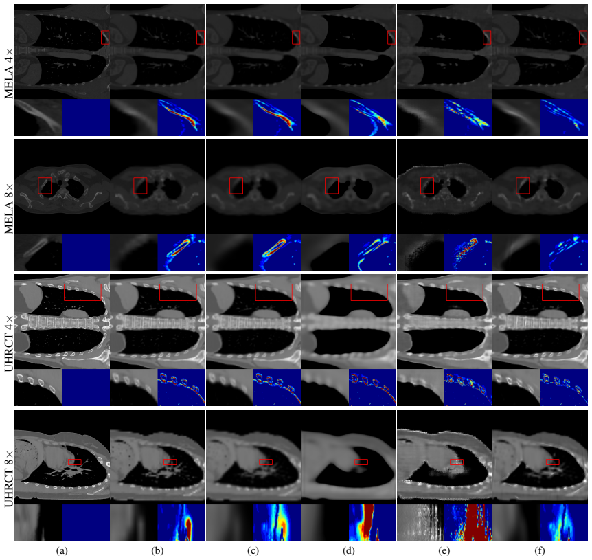
- On both public datasets the method outperforms the zero-shot baseline on all metrics and the supervised ArSSR on PSNR (with competitive SSIM), and the abstract reports expert evaluation supporting clinical potential at 4x.
Reading between the lines
- A natural untested corollary: the two-stage split (2D projection SR plus signed-density 3D reconstruction) transfers to other tomographic modalities — PET, SPECT, interventional cone-beam CT — wherever paired HR volumes are scarce but 2D projections are cheap.
- Because the paper concedes its prior is trained on frontal and lateral views, per-angle gains are likely uneven: a per-projection PSNR curve against true HR projections would show where the prior stops helping, and PAS probably matters most at oblique angles where the prior is weakest.
- The signed-density mechanism is a general device: any 3DGS task that models differences — motion- or artifact-corrected imaging, change detection, subtraction studies — could adopt leaky-ReLU densities the same way.
- The residual-learning design invites a cheaper alternative reading: a large part of the volume-level gain may come from NAB-GS itself rather than the diffusion prior; replacing DDNM with cubic upsampling inside NAB-GS would isolate the prior's marginal contribution across anatomies.
Editorial analysis
A structured set of objections, weighed in public.
Referee Report
Summary. The paper proposes a zero-shot 3D CT super-resolution framework that first upsamples low-resolution 2D CT projections using a diffusion model trained on large-scale chest X-ray images, with a per-projection adaptive sampling (PAS) strategy, and then reconstructs the 3D volume with 3D Gaussian splatting. To allow residual learning between the upsampled LR projections and the diffusion-enhanced projections, the authors introduce NAB-GS, which extends 3DGS by allowing negative Gaussian densities via a leaky ReLU activation. The method is evaluated on MELA and UHRCT datasets against traditional interpolation, supervised ArSSR, and zero-shot CuNeRF, and is supported by ablations on the diffusion starting step, activation function, negative slope, and reconstruction backbones.
Significance. If the reported results are robust, the paper makes a useful contribution: it provides a practical way to inject external 2D X-ray priors into 3D CT SR without paired HR CT volumes, and NAB-GS is a technically interesting extension of 3DGS that enables signed residual learning. The ablations are reasonably thorough and the pipeline components are clearly described. However, the strength of the 3D claim depends on the transferability of a chest X-ray diffusion prior to CT projections from other anatomies and across all angles, and the current evaluation provides only weak direct evidence for that transfer. The hyperparameter selection and small test sets further limit the confidence in the quantitative conclusions.
major comments (4)
- [Supplementary §9.3, Table 9; §5.3] The direct evidence for the 2D diffusion prior is weaker than the 3D results imply. At 8×, the proposed DDNM+PAS upsampling has SSIM 0.9107, below cubic (0.9180) and trilinear (0.9205); at 4× it is below trilinear (0.9643 vs 0.9652). Yet Table 1 reports that the full pipeline improves 3D SSIM over interpolation at both scales (e.g., MELA 8×: 0.9153 vs 0.9005 cubic). Because the only external information entering the 3D reconstruction is these 2D projections, the origin of the 3D gain is not established. Please report per-angle or angular-bin 2D metrics and an analysis of how much of the final 3D improvement is attributable to genuine projection enhancement versus hallucinated oblique-angle content fitted by NAB-GS.
- [§5.1, Supplementary §9.1] Hyperparameters appear to be selected on the evaluation data. τthr is set to 7 (4×) and 11 (8×); γ is tuned separately for MELA 4×/8× and UHRCT 4×/8× (0.09/0.003/0.03/0.003) after inspecting PSNR/SSIM on the test volumes (Tables 6–7). With only 20 and 10 test volumes and no error bars or significance tests, the reported margins over cubic (0.13–0.80 dB PSNR; 0.013–0.024 SSIM) may not be stable. Section 6 also concedes that the optimal γ varies with dataset. Please provide a validation-based hyperparameter selection protocol, report per-volume metrics with variance, and run significance tests (or state uncertainty).
- [§6, §5.1] The paper acknowledges that the diffusion prior is 'limited to frontal and lateral views' and that enhancement across 0–180° is non-trivial, yet the pipeline runs the same prior on 100 projections uniformly spaced between 0° and 180°. PAS (Eq. 10) only chooses a starting timestep; it does not add missing high-frequency content for off-distribution angles. No angle-resolved analysis is provided, so the central premise—that an external 2D X-ray prior transfers to all projection angles—remains unsupported. At minimum, report metrics broken down by angular bin and the distribution of tstart across angles.
- [Abstract, §5] The abstract states that 'expert evaluations present the framework's clinical potential at 4x', but no expert evaluation protocol, results, or statistical analysis is included in the main text or supplementary materials. This claim is either missing support or should be removed from the abstract.
minor comments (5)
- [§1] Typo: 'resulting in to a degradation' should be 'resulting in a degradation'.
- [Title] The arXiv title ('Zero-shot CT Super-Resolution using Diffusion-based 2D Projection Priors and Signed 3D Gaussians') and the main-text title ('Zero-shot Volumetric CT Super-Resolution using 3D Gaussian Splatting with Upsampled 2D X-ray Projection Priors') should be aligned.
- [§5.1] The sentence 'Note that unsupervised learning only uses a testset' is ambiguous. Clarify whether any validation split is used for selecting PAS thresholds and γ, and whether the reported numbers are on the same volumes used for hyperparameter tuning.
- [Eq. (4)] The notation for µi is unclear: 'µi is the covariance-related factor,(2π|Σi/Σ̂i|1/2)' appears to have a missing 'where' and possible missing parentheses. Please rewrite.
- [Table 1] The table caption does not state the number of test volumes (20 for MELA, 10 for UHRCT). Please add this information and report per-volume standard deviations.
Circularity Check
No significant circularity: the derivation chain is driven by an external diffusion prior and independent reconstruction baselines; noted test-set hyperparameter tuning is a rigor concern, not a circular step.
full rationale
The paper's central derivation is not circular. The 2D projection SR uses an unconditional DDPM trained on external X-ray datasets (ChestX-ray14, CheXpert), then applies DDNM with data consistency (Eq. 6: x̂0|t = A†y + (I − A†A)x0|t), so the upsampled projections are constrained by the LR projections and the external prior. The 3D reconstruction via NAB-GS fits Gaussian residuals between the upsampled LR volume and the diffusion-upsampled projections (Section 4.2); this is a supervised fitting to externally generated targets, not a self-consistency loop. No equation in the paper reduces to another by construction, and no load-bearing claim is justified only by a self-citation: references to DDNM, R2-GS, CuNeRF, ArSSR, and the diffusion literature are all prior external work. The per-dataset/per-resolution selection of the leaky-ReLU slope γ (Supplementary Tables 6–7) and the PAS threshold τthr is hyperparameter tuning; even if performed on evaluation data, it is a model-selection weakness that can overstate absolute gains, not a fitted parameter renamed as a prediction. Similarly, the modest 2D projection gains and lower SSIM at 8× (Table 9) raise a substantive question about whether the diffusion prior transfers across the full 0–180° angular range, but that is a correctness/validity concern about the external prior, not circularity. The framework is self-contained against external benchmarks and the reported improvements are not forced by the paper's own definitions.
Assumptions & free parameters
free parameters (4)
- negative slope gamma of leaky ReLU =
0.09 (MELA 4x), 0.003 (MELA 8x), 0.03 (UHRCT 4x), 0.003 (UHRCT 8x)
- PAS threshold tau_thr =
7 (4x), 11 (8x)
- Loss weights lambda1, lambda2 =
0.5, 0.05
- DDNM noise level sigma_y =
0.0015
assumptions (5)
- domain assumption The 2D X-ray diffusion prior transfers to CT projections from other anatomies and scanners
- domain assumption The downsampling degradation (sinc interpolation with Gaussian smoothing) accurately simulates low-resolution CT
- domain assumption The radiative Gaussian model from R2-GS adequately represents CT density fields
- ad hoc to paper Negative density values in the Gaussian field yield a stable optimization for residual learning
- domain assumption Diffusion-upsampled projections are reliable targets for the residual loss
Cite this review
Pith. "Pith review of Zero-shot CT Super-Resolution using Diffusion-based 2D Projection Priors and Signed 3D Gaussians." pith.science (2026). https://pith.science/paper/3SN4IJ3Z
@misc{pith2026250815151,
  author       = {Pith},
  title        = {Pith review of: Zero-shot CT Super-Resolution using Diffusion-based 2D Projection Priors and Signed 3D Gaussians},
  year         = {2026},
  howpublished = {\url{https://pith.science/paper/3SN4IJ3Z}},
  note         = {Machine review of arXiv:2508.15151}
}
read the original abstract
Computed tomography (CT) is important in clinical diagnosis, but acquiring high-resolution (HR) CT is constrained by radiation exposure risks. While deep learning-based super-resolution (SR) methods have shown promise for reconstructing HR CT from low-resolution (LR) inputs, supervised approaches require paired datasets that are often unavailable. Zero-shot methods address this limitation by operating on single LR inputs; however, they frequently fail to recover fine structural details due to limited LR information within individual volumes. To overcome these limitations, we propose a novel zero-shot 3D CT SR framework that integrates diffusion-based upsampled 2D projection priors into the 3D reconstruction process. Specifically, our framework consists of two stages: (1) LR CT projection SR, training a diffusion model on abundant X-ray data to upsample LR projections, thereby enhancing the scarce information inherent in the LR inputs. (2) 3D CT volume reconstruction, using 3D Gaussian splatting with our novel Negative Alpha Blending (NAB-GS), which models positive and negative Gaussian densities to learn signed residuals between diffusion-generated HR and upsampled LR projections. Our framework demonstrates superior quantitative and qualitative performance on two public datasets, and expert evaluations present the framework's clinical potential at 4x.
Figures


Figures from the paper (4 more)


Forward citations
Cited by 2 Pith papers
-
K-space Gaussian Representation for Parallel MRI
K-space Gaussian Representation (KGR) fits measured multi-coil k-space with shared Gabor-Gaussian primitives and refines the result with Hankel low-rank projection, improving accelerated MRI reconstruction over baseli...
-
K-space Gaussian Representation for Parallel MRI
KGR fits shared Gabor-Gaussian primitives directly in multi-coil k-space and applies low-rank projection, consistently outperforming representative parallel-MRI baselines on brain datasets.
Reference graph
Works this paper leans on
-
[1]
, " * write output.state after.block = add.period write newline
ENTRY address archivePrefix author booktitle chapter edition editor eid eprint howpublished institution isbn journal key month note number organization pages publisher school series title type volume year label extra.label sort.label short.list INTEGERS output.state before.all mid.sentence after.sentence after.block FUNCTION init.state.consts #0 'before.a...
-
[2]
write newline
" write newline "" before.all 'output.state := FUNCTION n.dashify 't := "" t empty not t #1 #1 substring "-" = t #1 #2 substring "--" = not "--" * t #2 global.max substring 't := t #1 #1 substring "-" = "-" * t #2 global.max substring 't := while if t #1 #1 substring * t #2 global.max substring 't := if while FUNCTION word.in bbl.in capitalize " " * FUNCT...
-
[3]
Andersen, A. H.; and Kak, A. C. 1984. Simultaneous algebraic reconstruction technique (SART): a superior implementation of the ART algorithm. Ultrasonic imaging, 6(1): 81--94
work page 1984
-
[4]
Biguri, A.; Dosanjh, M.; Hancock, S.; and Soleimani, M. 2016. TIGRE : a MATLAB-GPU toolbox for CBCT image reconstruction. Biomed. Phys. Eng. Express (BPEE), 2(5): 055010
work page 2016
-
[5]
Cai, Y.; Wang, J.; Yuille, A.; Zhou, Z.; and Wang, A. 2024. Structure-aware sparse-view x-ray 3d reconstruction. In Proc. Comput. Vis. Pattern Recognit. (CVPR) , 11174--11183
work page 2024
-
[6]
Chen, X.; Wang, X.; Zhou, J.; Qiao, Y.; and Dong, C. 2023 a . Activating more pixels in image super-resolution transformer. In Proc. Comput. Vis. Pattern Recognit. (CVPR) , 22367--22377
work page 2023
-
[7]
Chen, Z.; Yang, L.; Lai, J.-H.; and Xie, X. 2023 b . Cu NeRF : Cube-based neural radiance field for zero-shot medical image arbitrary-scale super resolution. In Proc. Comput. Vis. Pattern Recognit. (CVPR) , 21185--21195
work page 2023
-
[8]
Chu, Y.; Zhou, L.; Luo, G.; Qiu, Z.; and Gao, X. 2023. Topology-Preserving Computed Tomography Super-Resolution Based on Dual-Stream Diffusion Model. In Proc. Int'l Conf. Med. Image Comput. Comput. Assist. Interv. (MICCAI) , 260--270
work page 2023
Show all 40 references
-
[9]
C.; He, K.; and Tang, X
Dong, C.; Loy, C. C.; He, K.; and Tang, X. 2015. Image super-resolution using deep convolutional networks. IEEE Trans. Pattern Anal. Mach. Intell. (TPAMI) , 38(2): 295--307
2015
-
[10]
C.; Yan, K.; Yao, J.; Chen, X.; Liu, Z.; Lu, L.; et al
Fang, W.; Tang, Y.; Guo, H.; Yuan, M.; Mok, T. C.; Yan, K.; Yao, J.; Chen, X.; Liu, Z.; Lu, L.; et al. 2024. Cycle INR : Cycle Implicit Neural Representation for Arbitrary-Scale Volumetric Super-Resolution of Medical Data. In Proc. Comput. Vis. Pattern Recognit. (CVPR) , 11631--11641
2024
-
[11]
A.; Davis, L
Feldkamp, L. A.; Davis, L. C.; and Kress, J. W. 1984. Practical cone-beam algorithm. Journal of the Optical Society of America A, 1(6): 612--619
1984
-
[12]
L.; Louis, N.; Zbijewski, W.; Vaishnav, J.; Clark, K.; and Nicolella, D
Frazer, L. L.; Louis, N.; Zbijewski, W.; Vaishnav, J.; Clark, K.; and Nicolella, D. P. 2024. Super-resolution of clinical CT : Revealing microarchitecture in whole bone clinical CT image data. Bone, 185: 117115
2024
-
[13]
Ho, J.; Jain, A.; and Abbeel, P. 2020. Denoising diffusion probabilistic models. Adv. Neural Inf. Process. Syst. (NeurIPS) , 33: 6840--6851
2020
-
[14]
Kawar, B.; Elad, M.; Ermon, S.; and Song, J. 2022. Denoising diffusion restoration models. Adv. Neural Inf. Process. Syst. (NeurIPS) , 35: 23593--23606
2022
-
[15]
Kerbl, B.; Kopanas, G.; Leimk \"u hler, T.; and Drettakis, G. 2023. 3D Gaussian splatting for real-time radiance field rendering. ACM Trans. on Graph. (ToG) , 42(4): 139--1
2023
-
[16]
Ledig, C.; Theis, L.; Husz \'a r, F.; Caballero, J.; Cunningham, A.; Acosta, A.; Aitken, A.; Tejani, A.; Totz, J.; Wang, Z.; et al. 2017. Photo-realistic single image super-resolution using a generative adversarial network. In Proc. Comput. Vis. Pattern Recognit. (CVPR) , 4681--4690
2017
-
[17]
Liang, J.; Cao, J.; Sun, G.; Zhang, K.; Van Gool, L.; and Timofte, R. 2021. Swin IR : Image restoration using swin transformer. In Proc. of Int'l Conf. on Comput. Vis. (ICCV) , 1833--1844
2021
-
[18]
Liu, G.; Sun, H.; Li, J.; Yin, F.; and Yang, Y. 2023. Accelerating diffusion models for inverse problems through shortcut sampling. arXiv preprint arXiv:2305.16965
2023 arXiv
-
[19]
Lu, Z.; Li, J.; Liu, H.; Huang, C.; Zhang, L.; and Zeng, T. 2022. Transformer for single image super-resolution. In Proc. Comput. Vis. Pattern Recognit. (CVPR) , 457--466
2022
-
[20]
R.; and Semelka, R
Martin, D. R.; and Semelka, R. C. 2006. Health effects of ionising radiation from diagnostic CT . The Lancet, 367(9524): 1712--1714
2006
-
[21]
H.; Yu, L.; Kofler, J
McCollough, C. H.; Yu, L.; Kofler, J. M.; Leng, S.; Zhang, Y.; Li, Z.; and Carter, R. E. 2015. Degradation of CT low-contrast spatial resolution due to the use of iterative reconstruction and reduced dose levels. Radiology, 276(2): 499--506
2015
-
[22]
Q.; and Dhariwal, P
Nichol, A. Q.; and Dhariwal, P. 2021. Improved denoising diffusion probabilistic models. In International conference on machine learning, 8162--8171. PMLR
2021
-
[23]
Peng, C.; Lin, W.-A.; Liao, H.; Chellappa, R.; and Zhou, S. K. 2020. Saint: Spatially aware interpolation network for medical slice synthesis. In Proc. Comput. Vis. Pattern Recognit. (CVPR) , 7750--7759
2020
-
[24]
Rombach, R.; Blattmann, A.; Lorenz, D.; Esser, P.; and Ommer, B. 2022. High-resolution image synthesis with latent diffusion models. In Proc. Comput. Vis. Pattern Recognit. (CVPR) , 10684--10695
2022
-
[25]
J.; and Norouzi, M
Saharia, C.; Ho, J.; Chan, W.; Salimans, T.; Fleet, D. J.; and Norouzi, M. 2022. Image super-resolution via iterative refinement. IEEE Trans. Pattern Anal. Mach. Intell. (TPAMI) , 45(4): 4713--4726
2022
-
[26]
Shocher, A.; Cohen, N.; and Irani, M. 2018. Zero-shot super-resolution using deep internal learning. In Proc. Comput. Vis. Pattern Recognit. (CVPR) , 3118--3126
2018
-
[27]
Y.; and Pan, X
Sidky, E. Y.; and Pan, X. 2008. Image reconstruction in circular cone-beam computed tomography by constrained, total-variation minimization. Physics in Medicine & Biology, 53(17): 4777
2008
-
[28]
Sitzmann, V.; Martel, J.; Bergman, A.; Lindell, D.; and Wetzstein, G. 2020. Implicit neural representations with periodic activation functions. Adv. Neural Inf. Process. Syst. (NeurIPS) , 33: 7462--7473
2020
-
[29]
W.; Cho, S.; and Cho, N
Soh, J. W.; Cho, S.; and Cho, N. I. 2020. Meta-transfer learning for zero-shot super-resolution. In Proc. Comput. Vis. Pattern Recognit. (CVPR) , 3516--3525
2020
-
[30]
Song, J.; Meng, C.; and Ermon, S. 2020. Denoising diffusion implicit models. arXiv preprint arXiv:2010.02502
2020 arXiv
-
[31]
Wang, J.; Wang, R.; Tao, R.; and Zheng, G. 2022. UASSR : Unsupervised arbitrary scale super-resolution reconstruction of single anisotropic 3D images via disentangled representation learning. In Proc. Int'l Conf. Med. Image Comput. Comput. Assist. Interv. (MICCAI) , 453--462
2022
-
[32]
C.; and Wen, B
Wang, Y.; Yang, W.; Chen, X.; Wang, Y.; Guo, L.; Chau, L.-P.; Liu, Z.; Qiao, Y.; Kot, A. C.; and Wen, B. 2024. Sin SR : Diffusion-based image super-resolution in a single step. In Proc. Comput. Vis. Pattern Recognit. (CVPR) , 25796--25805
2024
-
[33]
Wang, Y.; Yu, J.; and Zhang, J. 2022. Zero-shot image restoration using denoising diffusion null-space model. arXiv preprint arXiv:2212.00490
2022 arXiv
-
[34]
C.; Sheikh, H
Wang, Z.; Bovik, A. C.; Sheikh, H. R.; and Simoncelli, E. P. 2004. Image quality assessment: from error visibility to structural similarity. IEEE Trans. Image Process. (TIP) , 13(4): 600--612
2004
-
[35]
Wu, Q.; Li, Y.; Sun, Y.; Zhou, Y.; Wei, H.; Yu, J.; and Zhang, Y. 2022. An arbitrary scale super-resolution approach for 3d MR images via implicit neural representation. IEEE J. Biomed. Health Inform. (JBHI), 27(2): 1004--1015
2022
-
[36]
W.; and Zhang, R
Yu, P.; Zhang, H.; Kang, H.; Tang, W.; Arnold, C. W.; and Zhang, R. 2022. RPLHR-CT dataset and transformer baseline for volumetric super-resolution from CT scans. In Proc. Int'l Conf. Med. Image Comput. Comput. Assist. Interv. (MICCAI) , 344--353
2022
-
[37]
J.; Cai, Y.; Cao, J.; Zhang, Y.; and Li, H
Zha, R.; Lin, T. J.; Cai, Y.; Cao, J.; Zhang, Y.; and Li, H. 2024. R ^2 - G aussian: Rectifying Radiative Gaussian Splatting for Tomographic Reconstruction. Adv. Neural Inf. Process. Syst. (NeurIPS)
2024
-
[38]
Zha, R.; Zhang, Y.; and Li, H. 2022. NAF : N eural attenuation fields for sparse-view CBCT reconstruction. In Proc. Int'l Conf. Med. Image Comput. Comput. Assist. Interv. (MICCAI) , 442--452
2022
-
[39]
Zhang, Y.; Tian, Y.; Kong, Y.; Zhong, B.; and Fu, Y. 2018. Residual dense network for image super-resolution. In Proc. Comput. Vis. Pattern Recognit. (CVPR) , 2472--2481
2018
-
[40]
Zhao, K.; Pang, K.; Hung, A. L. Y.; Zheng, H.; Yan, R.; and Sung, K. 2024. Mri super-resolution with partial diffusion models. IEEE Transactions on Medical Imaging (TMI)
2024
Reviewed August 5, 2026 · model on record in the stance chip above.
Discussion (0). Sign in to comment.