DB-FGA-Net: Dual Backbone Frequency Gated Attention Network for Multi-Class Brain Tumor Classification with Grad-CAM Interpretability
Pith reviewed 2026-05-18 04:04 UTC · model grok-4.3
The pith
A dual-backbone network with frequency-gated attention classifies brain tumors from MRI at up to 99.24 percent accuracy without data augmentation.
A machine-rendered reading of the paper's core claim, the machinery that carries it, and where it could break.
Core claim
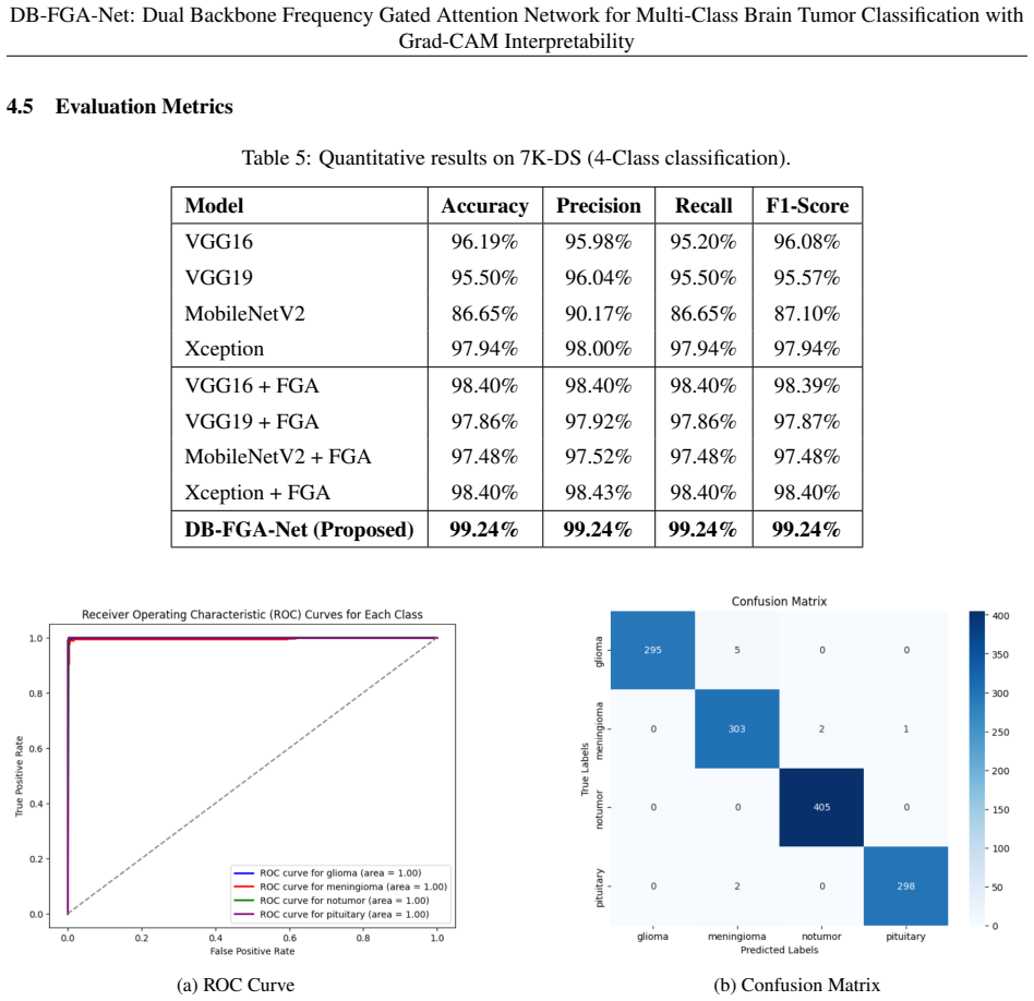
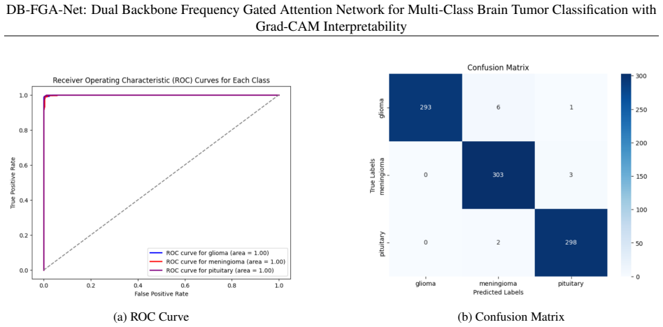
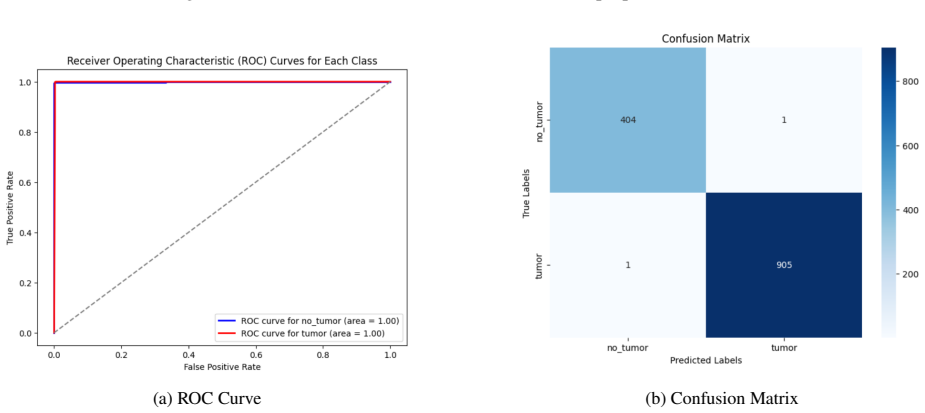
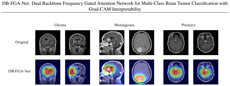
The central claim is that combining VGG16 and Xception backbones with a Frequency-Gated Attention Block lets the system capture both fine local details and broader context in MRI images, delivering 99.24 percent accuracy on four-class brain tumor classification without data augmentation, 98.68 percent on three-class and 99.85 percent on two-class versions of the same data, and 95.77 percent accuracy when tested on an independent collection while also supplying Grad-CAM visualizations of the relevant tumor areas.
What carries the argument
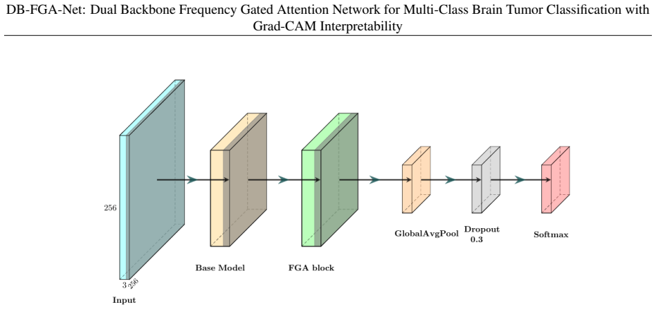
The Frequency-Gated Attention Block, which applies frequency-domain processing to selectively emphasize complementary features extracted from the paired VGG16 and Xception backbones.
If this is right
- Classification accuracy remains high across the two-class, three-class, and four-class tumor typing problems on the primary dataset.
- The model maintains competitive performance when applied to an independent dataset, exceeding several baseline approaches under identical conditions.
- Grad-CAM heatmaps make the regions used for each decision visible, supporting clinical review of the output.
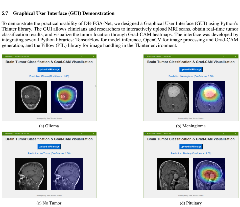
- A graphical user interface delivers real-time classification together with tumor localization maps for immediate use.
Where Pith is reading between the lines
- Success without augmentation suggests that frequency-domain attention can reduce dependence on large-scale labeled medical image collections.
- The same dual-backbone and gating pattern may transfer to classification tasks involving other scan types such as CT or ultrasound.
- The localization maps could let physicians quickly compare the model's focus area against their own visual assessment during diagnosis.
- Prospective evaluation on live patient streams from multiple centers would be a direct way to check readiness for everyday hospital deployment.
Load-bearing premise
The two MRI collections used for development and testing already contain enough of the image variability, scanner differences, and tumor appearances that occur in routine clinical neuro-oncology.
What would settle it
Testing the trained model on a fresh set of MRI scans gathered from additional hospitals or different scanner models and observing a substantial drop in accuracy would directly test whether the reported generalization holds.
Figures


read the original abstract
Brain tumors are a challenging problem in neuro-oncology, where early and precise diagnosis is important for successful treatment. Deep learning-based brain tumor classification methods often rely on heavy data augmentation which can limit generalization and trust in clinical applications. In this paper, we propose a double-backbone network integrating VGG16 and Xception with a Frequency-Gated Attention (FGA) Block to capture complementary local and global features. Our model achieves highly competitive performance without augmentation which demonstrates robustness to variably sized and distributed datasets. For further transparency, Grad-CAM is integrated to visualize the tumor regions based on which the model is giving prediction, bridging the gap between model prediction and clinical interpretability. The proposed framework achieves 99.24% accuracy on the 7K-DS dataset for the 4-class setting, along with 98.68% and 99.85% in the 3-class and 2-class settings, respectively. On the independent 3K-DS dataset, the model generalizes with 95.77% accuracy, outperforming several baseline methods under the same experimental setting. To further support clinical usability, we developed a graphical user interface (GUI) that provides real-time classification and Grad-CAM-based tumor localization. These findings suggest that augmentation-free, interpretable, and deployable deep learning models such as DB-FGA-Net hold strong potential for reliable clinical translation in brain tumor diagnosis.
Editorial analysis
A structured set of objections, weighed in public.
Referee Report
Summary. The manuscript proposes DB-FGA-Net, a dual-backbone architecture combining VGG16 and Xception with a Frequency-Gated Attention (FGA) block to classify brain tumors from MRI scans into 2-, 3-, or 4-class settings. The central claims are that the model attains 99.24% accuracy (4-class), 98.68% (3-class), and 99.85% (2-class) on the 7K-DS dataset without data augmentation, generalizes to 95.77% accuracy on an independent 3K-DS dataset while outperforming baselines, and provides clinical utility via Grad-CAM visualizations and a real-time GUI.
Significance. If the experimental claims hold after clarification, the work would demonstrate that dual-backbone designs with frequency-gated attention can deliver competitive augmentation-free performance on brain-tumor MRI tasks, reducing reliance on augmentation that may introduce artifacts. Explicit credit is due for the integration of Grad-CAM for decision visualization and the provision of a deployable GUI, both of which directly support interpretability and potential clinical translation.
major comments (2)
- [§4.1] §4.1 (Datasets): The descriptions of 7K-DS and 3K-DS provide no metadata on scanner field strength, acquisition parameters, slice thickness, multi-center sourcing, or patient demographics. This information is load-bearing for the no-augmentation generalization claim, because the 95.77% accuracy on the independent set could reflect limited distribution shift rather than robustness to real clinical variability.
- [§5] §5 (Experimental Results): Reported accuracies are given as single point estimates without the number of independent runs, standard deviations, or statistical significance tests against baselines. This undermines the claim that DB-FGA-Net 'outperforms several baseline methods under the same experimental setting,' as the magnitude and reliability of improvement cannot be assessed.
minor comments (2)
- [Figure 3] Figure 3 (Grad-CAM examples): Only a small number of slices are shown; adding representative cases from each tumor class and from both datasets would strengthen the interpretability section.
- [§3.2] Notation in §3.2: The frequency gating operation inside the FGA block is described in prose only; a compact equation or pseudocode would improve reproducibility.
Simulated Author's Rebuttal
We thank the referee for their insightful comments on our manuscript. We have carefully considered each point and provide detailed responses below, along with indications of revisions to the manuscript.
read point-by-point responses
-
Referee: [§4.1] §4.1 (Datasets): The descriptions of 7K-DS and 3K-DS provide no metadata on scanner field strength, acquisition parameters, slice thickness, multi-center sourcing, or patient demographics. This information is load-bearing for the no-augmentation generalization claim, because the 95.77% accuracy on the independent set could reflect limited distribution shift rather than robustness to real clinical variability.
Authors: We agree that additional metadata on the datasets would provide better context for interpreting the generalization results. The 7K-DS and 3K-DS are standard public datasets used in brain tumor classification literature, but they do not include detailed scanner field strength, acquisition parameters, or patient demographics in their releases. In the revised manuscript, we have expanded Section 4.1 to explicitly state the available information about the datasets and added a discussion on the limitations regarding distribution shift. This clarifies that while the independent dataset demonstrates generalization beyond the training distribution, further validation on multi-center data with full metadata would be valuable for clinical deployment. revision: partial
-
Referee: [§5] §5 (Experimental Results): Reported accuracies are given as single point estimates without the number of independent runs, standard deviations, or statistical significance tests against baselines. This undermines the claim that DB-FGA-Net 'outperforms several baseline methods under the same experimental setting,' as the magnitude and reliability of improvement cannot be assessed.
Authors: We acknowledge that single-point estimates do not fully convey the reliability of the results. To address this, we have re-run the experiments with 5 independent trials using different random seeds for initialization and data shuffling. The revised Section 5 now includes mean accuracy, precision, recall, and F1-score with standard deviations for DB-FGA-Net and all baselines. We have also performed statistical significance testing using paired t-tests, reporting p-values to confirm that the improvements are statistically significant. Updated tables and text reflect these changes. revision: yes
Circularity Check
Empirical ML paper with independent held-out evaluation
full rationale
This is a standard supervised deep-learning application paper that trains the proposed DB-FGA-Net on the 7K-DS dataset and measures accuracy on held-out test splits plus an independent 3K-DS corpus. No derivation chain, equations, or first-principles results are offered; the headline numbers (99.24 % 4-class, 95.77 % on 3K-DS) are direct empirical measurements rather than quantities obtained by fitting parameters that are then renamed as predictions. The architecture description, Grad-CAM usage, and GUI are likewise implementation details, not circular reductions. The paper is therefore self-contained against external benchmarks.
Axiom & Free-Parameter Ledger
axioms (1)
- domain assumption The 7K-DS and 3K-DS MRI datasets are representative of clinical brain tumor imaging variability and distribution shifts.
invented entities (1)
-
Frequency-Gated Attention (FGA) Block
no independent evidence
Lean theorems connected to this paper
-
IndisputableMonolith/Foundation/AlphaDerivationExplicit.leanalphaInverseRS unclear?
unclearRelation between the paper passage and the cited Recognition theorem.
Frequency-Gated Attention (FGA) block ... transforms X into the frequency domain using a 2D Fast Fourier Transform (FFT).
-
IndisputableMonolith/Cost/FunctionalEquation.leanwashburn_uniqueness_aczel unclear?
unclearRelation between the paper passage and the cited Recognition theorem.
achieves 99.24% accuracy ... without any augmentation which demonstrates robustness to variably sized and distributed datasets.
What do these tags mean?
- matches
- The paper's claim is directly supported by a theorem in the formal canon.
- supports
- The theorem supports part of the paper's argument, but the paper may add assumptions or extra steps.
- extends
- The paper goes beyond the formal theorem; the theorem is a base layer rather than the whole result.
- uses
- The paper appears to rely on the theorem as machinery.
- contradicts
- The paper's claim conflicts with a theorem or certificate in the canon.
- unclear
- Pith found a possible connection, but the passage is too broad, indirect, or ambiguous to say the theorem truly supports the claim.
Reference graph
Works this paper leans on
-
[1]
Jon A. Mukand, Dilshad D. Blackinton, Michael G. Crincoli, James J. Lee, Bernadette B. Santos,Incidence of Neurologic Deficits and Rehabilitation of Patients with Brain Tumors, American Journal of Physical Medicine & Rehabilitation, vol. 80, no. 5, pp. 346–350, 2001. DOI:10.1097/00002060−200105000−00006
- [2]
- [3]
- [4]
- [5]
-
[6]
Stieg,Early Detection Can Be Key to Surviving a Brain Tumor, 2016
Philip E. Stieg,Early Detection Can Be Key to Surviving a Brain Tumor, 2016. [Online]. Available: https://neurosurgery.weillcornell.org/about-us/blog/ early-detection-can-be-key-surviving-brain-tumor
work page 2016
-
[7]
MRI: What’s the Difference?, 2024
MD Anderson Cancer Center,CT Scan vs. MRI: What’s the Difference?, 2024. [Online]. Available:https://www. mdanderson.org/cancerwise/ct-scan-vs-mri--what-is-the-difference.h00-159616278.html
work page 2024
-
[8]
Geert Litjens, Thijs Kooi, Babak E. Bejnordi, Arnaud A.A. Setio, Francesco Ciompi, Mohsen Ghafoorian, Jeroen A.W.M. van der Laak, Bram van Ginneken, Clara I. Sánchez,A Survey on Deep Learning in Medical Image Analysis, Medical Image Analysis, vol. 42, pp. 60–88, 2017. DOI:10.1016/j.media.2017.07.005
- [9]
-
[10]
Dinggang Shen, Guorong Wu, Heung-Il Suk,Deep Learning in Medical Image Analysis, Annual Review of Biomedical Engineering, vol. 19, pp. 221–248, 2017. DOI:10.1146/annurev−bioeng−071516−044442
-
[11]
Ramazan ˙Incir, Ferhat Bozkurt,Improving brain tumor classification with combined convolutional neu- ral networks and transfer learning, Knowledge-Based Systems, vol. 299, p. 111981, 2024. DOI: 10.1016/j.knosys.2024.111981. 23 DB-FGA-Net: Dual Backbone Frequency Gated Attention Network for Multi-Class Brain Tumor Classification with Grad-CAM Interpretability
-
[12]
R. Preetha, M. Jasmine Pemeena Priyadarsini, J. S. Nisha,Hybrid 3B Net and EfficientNetB2 Model for Multi-Class Brain Tumor Classification, IEEE Access, vol. 13, pp. 63465–63485, 2025. DOI: 10.1109/ACCESS.2025.3558411
-
[13]
Rizal Dwi Prayogo, Nur Hamid, Hidetaka Nambo,Hybrid CNN-Based Transfer Learning Enhances Brain Tumor Classification on MRI Images, IEEE Access, vol. 13, pp. 116654–116668, 2025. DOI: 10.1109/ACCESS.2025.3584376
-
[14]
Adnan Saeed, Khurram Shehzad, Shahzad Sarwar Bhatti, Saim Ahmed, Ahmad Taher Azar,GGLA-NeXtE2NET: A Dual-Branch Ensemble Network With Gated Global-Local Attention for Enhanced Brain Tumor Recognition, IEEE Access, vol. 13, pp. 7234–7257, 2025. DOI:10.1109/ACCESS.2025.3525518
-
[15]
Bedriye Dogan, Hursit Burak Mutlu, Muhammed Yildirim, Sercan Yalcin, Serpil Aslan, Niranjana Sampathila, Ozal Yildirim, Edward J. Ciaccio, Ru-San Tan, U. Rajendra Acharya,Content-Based Brain Magnetic Resonance Image Retrieval and Classification With the Proposed Deep Learning and Tissue-Based System, IEEE Access, vol. 13, pp. 122684–122697, 2025. DOI:10.1...
-
[16]
Hussein Alshaari, Saeed Alqahtani,Deep-EFNet: An Optimized EfficientNetB0 Architecture With Dual Regular- ization for Scalable Multi-Class Brain Tumor Classification in MRI, IEEE Access, vol. 13, pp. 85682–85697,
-
[17]
DOI:10.1109/ACCESS.2025.3567919
-
[18]
Pratikkumar Chauhan, Munindra Lunagaria, Deepak Verma, Krunal Vaghela, Ganshyam Tejani, Sunil Sharma, Ahmed Khan,PBVit: A Patch-Based Vision Transformer for Enhanced Brain Tumor Detection, IEEE Access, vol. PP, pp. 1–1, 2024. DOI:10.1109/ACCESS.2024.3521002
-
[19]
Jyotismita Chaki, Marcin Wozniak,Brain Tumor Categorization and Retrieval Using Deep Brain Incep Res Architecture Based Reinforcement Learning Network, IEEE Access, vol. PP, pp. 1–1, 2023. DOI: 10.1109/ACCESS.2023.3334434
-
[20]
Anees Tariq, Muhammad Munwar Iqbal, Muhammad Javed Iqbal, Iftikhar Ahmad,Transforming Brain Tumor Detection Empowering Multi-Class Classification With Vision Transformers and EfficientNetV2, IEEE Access, vol. 13, pp. 63857–63876, 2025. DOI:10.1109/ACCESS.2025.3555638
-
[21]
Sivakumar, Ahmad Raza Khan, Syed Umar, R
N. Sivakumar, Ahmad Raza Khan, Syed Umar, R. N. Ravikumar, I. Bremnavas, Munindra Lunagaria, Krunal Vaghela, Ghanshyam G. Tejani, Sunil Kumar Sharma,A Hybrid Brain Tumor Classification Using FL With FedAvg and FedProx for Privacy and Robustness Across Heterogeneous Data Sources, IEEE Access, vol. 13, pp. 57705–57719, 2025. DOI:10.1109/ACCESS.2025.3549440
-
[22]
Ayesha Younis, Li Qiang, Zargaam Afzal, Mohammed Adamu, Halima Bello Kawuwa, Fida Hussain, Hamid Hussain,Abnormal Brain Tumors Classification Using ResNet50 and Its Comprehensive Evaluation, IEEE Access, vol. PP, pp. 1–1, 2024. DOI:10.1109/ACCESS.2024.3403902
-
[25]
DOI: 10.6084/m9.f igshare.1512427.v8
Jun Cheng,Brain Tumor Dataset, figshare, 2017. DOI: 10.6084/m9.f igshare.1512427.v8. [Online]. Available: https://doi.org/10.6084/m9.figshare.1512427.v8
-
[26]
Jyotismita Chaki,Brain Tumor MRI Dataset, IEEE Dataport, 2023. DOI: 10.21227/1jny−g144 . [Online]. Available:https://dx.doi.org/10.21227/1jny-g144
-
[27]
DOI: 10.34740/KAGGLE/DSV /12745533
Sartaj Bhuvaji, Ankita Kadam, Prajakta Bhumkar, Sameer Dedge, Swati Kanchan,Brain Tumor Classi- fication (MRI), Kaggle, 2025. DOI: 10.34740/KAGGLE/DSV /12745533 . [Online]. Available: https: //www.kaggle.com/dsv/12745533
- [28]
-
[29]
Ramprasaath R. Selvaraju, Michael Cogswell, Abhishek Das, Ramakrishna Vedantam, Devi Parikh, Dhruv Batra, Grad-CAM: Visual Explanations from Deep Networks via Gradient-based Localization, Proceedings of the IEEE International Conference on Computer Vision (ICCV), pp. 618–626, 2017. DOI:10.1109/ICCV.2017.74. 24 DB-FGA-Net: Dual Backbone Frequency Gated Att...
-
[31]
Adam: A Method for Stochastic Optimization
[Online]. Available:https://arxiv.org/abs/1412.6980
work page internal anchor Pith review Pith/arXiv arXiv
-
[32]
Ashish Vaswani, Noam Shazeer, Niki Parmar, Jakob Uszkoreit, Llion Jones, Aidan N. Gomez, Lukasz Kaiser, Illia Polosukhin,Attention is All You Need, Advances in Neural Information Processing Systems, vol. 30, pp. 5998–6008, 2017
work page 2017
-
[37]
Zhuo Xu, Yali Wang, Yu Li, Yandong Zhang,Frequency Attention Network for Image Classification, IEEE Transactions on Image Processing, vol. 29, pp. 6545–6556, 2020. DOI:10.1109/T IP.2020.2995054
work page doi:10.1109/t 2020
- [38]
-
[39]
DOI:10.1007/978−3−030−87749−9 1
-
[40]
Nashaat M. Hussain Hassan, Wadii Boulila,Efficient Approach for Brain Tumor Detection and Classification Using Fuzzy Thresholding and Deep Learning Algorithms, IEEE Access, vol. 13, pp. 78808–78832, 2025. DOI: 10.1109/ACCESS.2025.3566332
-
[41]
Very Deep Convolutional Networks for Large-Scale Image Recognition
Karen Simonyan, Andrew Zisserman,Very Deep Convolutional Networks for Large-Scale Image Recognition, arXiv preprint arXiv:1409.1556, 2014. [Online]. Available:http://arxiv.org/abs/1409.1556
work page internal anchor Pith review Pith/arXiv arXiv 2014
-
[43]
François Chollet,Xception: Deep Learning with Depthwise Separable Convolutions, Proceedings of the IEEE Conference on Computer Vision and Pattern Recognition (CVPR), pp. 1251–1258, 2017. DOI: 10.1109/CV P R.2017.195. 25
work page doi:10.1109/cv 2017
discussion (0)
Sign in with ORCID, Apple, or X to comment. Anyone can read and Pith papers without signing in.