Hide-and-Seek Attribution: Weakly Supervised Segmentation of Vertebral Metastases in CT
Pith reviewed 2026-05-17 00:19 UTC · model grok-4.3
The pith
Vertebra-level labels suffice for accurate segmentation of vertebral metastases in CT.
A machine-rendered reading of the paper's core claim, the machinery that carries it, and where it could break.
Core claim
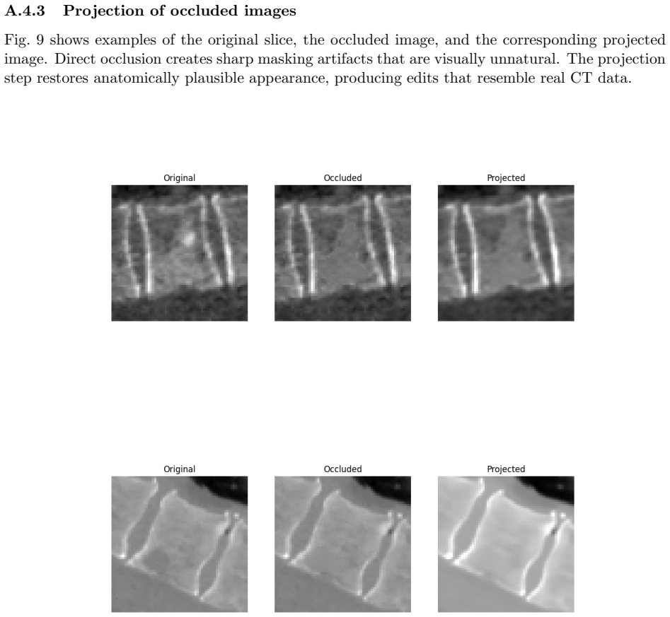
Hide-and-Seek Attribution isolates the malignant contribution of each candidate lesion by revealing it individually while hiding all others, projecting the edited image back to the data manifold with the diffusion autoencoder, and quantifying the effect on a latent-space classifier, thereby converting vertebra-level labels into reliable segmentations without any mask supervision during training.
What carries the argument
Hide-and-Seek Attribution: the process of selectively revealing one suspect lesion while hiding the others, followed by manifold projection via the diffusion autoencoder and latent classification to measure its isolated contribution to malignancy.
If this is right
- Lesion masks can be generated from cheap vertebra-level labels instead of expensive voxel annotations.
- Both lytic and blastic lesions that resemble benign changes receive accurate segmentations.
- The approach exceeds the performance of existing baselines that use the same weak supervision.
- Generative editing combined with selective occlusion supports weakly supervised medical segmentation.
Where Pith is reading between the lines
- The hide-and-seek principle could extend to other weakly supervised tasks where generative models can produce plausible healthy or normal versions of images.
- Manifold projection after selective occlusion may help disentangle subtle pathological signals from normal anatomical variation in additional imaging domains.
- Routine clinical reports that already contain vertebra-level tags could be leveraged to train detailed segmentation models at scale.
Load-bearing premise
The diffusion autoencoder must produce faithful healthy edits of vertebrae, and selectively revealing one candidate while hiding the rest must isolate the true malignant contribution without interference from normal image structure or model artifacts.
What would settle it
On a held-out case containing both confirmed metastases and degenerative changes, check whether the final segmentation assigns high attribution scores to non-malignant regions when revealed alone or low scores to verified lesions.
Figures


read the original abstract
Accurate segmentation of vertebral metastasis in CT is clinically important yet difficult to scale, as voxel-level annotations are scarce and both lytic and blastic lesions often resemble benign degenerative changes. We introduce a 2D weakly supervised method trained solely on vertebra-level healthy/malignant labels, without any lesion masks. The method combines a Diffusion Autoencoder (DAE) that produces a classifier-guided healthy edit of each vertebra with pixel-wise difference maps that propose suspect candidate lesions. To determine which regions truly reflect malignancy, we introduce Hide-and-Seek Attribution: each candidate is revealed in turn while all others are hidden, the edited image is projected back to the data manifold by the DAE, and a latent-space classifier quantifies the isolated malignant contribution of that component. High-scoring regions form the final lytic or blastic segmentation. On held-out radiologist annotations, we achieve strong blastic/lytic performance despite no mask supervision (F1: 0.91/0.85; Dice: 0.87/0.78), exceeding baselines (F1: 0.79/0.67; Dice: 0.74/0.55). These results show that vertebra-level labels can be transformed into reliable lesion masks, demonstrating that generative editing combined with selective occlusion supports accurate weakly supervised segmentation in CT.
Editorial analysis
A structured set of objections, weighed in public.
Referee Report
Summary. The manuscript presents a 2D weakly supervised segmentation method for vertebral metastases in CT that relies solely on vertebra-level healthy/malignant labels. A Diffusion Autoencoder generates classifier-guided healthy edits of each vertebra; pixel-wise difference maps propose candidate lesions; Hide-and-Seek Attribution then reveals one candidate at a time, projects the edited image back onto the data manifold via the DAE, and uses a latent-space classifier to score the isolated malignant contribution. Final segmentations are formed from high-scoring regions. On held-out radiologist annotations the method reports F1 scores of 0.91/0.85 and Dice scores of 0.87/0.78 for blastic/lytic lesions, outperforming the cited baselines.
Significance. If the core assumptions hold, the work demonstrates that generative editing combined with selective occlusion can convert coarse labels into usable lesion masks, addressing a practical bottleneck in scaling metastasis segmentation. The approach is technically distinctive in its use of manifold projection for attribution and could influence future weakly supervised pipelines in medical imaging, provided the reported gains prove robust.
major comments (3)
- [§3.3] §3.3 (Hide-and-Seek Attribution): the procedure assumes that DAE healthy edits faithfully remove lesions while preserving anatomy and that selective revelation isolates malignant contribution without residual structural cues or projection artifacts; no quantitative fidelity metrics (e.g., lesion-removal success rate or reconstruction PSNR on edited vs. original images) or failure-case analysis are supplied, which directly underpins the isolation claim.
- [§4.2] §4.2 and Table 2: performance is reported on held-out annotations with F1/Dice gains over baselines, yet no ablation removing the hide-and-seek step, no multi-lesion interaction controls, and no statistical significance tests or confidence intervals are provided; without these it is unclear whether the gains arise from the attribution mechanism or from correlated but non-causal image features.
- [§5.1] §5.1: the latent classifier is trained on DAE encodings of edited images, but the manuscript does not report how the classifier was validated for sensitivity to the specific edit patterns produced by the hide-and-seek procedure, leaving open the possibility that classification scores reflect manifold projection artifacts rather than true malignancy.
minor comments (2)
- [Figure 3] Figure 3: the qualitative examples of difference maps and final segmentations would benefit from side-by-side comparison with the DAE-edited images to illustrate the attribution effect.
- [Abstract] Abstract and §2: the baseline methods are referenced only by name; a brief description of their supervision level and architecture would help readers assess the fairness of the comparison.
Simulated Author's Rebuttal
We thank the referee for the constructive and detailed feedback. We address each major comment point by point below, indicating where revisions will be incorporated to improve the manuscript.
read point-by-point responses
-
Referee: [§3.3] §3.3 (Hide-and-Seek Attribution): the procedure assumes that DAE healthy edits faithfully remove lesions while preserving anatomy and that selective revelation isolates malignant contribution without residual structural cues or projection artifacts; no quantitative fidelity metrics (e.g., lesion-removal success rate or reconstruction PSNR on edited vs. original images) or failure-case analysis are supplied, which directly underpins the isolation claim.
Authors: We agree that quantitative validation of the DAE edit fidelity is necessary to support the isolation claim. In the revised manuscript we will add PSNR and SSIM metrics comparing original and edited vertebrae, a lesion-removal success rate obtained from radiologist review on a 50-vertebra subset, and a failure-case analysis section discussing residual artifacts and incomplete removals. revision: yes
-
Referee: [§4.2] §4.2 and Table 2: performance is reported on held-out annotations with F1/Dice gains over baselines, yet no ablation removing the hide-and-seek step, no multi-lesion interaction controls, and no statistical significance tests or confidence intervals are provided; without these it is unclear whether the gains arise from the attribution mechanism or from correlated but non-causal image features.
Authors: We accept that ablations and statistical controls are required to attribute gains specifically to Hide-and-Seek Attribution. The revision will include an ablation that disables the hide-and-seek step, an analysis of multi-lesion cases, paired statistical significance tests, and 95% confidence intervals added to Table 2. revision: yes
-
Referee: [§5.1] §5.1: the latent classifier is trained on DAE encodings of edited images, but the manuscript does not report how the classifier was validated for sensitivity to the specific edit patterns produced by the hide-and-seek procedure, leaving open the possibility that classification scores reflect manifold projection artifacts rather than true malignancy.
Authors: We acknowledge the need to demonstrate that the latent classifier responds to malignancy rather than projection artifacts. We will add sensitivity experiments that perturb edit patterns, compare scores on real versus synthetic edits, and report the classifier's cross-validation performance in the revised §5.1. revision: yes
Circularity Check
No significant circularity in the hide-and-seek attribution pipeline
full rationale
The paper introduces a weakly supervised segmentation method that relies on an external diffusion autoencoder for healthy edits and a separate latent-space classifier for attribution. The derivation chain consists of a sequence of independent processing steps (difference maps, selective occlusion, manifold projection, and classification) whose outputs are evaluated empirically on held-out radiologist annotations. No equations or procedural descriptions reduce the final segmentation masks to a fitted parameter, self-referential definition, or load-bearing self-citation by construction. The reported F1 and Dice scores are external benchmarks rather than tautological outputs of the input vertebra-level labels.
Axiom & Free-Parameter Ledger
axioms (1)
- domain assumption Diffusion autoencoders produce accurate healthy edits of vertebrae that remove only malignant features.
invented entities (1)
-
Hide-and-Seek Attribution
no independent evidence
Lean theorems connected to this paper
-
IndisputableMonolith/Cost/FunctionalEquationwashburn_uniqueness_aczel unclear?
unclearRelation between the paper passage and the cited Recognition theorem.
The method combines a Diffusion Autoencoder (DAE) that produces a classifier-guided healthy edit of each vertebra with pixel-wise difference maps that propose suspect candidate lesions... Hide-and-Seek Attribution: each candidate is revealed in turn while all others are hidden, the edited image is projected back to the data manifold by the DAE, and a latent-space classifier quantifies the isolated malignant contribution
What do these tags mean?
- matches
- The paper's claim is directly supported by a theorem in the formal canon.
- supports
- The theorem supports part of the paper's argument, but the paper may add assumptions or extra steps.
- extends
- The paper goes beyond the formal theorem; the theorem is a base layer rather than the whole result.
- uses
- The paper appears to rely on the theorem as machinery.
- contradicts
- The paper's claim conflicts with a theorem or certificate in the canon.
- unclear
- Pith found a possible connection, but the passage is too broad, indirect, or ambiguous to say the theorem truly supports the claim.
Reference graph
Works this paper leans on
-
[1]
Explaining image classifiers by removing input features using generative models
Chirag Agarwal and Anh Nguyen. Explaining image classifiers by removing input features using generative models. InProceedings of the Asian Conference on Computer Vision (ACCV), November 2020
work page 2020
-
[2]
Applying multiple instance learning for breast cancer lesion detection in mammography images
Nedra Amara and Said Gattoufi. Applying multiple instance learning for breast cancer lesion detection in mammography images. InProceedings of the 10th International Conference on Information and Communication Technologies for Ageing Well and e-Health - ICT4AWE, pages 93–97. INSTICC, SciTePress, 2024
work page 2024
-
[3]
Matan Atad, Vitalii Dmytrenko, Yitong Li, Xinyue Zhang, Matthias Keicher, Jan Kirschke, Bene Wiestler, Ashkan Khakzar, and Nassir Navab. Chexplaining in style: Counterfactual explanations for chest x-rays using stylegan.arXiv arXiv:2207.07553, 2022
-
[4]
Kirschke, and Matthias Keicher
Matan Atad, David Schinz, Hendrik Moeller, Robert Graf, Benedikt Wiestler, Daniel Rueck- ert, Nassir Navab, Jan S. Kirschke, and Matthias Keicher. Counterfactual explanations for medical image classification and regression using diffusion autoencoder.Machine Learning for Biomedical Imaging (Melba), 2(iMIMIC 2023 Special Issue):2024:024, 2024
work page 2023
-
[5]
Pathophysiology of bone metastases
James Berenson, Lakshmi Rajdev, and Michael Broder. Pathophysiology of bone metastases. Cancer Biology & Therapy, 5(9):1078–1081, 2006
work page 2006
-
[6]
C. Y. Chang, C. Buckless, K. J. Yeh, and M. Torriani. Automated detection and segmentation of sclerotic spinal lesions on body CTs using a deep convolutional neural network.Skeletal Radiology, 51(2):391–399, 2022
work page 2022
-
[7]
Grad-cam++: Generalized gradient-based visual explanations for deep convolutional networks
Aditya Chattopadhay, Anirban Sarkar, Prantik Howlader, and Vineeth N Balasubramanian. Grad-cam++: Generalized gradient-based visual explanations for deep convolutional networks. In2018 IEEE Winter Conference on Applications of Computer Vision (WACV), pages 839– 847, 2018
work page 2018
-
[8]
C-cam: Causal cam for weakly supervised semantic segmentation on medical image
Zhang Chen, Zhiqiang Tian, Jihua Zhu, Ce Li, and Shaoyi Du. C-cam: Causal cam for weakly supervised semantic segmentation on medical image. InProceedings of the IEEE/CVF Conference on Computer Vision and Pattern Recognition (CVPR), pages 11676–11685, June 2022
work page 2022
-
[9]
Junlong Cheng, Jin Ye, Zhongying Deng, Jianpin Chen, Tianbin Li, Haoyu Wang, Yanzhou Su, Ziyan Huang, Jilong Chen, Lei Jiangand Hui Sun, Junjun He, Shaoting Zhang, Min Zhu, and Yu Qiao. Sam-med2d, 2023
work page 2023
-
[10]
Jiri Chmelik, Roman Jakubicek, Petr Walek, Jiri Jan, Petr Ourednicek, Lukas Lambert, Elena Amadori, and Giampaolo Gavelli. Deep convolutional neural network-based segmentation and classification of difficult to define metastatic spinal lesions in 3d ct data.Medical Image Analysis, 49:76–88, 2018
work page 2018
-
[11]
E. Edelmers, A. N ¸ ikul ¸ins, K. L. Spr¯ udˇ za, P. Stapulone, N. S. P¯ uce, E. Skrebele, E. E. Si¸ nicina, V. C¯ ırule, A. Kazuˇ sa, and K. Boloˇ cko. AI-assisted detection and localization of spinal metastatic lesions.Diagnostics, 14(21):2458, 2024. 10
work page 2024
-
[12]
E.A. Eisenhauer, P. Therasse, J. Bogaerts, L.H. Schwartz, D. Sargent, R. Ford, J. Dancey, S. Arbuck, S. Gwyther, M. Mooney, L. Rubinstein, L. Shankar, L. Dodd, R. Kaplan, D. La- combe, and J. Verweij. New response evaluation criteria in solid tumours: Revised recist guideline (version 1.1).European Journal of Cancer, 45(2):228–247, 2009
work page 2009
-
[13]
Sarah C Foreman, David Schinz, Malek El Husseini, Sophia S Goller, J¨ urgen Weißinger, Anna- Sophia Dietrich, Martin Renz, Marie-Christin Metz, Georg C Feuerriegel, Benedikt Wiestler, et al. Deep learning to differentiate benign and malignant vertebral fractures at multidetector ct.Radiology, 310(3):e231429, 2024
work page 2024
-
[14]
Daryl R. Fourney, Evan M. Frangou, Timothy C. Ryken, Christian P. Dipaola, Christopher I. Shaffrey, Sigurd H. Berven, Mark H. Bilsky, James S. Harrop, Michael G. Fehlings, Stefano Boriani, Dean Chou, Meic H. Schmidt, David W. Polly, Roberto Biagini, Shane Burch, Mark B. Dekutoski, Aruna Ganju, Peter C. Gerszten, Ziya L. Gokaslan, Michael W. Groff, Norbert...
work page 2011
-
[15]
Ziba Gandomkar, Pek Lan Khong, Amanda Punch, and Sarah Lewis. Using occlusion-based saliency maps to explain an artificial intelligence tool in lung cancer screening: Agreement between radiologists, labels, and visual prompts.Journal of Digital Imaging, 35(5):1164–1175, 2022
work page 2022
-
[16]
Xueqi Guo, Yoshihisa Shinagawa, Sepehr Farhand, Halid Yerebakan, Kritika Iyer, Matthias Wolf, and Gerardo Hermosillo Valadez. Unsupervised abnormality segmentation in chest ct with anatomy-guided latent diffusion model and adaptive thresholding. InProceedings of the IEEE/CVF International Conference on Computer Vision (ICCV) Workshops, pages 1017– 1024, O...
work page 2025
-
[17]
Limai Jiang, Ruitao Xie, Bokai Yang, Juan He, Huazhen Huang, Yi Pan, and Yunpeng Cai. Weakly supervised lesion localization and attribution for oct images with a guided counterfac- tual explainer model.Expert Systems with Applications, 287:128129, 2025
work page 2025
-
[18]
Peng-Tao Jiang, Chang-Bin Zhang, Qibin Hou, Ming-Ming Cheng, and Yunchao Wei. Lay- ercam: Exploring hierarchical class activation maps for localization.IEEE Transactions on Image Processing, 30:5875–5888, 2021
work page 2021
-
[19]
Berg, Wan-Yen Lo, Piotr Dollar, and Ross Girshick
Alexander Kirillov, Eric Mintun, Nikhila Ravi, Hanzi Mao, Chloe Rolland, Laura Gustafson, Tete Xiao, Spencer Whitehead, Alexander C. Berg, Wan-Yen Lo, Piotr Dollar, and Ross Girshick. Segment anything. InProceedings of the IEEE/CVF International Conference on Computer Vision (ICCV), pages 4015–4026, October 2023
work page 2023
-
[20]
Florian Kofler, Hendrik M¨ oller, Josef A. Buchner, Ezequiel de la Rosa, Ivan Ezhov, Marcel Rosier, Isra Mekki, Suprosanna Shit, Moritz Negwer, Rami Al-Maskari, Ali Ert¨ urk, Shankeeth Vinayahalingam, Fabian Isensee, Sarthak Pati, Daniel Rueckert, Jan S. Kirschke, Stefan K. Ehrlich, Annika Reinke, Bjoern Menze, Benedikt Wiestler, and Marie Piraud. Panopti...
work page 2023
-
[21]
Segment anything in medical images.Nature Communications, 15(1):654, 2024
Jun Ma, Yuting He, Feifei Li, Lin Han, Chenyu You, and Bo Wang. Segment anything in medical images.Nature Communications, 15(1):654, 2024. 11
work page 2024
-
[22]
Bone metastases: An overview.Oncology Reviews, 11(1):321, 2017
Filipa Macedo, Katia Ladeira, Filipa Pinho, Nadine Saraiva, Nuno Bonito, Luisa Pinto, and Francisco Goncalves. Bone metastases: An overview.Oncology Reviews, 11(1):321, 2017
work page 2017
-
[23]
Cf-seg: Counterfactuals meet segmentation
Raghav Mehta, Fabio De Sousa Ribeiro, Tian Xia, M´ elanie Roschewitz, Ainkaran Santhi- rasekaram, Dominic C Marshall, and Ben Glocker. Cf-seg: Counterfactuals meet segmentation. InInternational Conference on Medical Image Computing and Computer-Assisted Interven- tion, pages 117–127. Springer, 2025
work page 2025
-
[24]
Masataka Motohashi, Yuki Funauchi, Takuya Adachi, Tomoyuki Fujioka, Naoya Otaka, Yuka Kamiko, Takashi Okada, Ukihide Tateishi, Atsushi Okawa, Toshitaka Yoshii, and Shingo Sato. A new deep learning algorithm for detecting spinal metastases on computed tomography im- ages.Spine (Phila Pa 1976), 49(6):390–397, 2024
work page 1976
-
[25]
Mohammed Bany Muhammad and Mohammed Yeasin. Eigen-CAM: Visual explanations for deep convolutional neural networks.SN Computer Science, 2(1):47, January 2021
work page 2021
-
[26]
Eunsun Oh, Hyun-joo Kim, Jong Won Kwon, Young Cheol Yoon, and Hyun Su Kim. Differen- tiation between spinal subchondral bone metastasis with focal pathologic endplate fracture and oedematous schmorl’s node.Journal of Medical Imaging and Radiation Oncology, 66(7):913– 919, 2022
work page 2022
-
[27]
Koji Onoue, Mai Yakami, Maho Nishio, Keita Nakane, Tetsuya Aramaki, Masashi Yakami, Kazuhiro Fujimoto, Yasushi Nagata, and Shogo Nishio. Temporal subtraction CT with non- rigid image registration improves detection of bone metastases by radiologists: results of a large-scale observer study.Scientific Reports, 11:18422, 2021
work page 2021
-
[28]
Mamba-based weakly supervised medical image segmentation with cross-modal textual information
Zhen Pan, Wenhui Huang, and Yuanjie Zheng. Mamba-based weakly supervised medical image segmentation with cross-modal textual information. Inproceedings of Medical Image Com- puting and Computer Assisted Intervention – MICCAI 2025, volume LNCS 15967. Springer Nature Switzerland, September 2025
work page 2025
-
[29]
Diffusion autoencoders: Toward a meaningful and decodable representation
Konpat Preechakul, Nattanat Chatthee, Suttisak Wizadwongsa, and Supasorn Suwajanakorn. Diffusion autoencoders: Toward a meaningful and decodable representation. InIEEE Confer- ence on Computer Vision and Pattern Recognition (CVPR), 2022
work page 2022
-
[30]
Malika Sanhinova, Nazim Haouchine, Steve D. Pieper, William M. Wells III, Tracy A. Balboni, Alexander Spektor, Mai Anh Huynh, Jeffrey P. Guenette, Bryan Czajkowski, Sarah Caplan, Patrick Doyle, Heejoo Kang, David B. Hackney, and Ron N. Alkalay. Registration of longi- tudinal spine CTs for monitoring lesion growth. In Olivier Colliot and Jhimli Mitra, edit...
work page 2024
-
[31]
Selvaraju, Michael Cogswell, Abhishek Das, Ramakrishna Vedantam, Devi Parikh, and Dhruv Batra
Ramprasaath R. Selvaraju, Michael Cogswell, Abhishek Das, Ramakrishna Vedantam, Devi Parikh, and Dhruv Batra. Grad-cam: Visual explanations from deep networks via gradient- based localization. InProceedings of the IEEE International Conference on Computer Vision (ICCV), Oct 2017
work page 2017
-
[32]
Lubdha M. Shah and Karen L. Salzman. Imaging of spinal metastatic disease.International Journal of Surgical Oncology, 2011(1):769753, 2011
work page 2011
-
[33]
Tao Sheng, Tejas Sudharshan Mathai, Alexander Shieh, and Ronald M Summers. Weakly- supervised detection of bone lesions in ct.Proceedings of SPIE–The International Society for Optical Engineering, 12927:129270Q, 2024. 12
work page 2024
-
[34]
COIN: Counterfactual inpainting for weakly supervised semantic segmentation for medical images
Danila Shvetsov, Johannes Ariva, Maxim Domnich, Raul Vicente, and Daniil Fishman. COIN: Counterfactual inpainting for weakly supervised semantic segmentation for medical images. In Luca Longo, Sebastian Lapuschkin, and Christin Seifert, editors,Explainable Artificial Intelligence, volume 2155 ofCommunications in Computer and Information Science. Springer,...
work page 2024
-
[35]
Hristina Uzunova, Jan Ehrhardt, Timo Kepp, and Heinz Handels. Interpretable explanations of black box classifiers applied on medical images by meaningful perturbations using variational autoencoders. In Elsa D. Angelini and Bennett A. Landman, editors,Medical Imaging 2019: Image Processing, volume 10949, page 1094911. International Society for Optics and ...
work page 2019
-
[36]
Weakly-supervised segmentation for disease localization in chest x-ray images
Ostap Viniavskyi, Mariia Dobko, and Oles Dobosevych. Weakly-supervised segmentation for disease localization in chest x-ray images. In Martin Michalowski and Robert Moskovitch, editors,Artificial Intelligence in Medicine, pages 249–259. Springer International Publishing, 2020
work page 2020
-
[37]
Marc-Andr´ e Weber, Alberto Bazzocchi, and Iris-M N¨ obauer-Huhmann. Tumors of the spine: When can biopsy be avoided?Seminars in Musculoskeletal Radiology, 26(4):453–468, 2022
work page 2022
-
[38]
Julia Wolleb, Florentin Bieder, Robin Sandk¨ uhler, and Philippe C. Cattin. Diffusion models for medical anomaly detection. InMedical Image Computing and Computer Assisted Inter- vention – MICCAI 2022: 25th International Conference, Singapore, September 18–22, 2022, Proceedings, Part VIII, page 35–45. Springer-Verlag, 2022
work page 2022
-
[39]
Diffusion- guided weakly supervised semantic segmentation
Sung-Hoon Yoon, Hoyong Kwon, Jaeseok Jeong, Daehee Park, and Kuk-Jin Yoon. Diffusion- guided weakly supervised semantic segmentation. InComputer Vision – ECCV 2024: 18th European Conference, Milan, Italy, September 29–October 4, 2024, Proceedings, Part XLVIII, page 393–411. Springer-Verlag, 2024
work page 2024
-
[40]
Adding conditional control to text-to-image diffusion models
Lvmin Zhang, Anyi Rao, and Maneesh Agrawala. Adding conditional control to text-to-image diffusion models. InProceedings of the IEEE/CVF International Conference on Computer Vision (ICCV), pages 3836–3847, October 2023. 13 A Appendix A.1 Implementation details A.1.1 Hide-and-Seek attribution algorithm Alg. 1 illustrates the procedure for identifying lytic...
work page 2023
discussion (0)
Sign in with ORCID, Apple, or X to comment. Anyone can read and Pith papers without signing in.