Recognition: no theorem link
Maximum Likelihood Estimation Yields Accurate Line-of-Response Assignment for Positron + Prompt Gamma Ray Events in Multiplexed PET (mPET)
Pith reviewed 2026-05-10 17:50 UTC · model grok-4.3
The pith
Maximum likelihood estimation assigns the correct line of response for over 94 percent of positron plus prompt gamma events in multiplexed PET.
A machine-rendered reading of the paper's core claim, the machinery that carries it, and where it could break.
Core claim
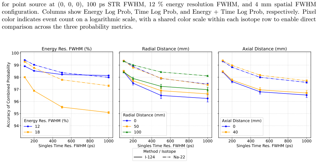
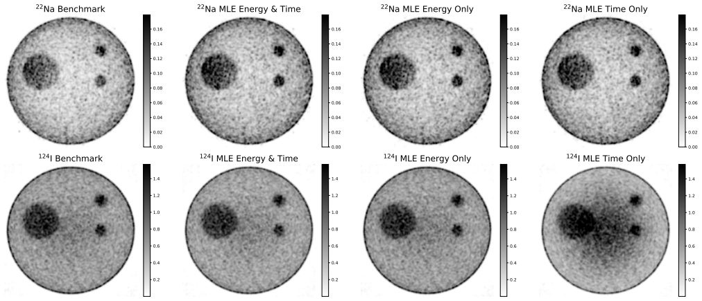
The authors propose a maximum likelihood estimation framework that uses spatial, timing, and energy information from detected photons to assign the most probable line of response to triple-coincidence events produced by beta-plus-gamma emitters, achieving over 96% accuracy for 22Na and 94% for 124I in simulations.
What carries the argument
maximum likelihood estimation framework leveraging spatial, timing, and energy information to select the most probable line of response among possible photon pairs in triple coincidences
Load-bearing premise
The simulations assume perfect knowledge of detector response, timing resolution, and energy spectra with no real-world imperfections or patient scatter.
What would settle it
A measurement on an actual PET scanner using 22Na sources where the fraction of correctly assigned lines of response falls below 90 percent.
Figures


read the original abstract
For accurate disease characterization using positron emission tomography (PET), it is desirable to image multiple radiotracers in a single scan. Conventional PET methods cannot do this due to the indistinguishable annihilation photons produced by different radiotracers. One approach is to label one radiotracer with a positron+prompt-gamma ($\beta^+\!\!-\!\!\gamma$) isotope producing triple coincidences, and another with a pure positron-emitting ($\beta^+$) isotope producing double coincidences. However, $\beta^+\!\!-\!\!\gamma$ emitters present challenges in correctly identifying the two annihilation photons, or equivalently, assigning the correct line-of-response (LOR) to triple-photon coincidence events. Here, we propose a maximum likelihood estimation (MLE) framework leveraging spatial, timing, and energy information to determine the most probable LOR. Simulation studies validated the method: simulations showed over 96\% and 94\% accuracy for LOR assignment of $\beta^+\!\!-\!\!\gamma$ emitters $^{22}$Na and $^{124}$I point sources, respectively. Furthermore, simulated phantom imaging of $^{22}$Na or $^{124}$I distributions alongside a $\beta^+$ emitter demonstrated that MLE LOR assignment achieved comparable image quality -- measured by contrast recovery coefficient (CRC) and cross-talk ratio (XR) -- to benchmark methods, where the prompt gamma was identified using an energy threshold ($\geq 650$ keV) for $^{22}$Na and as the highest-energy photon for $^{124}$I.
Editorial analysis
A structured set of objections, weighed in public.
Referee Report
Summary. The manuscript proposes a maximum likelihood estimation (MLE) framework leveraging spatial, timing, and energy information to assign the correct line-of-response (LOR) for triple-coincidence events from β+-γ emitters (22Na and 124I) in multiplexed PET, enabling separation from pure β+ emitters. Monte Carlo simulation studies report LOR assignment accuracies exceeding 96% for 22Na and 94% for 124I point sources, with phantom imaging yielding contrast recovery coefficients (CRC) and cross-talk ratios (XR) comparable to benchmark methods (energy threshold ≥650 keV for 22Na; highest-energy photon for 124I).
Significance. If validated, the approach could advance multiplexed PET by accurately identifying annihilation photons amid prompt gammas, supporting simultaneous multi-tracer imaging for improved disease characterization. The simulation results provide quantitative metrics on assignment accuracy and image quality under idealized conditions, with the first-principles MLE construction (no fitted parameters) as a methodological strength. However, the lack of real-scanner data and sensitivity analysis limits the assessed impact to potential rather than demonstrated applicability.
major comments (3)
- Methods: The likelihood model is presented only at an abstract level without explicit equations defining the joint probability over spatial, timing, and energy observables or the derivation of the MLE assignment rule. This is load-bearing for assessing whether the reported accuracies follow directly from the model or incorporate unstated approximations.
- Results (simulation studies): The >96% (22Na) and >94% (124I) LOR assignment accuracies, as well as the CRC/XR comparability, are obtained exclusively under the assumption of perfect detector response, timing resolution, and energy spectra. No perturbation analysis or sensitivity studies on deviations (e.g., finite energy resolution, timing jitter, or scatter) are reported, directly affecting the robustness of the central performance claims.
- Results (phantom imaging): Validation is confined to Monte Carlo simulations with no accompanying physical phantom or in vivo data from a real scanner. This leaves open whether unmodeled effects alter the posterior probabilities used for LOR assignment, undermining generalizability of the image-quality equivalence to benchmarks.
minor comments (1)
- The abstract should explicitly name the benchmark methods and any quantitative thresholds used for comparison to improve clarity for readers.
Simulated Author's Rebuttal
We thank the referee for the constructive review and for recognizing the potential of the MLE framework to advance multiplexed PET. We address each major comment point by point below, indicating the revisions we will incorporate.
read point-by-point responses
-
Referee: Methods: The likelihood model is presented only at an abstract level without explicit equations defining the joint probability over spatial, timing, and energy observables or the derivation of the MLE assignment rule. This is load-bearing for assessing whether the reported accuracies follow directly from the model or incorporate unstated approximations.
Authors: We agree that explicit equations are required for rigorous evaluation. In the revised manuscript we will expand the Methods section to present the full joint probability density P(spatial, timing, energy | LOR hypothesis) and derive the maximum-likelihood assignment rule from first principles, confirming that no fitted parameters or hidden approximations are used. revision: yes
-
Referee: Results (simulation studies): The >96% (22Na) and >94% (124I) LOR assignment accuracies, as well as the CRC/XR comparability, are obtained exclusively under the assumption of perfect detector response, timing resolution, and energy spectra. No perturbation analysis or sensitivity studies on deviations (e.g., finite energy resolution, timing jitter, or scatter) are reported, directly affecting the robustness of the central performance claims.
Authors: The reported figures were generated under idealized conditions to isolate the intrinsic performance of the MLE rule. We will add a dedicated sensitivity-analysis subsection that perturbs the model with realistic energy resolution, timing jitter, and scatter, quantifying the resulting degradation in assignment accuracy and image metrics. revision: yes
-
Referee: Results (phantom imaging): Validation is confined to Monte Carlo simulations with no accompanying physical phantom or in vivo data from a real scanner. This leaves open whether unmodeled effects alter the posterior probabilities used for LOR assignment, undermining generalizability of the image-quality equivalence to benchmarks.
Authors: The study is a simulation-based proof-of-concept. We will expand the Discussion to explicitly address how unmodeled real-scanner effects (detector inefficiencies, scatter, attenuation) could modify the posteriors, and we will state that experimental validation on physical phantoms is required for clinical translation. revision: partial
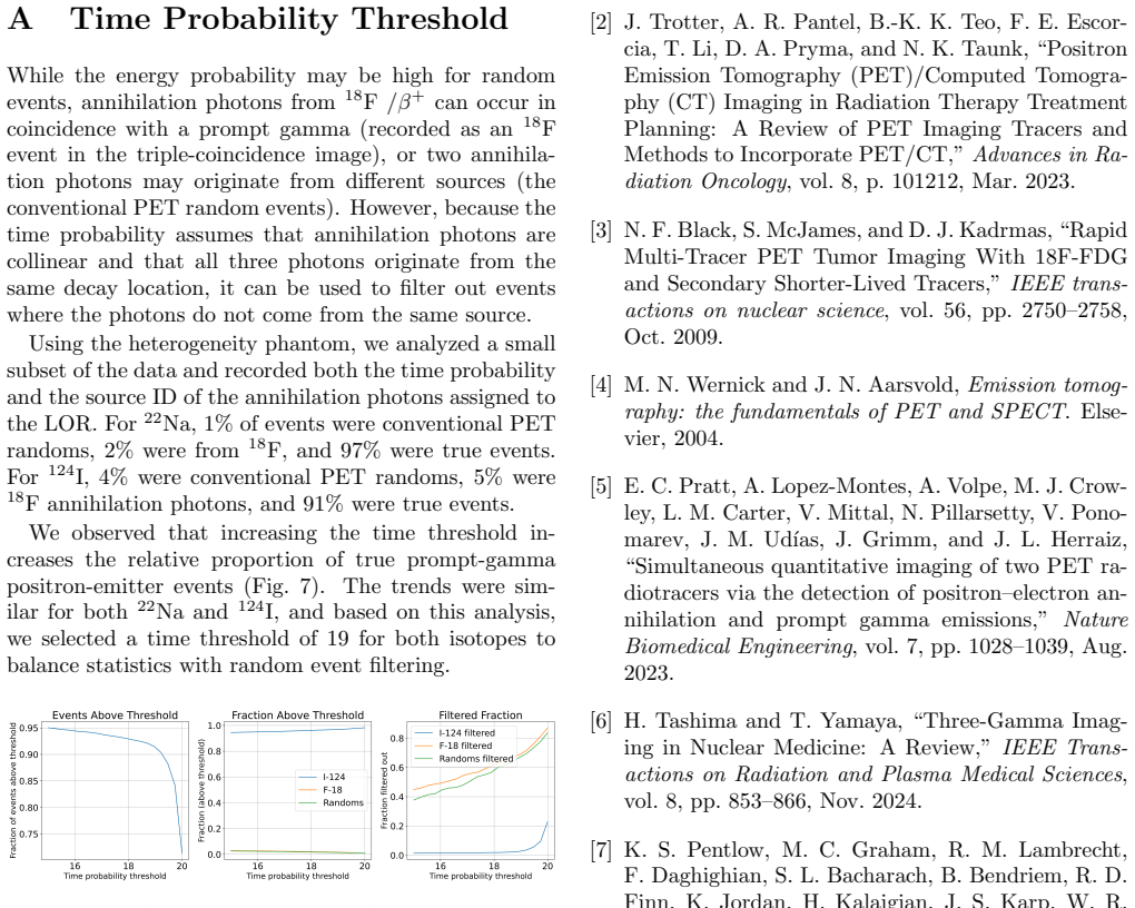
- Absence of physical phantom or in vivo data from a real scanner, which cannot be supplied without new experimental acquisitions outside the scope of the present simulation study.
Circularity Check
No circularity: MLE LOR assignment derived from first-principles detector physics
full rationale
The paper defines the maximum-likelihood estimator directly from the joint probability of spatial, timing, and energy observables for triple-coincidence events. No parameter is fitted to a subset of the target data and then re-used as a 'prediction'; no self-citation supplies a uniqueness theorem or ansatz that the present derivation depends upon; and the reported accuracies are simulation outputs, not algebraic identities. The derivation chain therefore remains independent of its own numerical results.
Axiom & Free-Parameter Ledger
axioms (1)
- domain assumption Detector response functions for position, time, and energy are known and stationary.
Reference graph
Works this paper leans on
-
[1]
Positron emis- sion tomography,
G. Muehllehner and J. S. Karp, “Positron emis- sion tomography,”Physics in Medicine & Biology, vol. 51, p. R117, June 2006
2006
-
[2]
Positron Emission Tomography (PET)/Computed Tomogra- phy (CT) Imaging in Radiation Therapy Treatment Planning: A Review of PET Imaging Tracers and Methods to Incorporate PET/CT,
J. Trotter, A. R. Pantel, B.-K. K. Teo, F. E. Escor- cia, T. Li, D. A. Pryma, and N. K. Taunk, “Positron Emission Tomography (PET)/Computed Tomogra- phy (CT) Imaging in Radiation Therapy Treatment Planning: A Review of PET Imaging Tracers and Methods to Incorporate PET/CT,”Advances in Ra- diation Oncology, vol. 8, p. 101212, Mar. 2023
2023
-
[3]
Rapid Multi-Tracer PET Tumor Imaging With 18F-FDG and Secondary Shorter-Lived Tracers,
N. F. Black, S. McJames, and D. J. Kadrmas, “Rapid Multi-Tracer PET Tumor Imaging With 18F-FDG and Secondary Shorter-Lived Tracers,”IEEE trans- actions on nuclear science, vol. 56, pp. 2750–2758, Oct. 2009
2009
-
[4]
M. N. Wernick and J. N. Aarsvold,Emission tomog- raphy: the fundamentals of PET and SPECT. Else- vier, 2004
2004
-
[5]
Simultaneous quantitative imaging of two PET ra- diotracers via the detection of positron–electron an- nihilation and prompt gamma emissions,
E. C. Pratt, A. Lopez-Montes, A. Volpe, M. J. Crow- ley, L. M. Carter, V. Mittal, N. Pillarsetty, V. Pono- marev, J. M. Ud´ ıas, J. Grimm, and J. L. Herraiz, “Simultaneous quantitative imaging of two PET ra- diotracers via the detection of positron–electron an- nihilation and prompt gamma emissions,”Nature Biomedical Engineering, vol. 7, pp. 1028–1039, Aug. 2023
2023
-
[6]
Three-Gamma Imag- ing in Nuclear Medicine: A Review,
H. Tashima and T. Yamaya, “Three-Gamma Imag- ing in Nuclear Medicine: A Review,”IEEE Trans- actions on Radiation and Plasma Medical Sciences, vol. 8, pp. 853–866, Nov. 2024
2024
-
[7]
Quantitative imag- ing of iodine-124 with PET,
K. S. Pentlow, M. C. Graham, R. M. Lambrecht, F. Daghighian, S. L. Bacharach, B. Bendriem, R. D. Finn, K. Jordan, H. Kalaigian, J. S. Karp, W. R. Robeson, and S. M. Larson, “Quantitative imag- ing of iodine-124 with PET,”Journal of Nuclear Medicine, vol. 37, pp. 1557–1562, Sept. 1996
1996
-
[8]
Dual-isotope PET using positron-gamma emitters,
A. Andreyev and A. Celler, “Dual-isotope PET using positron-gamma emitters,”Physics in Medicine & Biology, vol. 56, p. 4539, July 2011
2011
-
[9]
Methods for increasing the sensitivity of si- multaneous multi-isotope positron emission tomog- raphy,
E. Gonz´ alez, P. D. Olcott, M. Bieniosek, and C. S. Levin, “Methods for increasing the sensitivity of si- multaneous multi-isotope positron emission tomog- raphy,” in2011 IEEE Nuclear Science Symposium Conference Record, pp. 3597–3601, Oct. 2011. ISSN: 1082-3654
2011
-
[10]
Methodology for Quantitative Rapid Multi-Tracer PET Tumor Characterizations,
D. J. Kadrmas and J. M. Hoffman, “Methodology for Quantitative Rapid Multi-Tracer PET Tumor Characterizations,”Theranostics, vol. 3, pp. 757– 773, Oct. 2013
2013
-
[11]
Multiplexed imaging of ra- dionuclides,
G. Soultanidis, J. L. Herraiz, Z. A. Fayad, J. Grimm, and A. J. P. Teunissen, “Multiplexed imaging of ra- dionuclides,”Nature Biomedical Engineering, vol. 9, pp. 993–1006, July 2025. 11
2025
-
[12]
Triplexed PET: Experimental Results of Imaging Three Iso- topes Simultaneously in a Single PET Imaging Session,
S. J. Zou, M. N. Ullah, D. Innes, Y. Shah, G. Chinn, H. Houson, S. Lapi, and C. S. Levin, “Triplexed PET: Experimental Results of Imaging Three Iso- topes Simultaneously in a Single PET Imaging Session,” in2025 IEEE Nuclear Science Sympo- sium (NSS), Medical Imaging Conference (MIC) and Room Temperature Semiconductor Detector Confer- ence (RTSD), pp. 1–2,...
2025
-
[13]
Recovery and normalization of triple co- incidences in pet,
E. Lage, V. Parot, S. C. Moore, A. Sitek, J. M. Ud´ ıas, S. R. Dave, M.-A. Park, J. J. Vaquero, and J. L. Herraiz, “Recovery and normalization of triple co- incidences in pet,”Medical physics, vol. 42, no. 3, pp. 1398–1410, 2015
2015
-
[14]
Dual-radioisotope PET data acquisition and analysis,
R. S. Miyaoka, W. C. Hunter, A. Andreyev, L. Pierce, T. K. Lewellen, A. Celler, and P. E. Ki- nahan, “Dual-radioisotope PET data acquisition and analysis,” in2011 IEEE Nuclear Science Symposium Conference Record, pp. 3780–3783, Oct. 2011. ISSN: 1082-3654
2011
-
[15]
Positron emission tomography with additional gamma-ray detectors for multiple-tracer imaging,
T. Fukuchi, T. Okauchi, M. Shigeta, S. Yamamoto, Y. Watanabe, and S. Enomoto, “Positron emission tomography with additional gamma-ray detectors for multiple-tracer imaging,”Medical Physics, vol. 44, no. 6, pp. 2257–2266, 2017
2017
-
[16]
Si- multaneous micro-pet imaging of f-18 and i-124 with correction for triple-random coincidences,
S. C. Moore, S. Krishnamoorthy, E. Blankemeyer, S. D. Carlin, J. S. Karp, and S. D. Metzler, “Si- multaneous micro-pet imaging of f-18 and i-124 with correction for triple-random coincidences,” in15th International Meeting on Fully Three-Dimensional Image Reconstruction in Radiology and Nuclear Medicine, vol. 11072, pp. 79–83, SPIE, 2019
2019
-
[17]
Quantita- tive Imaging of 55CO and 18F-Labeled Tracers in a Single “Multiplexed
S. J. Zou, I. Lim, J. W. Foster, G. Chinn, H. A. Hou- son, S. E. Lapi, J. Rao, and C. S. Levin, “Quantita- tive Imaging of 55CO and 18F-Labeled Tracers in a Single “Multiplexed” Pet Imaging Session,” in2025 IEEE 22nd International Symposium on Biomedical Imaging (ISBI), pp. 1–5, Apr. 2025. ISSN: 1945- 8452
2025
-
[18]
Simul- taneous multi-isotope pet: A computational frame- work for line of response (lor) identification,
E. F. Hofgard, G. Chinn, and C. S. Levin, “Simul- taneous multi-isotope pet: A computational frame- work for line of response (lor) identification,” in 2020 IEEE Nuclear Science Symposium and Medi- cal Imaging Conference (NSS/MIC), pp. 1–2, 2020
2020
-
[19]
GATE: a simulation toolkit for PET and SPECT,
S. Janet al., “GATE: a simulation toolkit for PET and SPECT,”Physics in Medicine & Biology, vol. 49, p. 4543, Sept. 2004
2004
-
[20]
Petcoil: First results from a second- generation rf-penetrable tof-pet brain insert for si- multaneous pet/mri,
Q. Donget al., “Petcoil: First results from a second- generation rf-penetrable tof-pet brain insert for si- multaneous pet/mri,”Physics in Medicine and Biol- ogy, vol. 69, Sept. 2024
2024
-
[21]
Photo-Detectors for Time of Flight Positron Emission Tomography (ToF-PET),
V. C. Spanoudaki and C. S. Levin, “Photo-Detectors for Time of Flight Positron Emission Tomography (ToF-PET),”Sensors, vol. 10, pp. 10484–10505, Nov. 2010. Publisher: Molecular Diversity Preser- vation International
2010
-
[22]
Innovations in instrumen- tation for positron emission tomography,
E. Berg and S. R. Cherry, “Innovations in instrumen- tation for positron emission tomography,”Seminars in nuclear medicine, vol. 48, pp. 311–331, July 2018
2018
-
[23]
Fully 3d list-mode time-of-flight pet image recon- struction on gpus using cuda,
J.-y. Cui, G. Pratx, S. Prevrhal, and C. S. Levin, “Fully 3d list-mode time-of-flight pet image recon- struction on gpus using cuda,”Medical physics, vol. 38, no. 12, pp. 6775–6786, 2011
2011
-
[24]
Positron range in PET imaging: non-conventional isotopes,
L. Jødal, C. Le Loirec, and C. Champion, “Positron range in PET imaging: non-conventional isotopes,” Physics in Medicine & Biology, vol. 59, p. 7419, Nov. 2014
2014
-
[25]
Methods to Compensate the Time Walk Errors in Timing Measurements for PET Detectors,
S. Xie, X. Zhang, Q. Huang, Z. Gong, J. Xu, and Q. Peng, “Methods to Compensate the Time Walk Errors in Timing Measurements for PET Detectors,” IEEE Transactions on Radiation and Plasma Med- ical Sciences, vol. 4, pp. 555–562, Sept. 2020. Con- ference Name: IEEE Transactions on Radiation and Plasma Medical Sciences. 12
2020
discussion (0)
Sign in with ORCID, Apple, or X to comment. Anyone can read and Pith papers without signing in.