Recognition: 2 theorem links
· Lean TheoremBayesian Aneurysm Growth Detection via Surface Displacement Modeling
Pith reviewed 2026-05-10 18:15 UTC · model grok-4.3
The pith
A Bayesian model using normal-directed displacements and the surrounding vessel as reference detects aneurysm growth with AUC 0.86-0.87 and improves expert agreement over volume measures.
A machine-rendered reading of the paper's core claim, the machinery that carries it, and where it could break.
Core claim
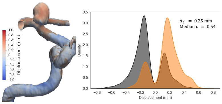
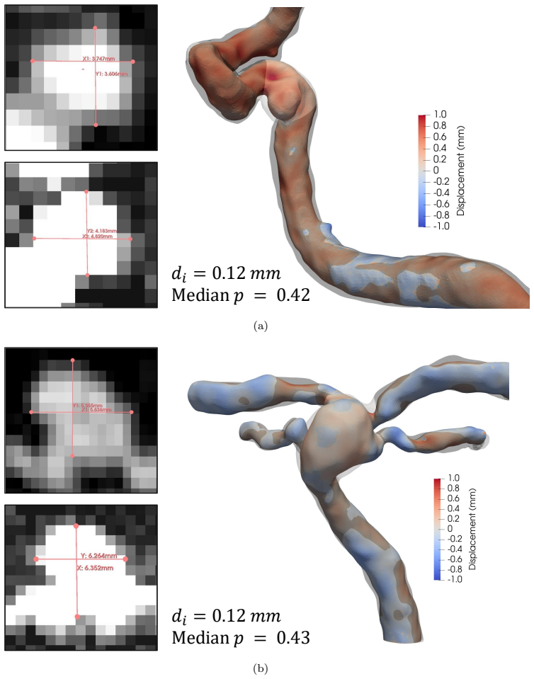
We show that a Bayesian displacement-based model using the surrounding vessel as an internal reference achieves strong discrimination of aneurysm growth (AUC 0.86-0.87) and improves agreement with expert labels (Cohen's kappa up to 0.66 vs. 0.35 for volumetric criteria), while providing calibrated posterior probabilities with uncertainty bounds. The method registers baseline and follow-up surfaces, computes normal-directed displacements, and summarizes change as the difference between mean aneurysm displacement and mean displacement on the surrounding non-aneurysmal vessel segment.
What carries the argument
Bayesian model of normal-directed surface displacements that subtracts mean aneurysm displacement from mean displacement on the adjacent vessel segment to isolate true growth from imaging and registration artifacts.
If this is right
- Calibrated posterior probabilities allow clinicians to set risk-adjusted thresholds for intervention or repeat imaging in borderline cases.
- Robustness to lower-expertise labels supports deployment across centers with varying rater experience.
- Probabilistic outputs with uncertainty bounds reduce reliance on binary volume or diameter rules that miss subtle surface change.
- Cross-sequence and cross-scanner performance enables consistent surveillance protocols without retraining per site.
Where Pith is reading between the lines
- The same displacement-difference approach could be tested on other longitudinal vascular datasets, such as aortic or carotid aneurysms, to check whether the internal-reference principle generalizes.
- High-uncertainty cases flagged by the model could be routed automatically for additional imaging sequences or higher-resolution acquisitions.
- Incorporating the posterior growth probability into existing rupture-risk calculators might improve patient-specific decision thresholds without requiring new clinical trials for every scanner type.
Load-bearing premise
The surrounding non-aneurysmal vessel segment experiences no meaningful structural change between the two scans, so its average displacement can cancel out shared imaging and processing errors.
What would settle it
A new cohort in which independent high-resolution imaging shows clear vessel-wall remodeling or movement in the reference segment and the model's AUC for growth detection falls below 0.75.
Figures


read the original abstract
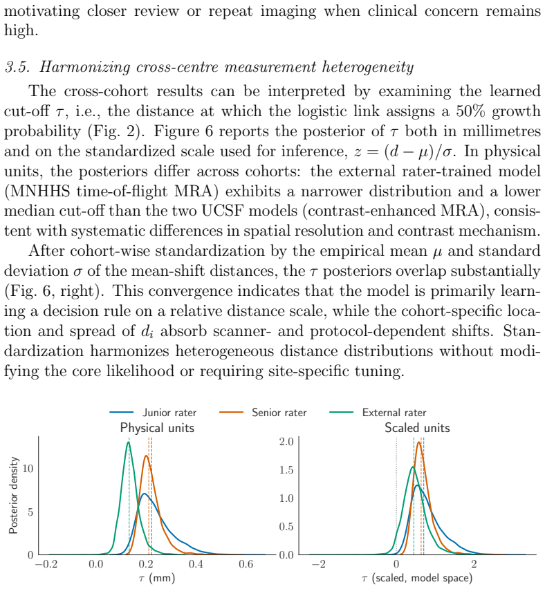
Clinical decisions for unruptured intracranial aneurysms depend on detecting growth on follow-up magnetic resonance angiography (MRA). Growth is typically judged from manual 2D diameters on few slices, which vary across clinicians and frequently miss subtle 3D change. Even with 3D segmentations, apparent differences can reflect resolution, segmentation, surface processing, or registration mismatch rather than true growth; most criteria remain heuristic and binary. We show that a Bayesian displacement-based model using the surrounding vessel as an internal reference achieves strong discrimination of aneurysm growth (AUC 0.86-0.87) and improves agreement with expert labels (Cohen's kappa up to 0.66 vs. 0.35 for volumetric criteria), while providing calibrated posterior probabilities with uncertainty bounds. The method registers baseline and follow-up surfaces, computes normal-directed displacements, and summarizes change as the difference between mean aneurysm displacement and mean displacement on the surrounding non-aneurysmal vessel segment. The vessel segment serves as an internal control for imaging and processing variability, assuming negligible structural change over the surveillance interval. We evaluate two cohorts spanning time-of-flight and contrast-enhanced longitudinal MRA studies: a public dataset labeled from neuroradiologist-provided measurements and an institutional dataset labeled by senior and junior raters. Performance is preserved when training on lower-expertise labels, indicating robustness to label variability. Calibrated probabilities may aid clinical decision-making in borderline cases, where high uncertainty can motivate repeat imaging. This framework provides interpretable probabilistic growth assessment from longitudinal MRA, reduces dependence on clinician expertise, and supports cross-center surveillance across scanners and angiography sequences.
Editorial analysis
A structured set of objections, weighed in public.
Referee Report
Summary. The manuscript proposes a Bayesian displacement-based model for detecting growth in unruptured intracranial aneurysms from longitudinal MRA. Surfaces are registered between baseline and follow-up scans, normal-directed displacements are computed, and growth is quantified as the difference between mean aneurysm displacement and mean displacement on the adjacent non-aneurysmal vessel segment (treated as an internal control). The method is evaluated on a public dataset and an institutional cohort, reporting AUC 0.86-0.87 for growth discrimination, Cohen's kappa up to 0.66 (vs. 0.35 for volumetric criteria), and calibrated posterior probabilities with uncertainty bounds.
Significance. If the central results hold after addressing the load-bearing assumption, the work would provide a more objective, probabilistic alternative to manual diameter or volumetric criteria for aneurysm surveillance. The internal-reference construction and uncertainty quantification are strengths that could reduce inter-observer variability and support decisions in borderline cases across different scanners and sequences.
major comments (1)
- [Abstract and Methods] Abstract and Methods (vessel reference construction): The growth metric is defined as the difference in mean normal-directed displacements between the aneurysm surface and the surrounding vessel segment. This construction is valid only under the assumption of negligible vessel change over the surveillance interval, yet no auxiliary analysis (vessel-only displacement statistics, correlation with interval length, or sensitivity to simulated vessel drift) is reported to bound possible violations. Because any systematic vessel displacement is subtracted from the aneurysm signal, this untested assumption is load-bearing for the reported AUC and kappa values.
minor comments (3)
- [Results] Results: Report AUC and kappa values with confidence intervals or p-values for the improvement over volumetric baselines to allow assessment of statistical significance.
- [Methods] Methods: Provide more detail on the surface registration algorithm, the exact definition of the vessel segment boundaries, and the Bayesian model hyperparameters to support reproducibility.
- [Figures] Figure captions: Ensure all figures showing posterior distributions or displacement maps include scale bars, color legends, and explicit indication of which surfaces correspond to aneurysm versus vessel.
Simulated Author's Rebuttal
We thank the referee for the constructive comment on the vessel reference construction. We address the concern directly below and will revise the manuscript to strengthen the supporting evidence for the central assumption.
read point-by-point responses
-
Referee: [Abstract and Methods] Abstract and Methods (vessel reference construction): The growth metric is defined as the difference in mean normal-directed displacements between the aneurysm surface and the surrounding vessel segment. This construction is valid only under the assumption of negligible vessel change over the surveillance interval, yet no auxiliary analysis (vessel-only displacement statistics, correlation with interval length, or sensitivity to simulated vessel drift) is reported to bound possible violations. Because any systematic vessel displacement is subtracted from the aneurysm signal, this untested assumption is load-bearing for the reported AUC and kappa values.
Authors: We agree that the assumption of negligible structural change in the non-aneurysmal vessel segment is load-bearing for the growth metric and the reported performance metrics. The original manuscript states the assumption explicitly but does not provide the auxiliary analyses suggested. In the revised manuscript we will add: (i) summary statistics of mean normal-directed displacements on the vessel-only segments for both cohorts, (ii) the correlation between vessel displacement and surveillance interval length, and (iii) a sensitivity analysis that injects controlled vessel drift and quantifies the resulting change in AUC and Cohen’s kappa. These additions will bound the possible impact of violations and increase confidence in the internal-reference approach. revision: yes
Circularity Check
No significant circularity; growth metric defined from physical reference assumption with external validation against expert labels
full rationale
The paper proposes a displacement-based growth metric by registering surfaces, computing normal-directed displacements, and taking the difference between mean aneurysm displacement and mean vessel-segment displacement. This construction rests on an explicit physical assumption (vessel stability as internal control) rather than defining the metric in terms of itself or fitting parameters to the target labels and then re-presenting the fit as a prediction. Evaluation uses independent expert labels on two separate cohorts to compute AUC and Cohen's kappa; no step reduces the reported performance to a tautology or to a self-citation chain. No uniqueness theorems, ansatzes smuggled via prior work, or renaming of known results are invoked. The Bayesian modeling step provides posterior probabilities and uncertainty but does not alter the external grounding of the performance claims.
Axiom & Free-Parameter Ledger
axioms (1)
- domain assumption The surrounding non-aneurysmal vessel segment undergoes negligible structural change over the surveillance interval
Lean theorems connected to this paper
-
IndisputableMonolith/Foundation/AbsoluteFloorClosure.leanabsolute_floor_iff_bare_distinguishability unclear?
unclearRelation between the paper passage and the cited Recognition theorem.
The vessel segment serves as an internal control for imaging and processing variability, assuming negligible structural change over the surveillance interval... d_i = a_i - v_i ... Bayesian soft-threshold model
-
IndisputableMonolith/Cost/FunctionalEquation.leanwashburn_uniqueness_aczel unclear?
unclearRelation between the paper passage and the cited Recognition theorem.
normal-directed displacements... logistic(s(d*_i - τ))
What do these tags mean?
- matches
- The paper's claim is directly supported by a theorem in the formal canon.
- supports
- The theorem supports part of the paper's argument, but the paper may add assumptions or extra steps.
- extends
- The paper goes beyond the formal theorem; the theorem is a base layer rather than the whole result.
- uses
- The paper appears to rely on the theorem as machinery.
- contradicts
- The paper's claim conflicts with a theorem or certificate in the canon.
- unclear
- Pith found a possible connection, but the passage is too broad, indirect, or ambiguous to say the theorem truly supports the claim.
Reference graph
Works this paper leans on
-
[1]
T. Inoue, H. Shimizu, M. Fujimura, A. Saito, T. Tominaga, Annual rup- ture risk of growing unruptured cerebral aneurysms detected by mag- netic resonance angiography: Clinical article, Journal of Neurosurgery 117 (1) (2012) 20–25. doi:10.3171/2012.4.JNS112225
-
[2]
W. A. Mehan, J. M. Romero, J. A. Hirsch, D. J. Sabbag, R. G. Gon- zalez, J. J. Heit, P. W. Schaefer, Unruptured intracranial aneurysms conservatively followed with serial CT angiography: could morphology and growth predict rupture?, Journal of NeuroInterventional Surgery 6 (10) (2014) 761–766. doi:10.1136/neurintsurg-2013-010944. 23
-
[3]
L. T. van der Kamp, G. J. E. Rinkel, D. Verbaan, R. van den Berg, W. P. Vandertop, Y. Murayama, T. Ishibashi, A. Lindgren, T. Koivisto, M. Teo, J. St George, R. Agid, I. Radovanovic, J. Moroi, K. Igase, I. R. van den Wijngaard, M. Rahi, J. Rinne, J. Kuhmonen, H. D. Boogaarts, G. K. C. Wong, J. M. Abrigo, A. Morita, Y. Shiokawa, K. A. M. Hacken- berg, N. E...
-
[4]
W. Brinjikji, Y.-Q. Zhu, G. Lanzino, H. Cloft, M. Murad, Z. Wang, D. Kallmes, Risk Factors for Growth of Intracranial Aneurysms: A Sys- tematic Review and Meta-Analysis, AJNR: American Journal of Neu- roradiology 37 (4) (2016) 615–620. doi:10.3174/ajnr.A4575
-
[5]
N. Etminan, G. J. Rinkel, Unruptured intracranial aneurysms: develop- ment, rupture and preventive management, Nature Reviews Neurology 12 (12) (2016) 699–713. doi:10.1038/nrneurol.2016.150
-
[6]
A. M. Algra, A. Lindgren, M. D. I. Vergouwen, J. P. Greving, I. C. van der Schaaf, T. P. C. van Doormaal, G. J. E. Rinkel, Procedural Clin- ical Complications, Case-Fatality Risks, and Risk Factors in Endovascu- lar and Neurosurgical Treatment of Unruptured Intracranial Aneurysms: A Systematic Review and Meta-analysis, JAMA Neurology 76 (3) (2019) 282–293...
-
[7]
B. G. Thompson, R. D. Brown, S. Amin-Hanjani, J. P. Broderick, K. M. Cockroft, E. S. Connolly, G. R. Duckwiler, C. C. Harris, V. J. Howard, S. C. C. Johnston, P. M. Meyers, A. Molyneux, C. S. Ogilvy, A. J. Ringer, J. Torner, on behalf of the American Heart Association Stroke Council, Council on Cardiovascular and Stroke Nursing, and Council on Epidemiolog...
-
[8]
N. Etminan, D. A. d. Sousa, C. Tiseo, R. Bourcier, H. Desal, A. Lind- gren, T. Koivisto, D. Netuka, S. Peschillo, S. L´ emeret, A. Lal, M. D. Vergouwen, G. J. Rinkel, European Stroke Organisation (ESO) guide- lines on management of unruptured intracranial aneurysms, European Stroke Journal 7 (3) (2022) V. doi:10.1177/23969873221099736. 24
-
[9]
D. Backes, M. D. Vergouwen, A. T. Tiel Groenestege, A. S. E. Bor, B. K. Velthuis, J. P. Greving, A. Algra, M. J. Wermer, M. A. van Walderveen, K. G. terBrugge, R. Agid, G. J. Rinkel, PHASES Score for Predic- tion of Intracranial Aneurysm Growth, Stroke 46 (5) (2015) 1221–1226. doi:10.1161/STROKEAHA.114.008198
-
[10]
D. Backes, G. J. Rinkel, J. P. Greving, B. K. Velthuis, Y. Murayama, H. Takao, T. Ishibashi, M. Igase, K. G. terBrugge, R. Agid, J. E. J¨ a¨ askel¨ ainen, A. E. Lindgren, T. Koivisto, M. Von Und Zu Fraun- berg, S. Matsubara, J. Moroi, G. K. Wong, J. M. Abrigo, K. Igase, K. Matsumoto, M. J. Wermer, M. A. Van Walderveen, A. Algra, M. D. Vergouwen, ELAPSS sc...
-
[11]
H. J. Kim, D. Y. Yoon, E. S. Kim, H. J. Lee, H. J. Jeon, J. Y. Lee, B.- M. Cho, Intraobserver and interobserver variability in CT angiography and MR angiography measurements of the size of cerebral aneurysms, Neuroradiology 59 (5) (2017) 491–497. doi:10.1007/s00234-017-1826-y
-
[12]
K. Timmins, H. Kuijf, M. Vergouwen, M. Otten, Y. Ruigrok, B. Velthuis, I. van der Schaaf, Reliability and Agreement of 2D and 3D Measure- ments on MRAs for Growth Assessment of Unruptured Intracranial Aneurysms, AJNR: American Journal of Neuroradiology 42 (9) (2021) 1598–1603. doi:10.3174/ajnr.A7186
-
[13]
A. Planinc, N. ˇSpegel, Z. Podobnik, U. ˇSinigoj, P. Skubic, J. H. Choi, W. Park, T. Robiˇ c, N. Tabor, L. Jarabek, ˇZ. ˇSpiclin, ˇZ. Bizjak, As- sessing accuracy and consistency in intracranial aneurysm sizing: hu- man expertise vs. artificial intelligence, Scientific Reports 14 (1) (2024) 16080. doi:10.1038/s41598-024-65825-4
-
[14]
A. Malhotra, X. Wu, H. P. Forman, H. K. Grossetta Nardini, C. C. Matouk, D. Gandhi, C. Moore, P. Sanelli, Growth and Rupture Risk of Small Unruptured Intracranial Aneurysms: A Systematic Review, Annals of Internal Medicine 167 (1) (2017) 26–33. doi:10.7326/M17- 0246
-
[15]
L. Boussel, V. Rayz, C. McCulloch, A. Martin, G. Acevedo- Bolton, M. Lawton, R. Higashida, W. S. Smith, W. L. Young, 25 D. Saloner, Aneurysm Growth Occurs at Region of Low Wall Shear Stress: Patient-Specific Correlation of Hemodynamics and Growth in a Longitudinal Study, Stroke 39 (11) (2008) 2997–3002. doi:10.1161/STROKEAHA.108.521617
-
[16]
A. Firouzian, R. Manniesing, C. T. Metz, S. Klein, B. K. Velthuis, G. J. E. Rinkel, A. Van Der Lugt, W. J. Niessen, Intracranial aneurysm growth quantification in CTA, San Diego, California, USA, 2012, p. 831448. doi:10.1117/12.910713
-
[17]
X. Liu, H. Haraldsson, Y. Wang, E. Kao, M. Ballweber, A. Mar- tin, C. McCulloch, F. Faraji, D. Saloner, for the UCSF Intracranial Aneurysm Monitoring Group, A Volumetric Metric for Monitoring In- tracranial Aneurysms: Repeatability and Growth Criteria in a Longitu- dinal MR Imaging Study, American Journal of Neuroradiology 42 (9) (2021) 1591–1597. doi:10....
-
[18]
ˇZ. Bizjak, ˇZ. ˇSpiclin, Aneurysm growth evaluation and detection: a computer-assisted follow-up MRA analysis, Scientific Reports 14 (1) (2024) 19609. doi:10.1038/s41598-024-70453-z
-
[19]
F. Goudarzian, K. Kondratiuk, V. L. Rayz, Predicting Cerebral Aneurysm Rupture, Neuroimaging Clinics of North America 35 (3) (2025) 333–347. doi:10.1016/j.nic.2025.05.002
-
[20]
J. H. Maki, M. R. Prince, F. J. Londy, T. L. Chenevert, The ef- fects of time varying intravascular signal intensity and k-space ac- quisition order on three-dimensional MR angiography image qual- ity, Journal of Magnetic Resonance Imaging 6 (4) (1996) 642–651. doi:10.1002/jmri.1880060413
-
[21]
Tsuruda, D
J. Tsuruda, D. Saloner, D. Norman, Artifacts associated with MR neu- roangiography., American Journal of Neuroradiology 13 (5) (1992) 1411
1992
-
[22]
W. E. Lorensen, H. E. Cline, Marching cubes: A high resolution 3D sur- face construction algorithm, SIGGRAPH Comput. Graph. 21 (4) (1987) 163–169. doi:10.1145/37402.37422
-
[23]
C. M. De Nys, E. S. Liang, M. Prior, M. A. Woodruff, J. I. Novak, A. R. Murphy, Z. Li, C. D. Winter, M. C. Allenby, Time-of-Flight MRA of In- tracranial Aneurysms with Interval Surveillance, Clinical Segmentation 26 and Annotations, Scientific Data 11 (1) (2024) 555. doi:10.1038/s41597- 024-03397-8
-
[24]
K. A. M. Hackenberg, A. Algra, R. Al-Shahi Salman, J. Fr¨ osen, D. Hasan, S. Juvela, D. Langer, P. Meyers, A. Morita, G. Rinkel, N. Etminan, the Unruptured Aneurysms and SAH CDE Project In- vestigators, J. I. Suarez, R. L. Macdonald, S. Amin-Hanjani, R. D. Brown, A. L. De Oliveira Manoel, C. P. Derdeyn, N. Etminan, E. Keller, P. D. LeRoux, S. Mayer, A. Mo...
-
[25]
M. D. Hoffman, A. Gelman, The No-U-Turn sampler: Adaptively setting path lengths in hamiltonian monte carlo, Journal of Machine Learning Research 15 (1) (2014) 1593–1623
2014
-
[26]
A. Hans, S. Bhattacharya, I. Bilionis, P. P. Vlachos, Stochastic volumet- ric reconstruction, in: 15th Int. Symp. on Particle Image Velocimetry- ISPIV, 2023
2023
-
[27]
A. Hans, S. Bhattacharya, K. Hao, P. Vlachos, I. Bilionis, Bayesian re- construction of 3d particle positions in high-seeding density flows, Mea- surement Science and Technology 35 (11) (2024) 116002. 27
2024
-
[28]
T. K. Koo, M. Y. Li, A Guideline of Selecting and Reporting Intraclass Correlation Coefficients for Reliability Research, Journal of Chiropractic Medicine 15 (2) (2016) 155–163. doi:10.1016/j.jcm.2016.02.012
-
[29]
A. Hans, A. M. Chaudhari, I. Bilionis, J. H. Panchal, Quantifying in- dividuals’ theory-based knowledge using probabilistic causal graphs: a bayesian hierarchical approach, in: International Design Engineering Technical Conferences and Computers and Information in Engineer- ing Conference, Vol. 83921, American Society of Mechanical Engineers, 2020, p. V003T03A014
2020
-
[30]
A. Hans, A. M. Chaudhari, I. Bilionis, J. H. Panchal, A bayesian hierar- chical model for extracting individuals’ theory-based causal knowledge, Journal of Computing and Information Science in Engineering 23 (3) (2023) 031011
2023
-
[31]
E. Bullitt, D. Zeng, B. Mortamet, A. Ghosh, S. R. Aylward, W. Lin, B. L. Marks, K. Smith, The effects of healthy ag- ing on intracerebral blood vessels visualized by magnetic reso- nance angiography, Neurobiology of Aging 31 (2) (2010) 290–300. doi:10.1016/j.neurobiolaging.2008.03.022
-
[32]
M. C. Brindise, S. Rothenberger, B. Dickerhoff, S. Schnell, M. Markl, D. Saloner, V. L. Rayz, P. P. Vlachos, Multi-modality cerebral aneurysm haemodynamic analysis:in vivo4D flow MRI,in vitrovolumet- ric particle velocimetry andin silicocomputational fluid dynamics, Journal of The Royal Society Interface 16 (158) (2019) 20190465. doi:10.1098/rsif.2019.0465
- [33]
-
[34]
W.-C. Hsu, M. Meuschke, A. F. Frangi, B. Preim, K. Lawonn, A survey of intracranial aneurysm detection and segmentation, Medical Image Analysis 101 (2025) 103493. doi:10.1016/j.media.2025.103493. 28
discussion (0)
Sign in with ORCID, Apple, or X to comment. Anyone can read and Pith papers without signing in.