Recognition: 2 theorem links
· Lean TheoremDual-Tuned 31P-1H Dual-Row Loop/Dipole 32-element Transceiver Array for Human Brain Spectroscopy at 9.4T
Pith reviewed 2026-05-10 18:01 UTC · model grok-4.3
The pith
A 32-element loop-dipole array provides full-brain coverage for dual-frequency 31P-1H spectroscopy at 9.4T.
A machine-rendered reading of the paper's core claim, the machinery that carries it, and where it could break.
Core claim
The authors constructed numerical models of dual-row arrays with loops for 31P and coaxial-end folded-end dipoles for 1H. Using multi-tissue voxel simulations to optimize circularly polarized excitation, they built and tested the 32-element array on phantom and human volunteer. Measurements confirmed full-brain imaging capability with reasonable signal-to-noise ratio and transmit performance at both resonance frequencies, outperforming a prior single-row loop design.
What carries the argument
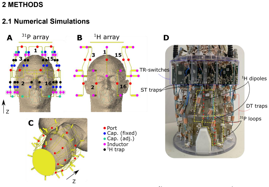
The 32-element dual-tuned transceiver array using a dual-row configuration with loops for 31P and dipoles for 1H in a tight-fit single layer.
If this is right
- The array supports full-brain 31P MRS including cerebellum and brainstem at 9.4T.
- It provides adequate 1H transmit and receive performance for localization and shimming.
- Performance matches or exceeds previous dual-tuned single-row loop arrays in coverage.
- The design can be adapted for use at 10.5T, 11.7T, and 14T.
Where Pith is reading between the lines
- This hybrid approach may simplify multi-nuclear experiments by integrating both frequencies in one setup.
- Enhanced coverage of deep brain structures could open new avenues for studying metabolic disorders affecting the brainstem.
- Success here indicates that similar element combinations might help overcome sensitivity challenges as MRI fields increase further.
Load-bearing premise
Numerical electromagnetic simulations using a multi-tissue voxel model accurately predict real-world transmit and receive performance in human subjects without significant discrepancies from unmodeled factors such as cable losses or subject-specific anatomy variations.
What would settle it
Direct in-vivo measurements on a volunteer that show substantially lower transmit efficiency or SNR in the cerebellum and brainstem than predicted by the simulations would disprove the performance claims.
Figures


read the original abstract
Purpose The goal of this work is to develop and evaluate a single-layer tight-fit 32-element double-tuned loop/dipole transceiver (TxRx) array for human brain 31P MRS at 9.4T, achieving reasonable transmit and receive performance and full-brain coverage at both frequencies. Methods First, we developed numerical models of dual-row TxRx arrays for 31P (loop array) and 1H (coaxial-end folded-end dipole array) frequencies at 9.4T. Next, a multi-tissue voxel model was used to simulate Tx-performance of the arrays and define optimal CP-mode excitation. Following this, the proposed array performance was evaluated by MR measurements both on a phantom and a healthy volunteer. Finally, we compared the proposed array to a previously reported dual-tuned single-row loop-based TxRx array. Results The developed 32-element double-tuned array demonstrated full-brain (including the cerebellum and brain stem) imaging capabilities, reasonable SNR and transmit performance at both frequencies at 9.4T. Conclusion As a proof of concept, we developed a 32-element double-tuned UHF tight-fit TxRx human head array coil for 31P MRS with sufficient 1H performance using a combination of loop and dipole array elements. The proposed array design could also be adapted to higher fields, i.e., 10.5T, 11.7T, and 14T.
Editorial analysis
A structured set of objections, weighed in public.
Referee Report
Summary. The manuscript describes the development of a 32-element dual-tuned 31P-1H transceiver array using a combination of loop and dipole elements in a dual-row configuration for human brain imaging and spectroscopy at 9.4T. Through numerical simulations with a multi-tissue model to optimize CP-mode excitation, followed by phantom and single-volunteer MR measurements, the authors claim that the array achieves full-brain coverage including the cerebellum and brainstem with reasonable transmit and receive performance at both frequencies, and compare it favorably to a prior single-row loop array.
Significance. If the performance claims hold, this work advances RF coil design for ultra-high-field multi-nuclear MRI/MRS by integrating loops and dipoles in a single-layer tight-fit array to achieve improved coverage at 9.4T while maintaining usable 1H imaging alongside 31P spectroscopy. The combined simulation-plus-phantom/volunteer validation approach and explicit comparison to prior hardware provide a concrete basis for assessing trade-offs in transmit efficiency and SNR.
major comments (2)
- [Results] Results section: The central claim of demonstrated full-brain coverage (including cerebellum and brain stem) with reasonable SNR and transmit performance rests on qualitative images and limited metrics from a single phantom and one healthy volunteer. No region-specific quantitative B1+ or SNR maps for deep structures, no multi-subject statistics, and no head-to-head comparison with the prior single-row array under matched conditions are reported, leaving the performance assertions only partially supported.
- [Methods] Methods and Results: The multi-tissue voxel model is used to define CP-mode excitation, yet no quantitative assessment of simulation-experiment agreement (e.g., predicted vs. measured B1+ in cerebellum) or sensitivity to unmodeled factors such as cable losses or inter-subject anatomy is provided, which directly affects confidence in the full-brain coverage claim.
minor comments (1)
- [Abstract] Abstract: The term 'coaxial-end folded-end dipole array' is ambiguous; clarify the exact dipole geometry (e.g., 'coaxially fed folded dipole elements').
Simulated Author's Rebuttal
We thank the referee for the constructive feedback on our manuscript describing the 32-element dual-tuned loop/dipole array. We have addressed the concerns about the strength of evidence for full-brain coverage by adding quantitative metrics and clarifications in the revised version.
read point-by-point responses
-
Referee: [Results] Results section: The central claim of demonstrated full-brain coverage (including cerebellum and brain stem) with reasonable SNR and transmit performance rests on qualitative images and limited metrics from a single phantom and one healthy volunteer. No region-specific quantitative B1+ or SNR maps for deep structures, no multi-subject statistics, and no head-to-head comparison with the prior single-row array under matched conditions are reported, leaving the performance assertions only partially supported.
Authors: We agree that the evidence is based on a single volunteer and phantom, which is typical for proof-of-concept hardware papers. In the revised manuscript we have extracted and reported region-specific B1+ and SNR values from the cerebellum and brainstem in the volunteer data to provide quantitative support. The comparison to the prior single-row loop array uses published results from that work; a matched-condition head-to-head scan was not possible with the available hardware, but we have clarified the setup differences and the coverage advantage shown in the images. Multi-subject statistics are beyond the scope of this initial study. revision: partial
-
Referee: [Methods] Methods and Results: The multi-tissue voxel model is used to define CP-mode excitation, yet no quantitative assessment of simulation-experiment agreement (e.g., predicted vs. measured B1+ in cerebellum) or sensitivity to unmodeled factors such as cable losses or inter-subject anatomy is provided, which directly affects confidence in the full-brain coverage claim.
Authors: We accept this criticism. The revised manuscript now includes a quantitative comparison of simulated versus measured B1+ distributions, with explicit values reported for the cerebellum. We have also added a sensitivity analysis to cable losses in the simulations and a brief discussion of inter-subject anatomical variability as a limitation of the model. revision: yes
Circularity Check
No significant circularity detected; claims rest on independent measurements
full rationale
The paper's chain proceeds from numerical EM modeling of the array geometry, through standard multi-tissue voxel simulations used only to select CP-mode excitation weights, to direct MR measurements on a phantom and one volunteer that independently assess SNR, B1+, and coverage. These measurements are not derived from or forced by the simulation inputs; they constitute separate empirical data. The comparison to a prior single-row array is a side-by-side reporting of measured results rather than a self-referential reduction. No equations, fitted parameters renamed as predictions, or load-bearing self-citations appear that would make the central performance claims equivalent to their own inputs by construction. The workflow is a conventional simulation-guided hardware validation and therefore self-contained against external benchmarks.
Axiom & Free-Parameter Ledger
free parameters (1)
- Array geometry and tuning parameters
axioms (1)
- domain assumption Multi-tissue voxel model accurately represents human head dielectric properties at 9.4T for transmit field prediction.
Lean theorems connected to this paper
-
IndisputableMonolith/Cost/*Jcost uniqueness / washburn_uniqueness_aczel unclear?
unclearRelation between the paper passage and the cited Recognition theorem.
The developed 32-element double-tuned array demonstrated full-brain... reasonable SNR and transmit performance at both frequencies at 9.4T.
What do these tags mean?
- matches
- The paper's claim is directly supported by a theorem in the formal canon.
- supports
- The theorem supports part of the paper's argument, but the paper may add assumptions or extra steps.
- extends
- The paper goes beyond the formal theorem; the theorem is a base layer rather than the whole result.
- uses
- The paper appears to rely on the theorem as machinery.
- contradicts
- The paper's claim conflicts with a theorem or certificate in the canon.
- unclear
- Pith found a possible connection, but the passage is too broad, indirect, or ambiguous to say the theorem truly supports the claim.
Reference graph
Works this paper leans on
-
[1]
Pros and cons of ultra-high-field MRI/MRS for human application
Ladd ME, Bachert P, Meyerspeer M, et al. Pros and cons of ultra-high-field MRI/MRS for human application. Prog Nucl Magn Reson Spectrosc. 2018;109:1-
2018
-
[2]
doi:10.1016/j.pnmrs.2018.06.001
-
[3]
Broadband proton decoupled natural abundance13 C NMR spectroscopy of humans at 1.5 T
Heerschap A, Luyten PR, Van Der Heyden JI, Oosterwaal LJMP, Hollander JAD. Broadband proton decoupled natural abundance13 C NMR spectroscopy of humans at 1.5 T. NMR Biomed. 1989;2(3):124-132. doi:10.1002/nbm.1940020309
-
[4]
Broadband proton decoupling in human31 p NMR spectroscopy
Luyten PR, Bruntink G, Sloff FM, et al. Broadband proton decoupling in human31 p NMR spectroscopy. NMR Biomed. 1989;1(4):177-183. doi:10.1002/nbm.1940010405
-
[5]
Efficient1 H to31 P polarization transfer on a clinical 3T MR system
Klomp DWJ, Wijnen JP , Scheenen TWJ, Heerschap A. Efficient1 H to31 P polarization transfer on a clinical 3T MR system. Magn Reson Med. 2008;60(6):1298-1305. doi:10.1002/mrm.21733
-
[6]
In Vivo13 C imaging enhanced by polarization transfer
Ueshima Y, Yamai S, Ikehira H, et al. In Vivo13 C imaging enhanced by polarization transfer. Magn Reson Med. 1990;15(1):158-164. doi:10.1002/mrm.1910150118 20
-
[7]
Transceiver-Phased Arrays for Human Brain Studies at 7 T
Avdievich NI. Transceiver-Phased Arrays for Human Brain Studies at 7 T. Appl Magn Reson. 2011;41(2-4):483-506. doi:10.1007/s00723-011-0280-y
-
[8]
Design of a nested eight‐channel sodium and four‐channel proton coil for 7T knee imaging
Brown R, Madelin G, Lattanzi R, et al. Design of a nested eight‐channel sodium and four‐channel proton coil for 7T knee imaging. Magn Reson Med. 2013;70(1):259-268. doi:10.1002/mrm.24432
-
[9]
A transmit-only/receive-only (TORO) RF system for high-field MRI/MRS applications
Barberi EA, Gati JS, Rutt BK, Menon RS. A transmit-only/receive-only (TORO) RF system for high-field MRI/MRS applications. Magn Reson Med. 2000;43(2):284-289. doi:10.1002/(SICI)1522-2594(200002)43:2%3C284::AID- MRM16%3E3.0.CO;2-C
-
[10]
Transceive surface coil array for magnetic resonance imaging of the human brain at 4 T
Pinkerton RG, Barberi EA, Menon RS. Transceive surface coil array for magnetic resonance imaging of the human brain at 4 T. Magn Reson Med. 2005;54(2):499-
2005
-
[11]
doi:10.1002/mrm.20583
-
[12]
Three‐layered radio frequency coil arrangement for sodium MRI of the human brain at 9.4 Tesla
Shajan G, Mirkes C, Buckenmaier K, Hoffmann J, Pohmann R, Scheffler K. Three‐layered radio frequency coil arrangement for sodium MRI of the human brain at 9.4 Tesla. Magn Reson Med. 2016;75(2):906-916. doi:10.1002/mrm.25666
-
[13]
Mirkes C, Shajan G, Chadzynski G, Buckenmaier K, Bender B, Scheffler K. 31P CSI of the human brain in healthy subjects and tumor patients at 9.4 T with a three-layered multi-nuclear coil: initial results. Magn Reson Mater Phys Biol Med. 2016;29(3):579-589. doi:10.1007/s10334-016-0524-9
-
[14]
A flexible nested sodium and proton coil array with wideband matching for knee cartilage MRI at 3T
Brown R, Lakshmanan K, Madelin G, et al. A flexible nested sodium and proton coil array with wideband matching for knee cartilage MRI at 3T. Magn Reson Med. 2016;76(4):1325-1334. doi:10.1002/mrm.26017
-
[15]
A nested phosphorus and proton coil array for brain magnetic resonance imaging and spectroscopy
Brown R, Lakshmanan K, Madelin G, Parasoglou P . A nested phosphorus and proton coil array for brain magnetic resonance imaging and spectroscopy. NeuroImage. 2016;124:602-611. doi:10.1016/j.neuroimage.2015.08.066
-
[16]
Avdievich NI, Ruhm L, Dorst J, Scheffler K, Korzowski A, Henning A. Double‐ tuned 31P/1H human head array with high performance at both frequencies for spectroscopic imaging at 9.4T. Magn Reson Med. 2020;84(2):1076-1089. doi:10.1002/mrm.28176
-
[17]
Avdievich NI, Giapitzakis IA, Pfrommer A, Shajan G, Scheffler K, Henning A. Decoupling of a double-row 16-element tight-fit transceiver phased array for human whole-brain imaging at 9.4 T. NMR Biomed. 2018;31(9):e3964. doi:10.1002/nbm.3964
-
[18]
Avdievich NI, Hoffmann J, Shajan G, et al. Evaluation of transmit efficiency and SAR for a tight fit transceiver human head phased array at 9.4 T. NMR Biomed. 2017;30(2). doi:10.1002/nbm.3680
-
[19]
A 7T 8 channel transmit-receive dipole array for head imaging: dipole element and coil evaluation
Chen G, Cloos M, Sodickson D, Wiggins G. A 7T 8 channel transmit-receive dipole array for head imaging: dipole element and coil evaluation. In: Proc. Intl. Soc. Mag. Reson. Med. 22. 2014:0621. 21
2014
-
[20]
Clément J, Gruetter R, Ipek Ö. A combined 32‐channel receive‐loops/8‐channel transmit‐dipoles coil array for whole‐brain MR imaging at 7T. Magn Reson Med. 2019;82(3):1229-1241. doi:10.1002/mrm.27808
-
[21]
Avdievich NI, Solomakha G, Ruhm L, Bause J, Scheffler K, Henning A. Bent folded‐end dipole head array for ultrahigh‐field MRI turns “dielectric resonance” from an enemy to a friend. Magn Reson Med. 2020;84(6):3453-3467. doi:10.1002/mrm.28336
-
[22]
Avdievich NI, Solomakha G, Ruhm L, Henning A, Scheffler K. Unshielded bent folded‐end dipole 9.4 T human head transceiver array decoupled using modified passive dipoles. Magn Reson Med. 2021;86(1):581-597. doi:10.1002/mrm.28711
-
[23]
9.4 T double‐tuned 13C/1H human head array using a combination of surface loops and dipole antennas
Avdievich NI, Solomakha G, Ruhm L, Henning A, Scheffler K. 9.4 T double‐tuned 13C/1H human head array using a combination of surface loops and dipole antennas. NMR Biomed. 2021;34(10). doi:10.1002/nbm.4577
-
[24]
Double-Row 16- element Folded-End Dipole Transceiver Array for Human Whole Brain Imaging at 9.4T
Nikulin AV, Bosch D, Solomakha G, Scheffler K, Avdievich NI. Double-Row 16- element Folded-End Dipole Transceiver Array for Human Whole Brain Imaging at 9.4T. NMR Biomed. 2023;n/a(n/a):e4981. doi:10.1002/nbm.4981
-
[25]
Transceiver 16‐Channel Coaxial‐End Dipole Array for Combined Head and C‐Spine MRI at 9.4 T
Solomakha GA, Glang F, May MW, et al. Transceiver 16‐Channel Coaxial‐End Dipole Array for Combined Head and C‐Spine MRI at 9.4 T. NMR Biomed. 2026;39(3):e70228. doi:10.1002/nbm.70228
-
[26]
Christ A, Kainz W, Hahn EG, et al. The Virtual Family—development of surface- based anatomical models of two adults and two children for dosimetric simulations. Phys Med Biol. 2010;55(2):N23-N38. doi:10.1088/0031- 9155/55/2/N01
-
[27]
Solomakha GA, Bosch D, Glang F, Scheffler K, Avdievich NI. Evaluation of coaxial dipole antennas as transceiver elements of human head array for ultra‐ high field MRI at 9.4T. Magn Reson Med. Published online November 27, 2023:mrm.29941. doi:10.1002/mrm.29941
-
[28]
Floating shield current suppression trap
Seeber DA, Jevtic J, Menon A. Floating shield current suppression trap. Concepts Magn Reson Part B Magn Reson Eng. 2004;21B(1):26-31. doi:10.1002/cmr.b.20008
-
[29]
Quick measurement of NMR-coil sensitivity with a dual- loop probe
Darrasse L, Kassab G. Quick measurement of NMR-coil sensitivity with a dual- loop probe. Rev Sci Instrum. 1993;64(7):1841-1844. doi:10.1063/1.1144020
-
[30]
Imaging of the active B1 field in vivo
Stollberger R, Wach P. Imaging of the active B1 field in vivo. Magn Reson Med. 1996;35(2):246-251. doi:10.1002/mrm.1910350217
-
[31]
Yarnykh VL. Actual flip‐angle imaging in the pulsed steady state: A method for rapid three‐dimensional mapping of the transmitted radiofrequency field. Magn Reson Med. 2007;57(1):192-200. doi:10.1002/mrm.21120 22
-
[32]
Image reconstruction in SNR units: A general method for SNR measurement†
Kellman P, McVeigh ER. Image reconstruction in SNR units: A general method for SNR measurement†. Magn Reson Med. 2005;54(6):1439-1447. doi:10.1002/mrm.20713
-
[33]
Rodgers CT, Robson MD. Receive array magnetic resonance spectroscopy: Whitened singular value decomposition (WSVD) gives optimal Bayesian solution. Magn Reson Med. 2010;63(4):881-891. doi:10.1002/mrm.22230
-
[34]
Modern Spectral Estimation: Theory and Application
Kay SM. Modern Spectral Estimation: Theory and Application. Prentice Hall; 1988
1988
-
[35]
Noise figure limits for circular loop MR coils
Kumar A, Edelstein WA, Bottomley PA. Noise figure limits for circular loop MR coils. Magn Reson Med. 2009;61(5):1201-1209. doi:10.1002/mrm.21948
-
[36]
Evaluation of short folded dipole antennas as receive elements of ultra-high-field human head array
Avdievich NI, Solomakha G, Ruhm L, Scheffler K, Henning A. Evaluation of short folded dipole antennas as receive elements of ultra-high-field human head array. Magn Reson Med. 2019;82(2):811-824. doi:10.1002/mrm.27754
-
[37]
A combined electric dipole and loop head coil for 7T head imaging
Chen G, Lakshmanan K, Sodickson D, Wiggins G. A combined electric dipole and loop head coil for 7T head imaging. In: Proc. Intl. Soc. Mag. Reson. Med. 23. 2015:3133
2015
-
[38]
Double-row dipole/loop combined array for human whole brain imaging at 7 T
Avdievich NI, Nikulin AV, Ruhm L, Magill AW, Henning A, Scheffler K. Double-row dipole/loop combined array for human whole brain imaging at 7 T. NMR Biomed. 2022;35(10):e4773. doi:10.1002/nbm.4773
-
[39]
Pfrommer A, Henning A. The ultimate intrinsic signal‐to‐noise ratio of loop‐ and dipole‐like current patterns in a realistic human head model. Magn Reson Med. 2018;80(5):2122-2138. doi:10.1002/mrm.27169 23 Supp. Table 1. Numerically calculated Tx efficiency (mean B 1+ over the brain, normalized to the square root of the stimulated power), pSAR10g, SAR-eff...
discussion (0)
Sign in with ORCID, Apple, or X to comment. Anyone can read and Pith papers without signing in.