Recognition: unknown
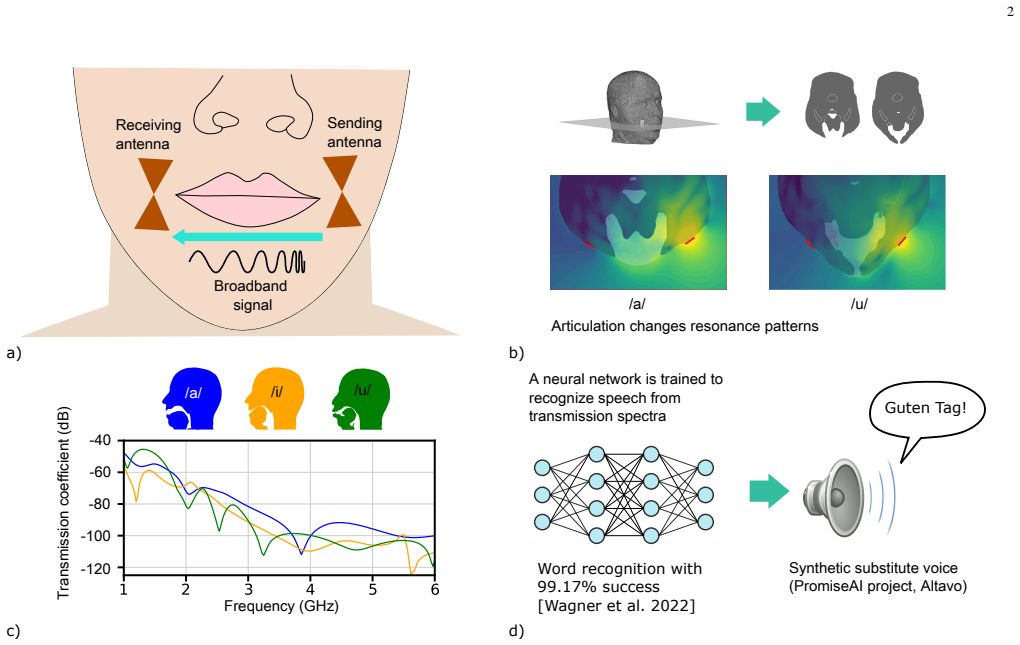
Articulatory movements influence electromagnetic wave transmission through the vocal tract
Pith reviewed 2026-05-10 00:51 UTC · model grok-4.3
The pith
Articulatory positions during vowel production create distinct electromagnetic transmission patterns through the head.
A machine-rendered reading of the paper's core claim, the machinery that carries it, and where it could break.
Core claim
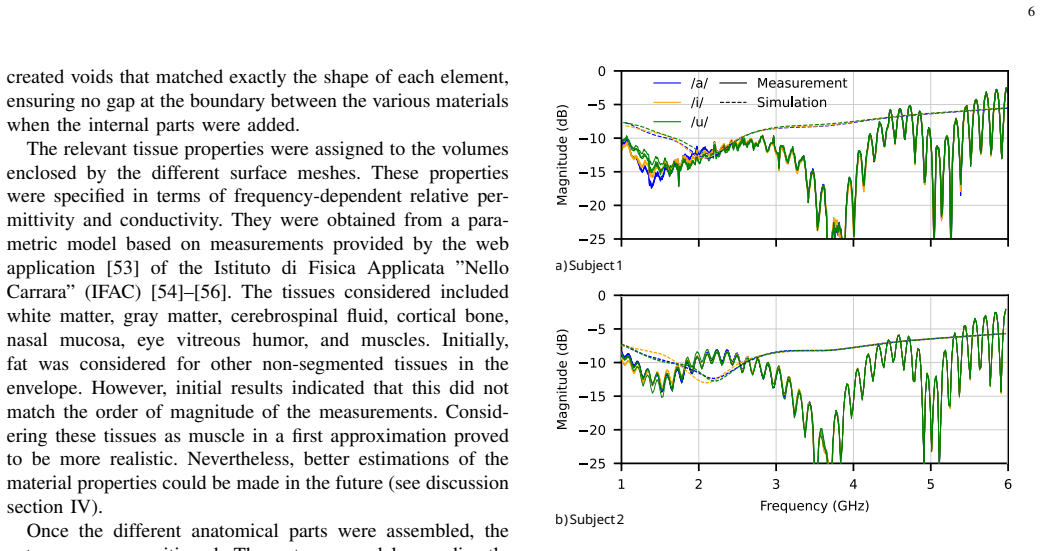
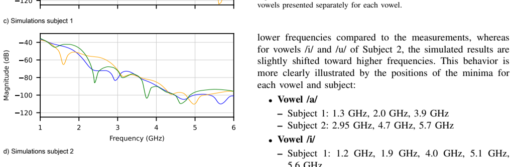
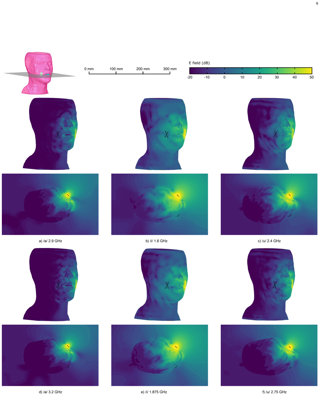
The transmission coefficient of electromagnetic waves through the head varies systematically with articulatory configuration: local minima and maxima appear at frequencies that depend on the vowel, these extrema correlate with spatial variations in the internal electric-field amplitude, the overall curve shape is set by resonance patterns combined with antenna location, and the degree of mouth opening controls the form of the scattering modes. The electric field inside the head follows a Mie-scattering pattern across the 1-6 GHz range. Direct comparison of the model with measurements on two subjects confirms that the same vowels yield similar coefficient patterns in both people.
What carries the argument
A realistic finite-element model derived from MRI images acquired during sustained vowel production, which computes the electromagnetic field distribution and the resulting transmission coefficient between antennas placed on the head.
If this is right
- The same vowel produces recognizably similar transmission patterns across different speakers.
- Local maxima and minima in the transmission coefficient directly reflect local increases or decreases in electric-field strength inside the head.
- The shape of the scattering modes changes when the mouth opening changes.
- The overall transmission curve is set by the combination of internal resonances and the fixed locations of the antennas.
- A validated numerical model now exists that can be used to explore other speech gestures without repeated physical measurements.
Where Pith is reading between the lines
- Designers of radio-based silent-speech sensors could use the model to optimize antenna placement and frequency bands for specific vowel contrasts before building hardware.
- Extending the same simulation approach to consonants or rapid sequences would test whether the transmission signatures remain distinct enough for real-time recognition.
- The observed Mie-scattering behavior suggests that similar geometry-driven effects may appear in other body regions where tissue boundaries create comparable size-to-wavelength ratios.
Load-bearing premise
The MRI-derived models correctly capture the exact shapes, tissue boundaries, and dielectric properties of the vocal tract and head at the moment each vowel is produced.
What would settle it
Acquire new MRI scans and scattering measurements for a third subject pronouncing a fourth vowel and check whether the simulated transmission-coefficient curve still reproduces the measured locations of peaks and dips within the observed level of agreement.
Figures


read the original abstract
This study experimentally validates a numerical model of electromagnetic propagation through the human head during the pronunciation of different vowels, with the goal of improving our understanding of the underlying physical phenomena. A realistic finite element model was created from magnetic resonance images acquired while pronouncing the vowels /a/, /i/, and /u/. The model was validated against scattering matrix measurements obtained from two subjects whose geometries were modeled. Despite several potential sources of discrepancy, the simulations and measurements showed good qualitative agreement, confirming the validity of the approach. Similar transmission coefficient patterns were observed across subjects for the same vowels. Within the investigated frequency range of (1-6 GHz), the electric field exhibited a Mie scattering pattern. Local minima and maxima in the transmission coefficient, characterizing different articulatory configurations, were correlated with local variations in the electric field amplitude. The transmission coefficient's shape results from an interplay between resonance patterns and antenna placement, while the degree of mouth opening influences the shape of scattering modes. Although technically challenging, this numerical approach proved effective for studying electromagnetic propagation in the human head. The resulting robust numerical model and improved understanding of the underlying physics are expected to facilitate the development of radio-frequency-based silent speech interfaces.
Editorial analysis
A structured set of objections, weighed in public.
Referee Report
Summary. The manuscript claims to experimentally validate a finite-element numerical model of electromagnetic wave propagation through the human vocal tract during vowel production. MRI-derived models for vowels /a/, /i/, and /u/ are compared against scattering-matrix measurements from two subjects in the 1-6 GHz range. Good qualitative agreement is reported despite potential discrepancies, with observations of Mie-like electric field patterns, correlations between transmission features and articulatory configurations, and the role of mouth opening in shaping scattering modes. The validated model is intended to support development of RF-based silent speech interfaces.
Significance. If the validation holds, this provides a subject-specific computational framework for analyzing how articulatory movements modulate EM transmission in the head, which could inform RF silent speech technologies. The direct comparison to independent experimental data across multiple vowels and subjects, plus the consistency of patterns between subjects, adds credibility and suggests broader applicability beyond individual anatomy.
major comments (2)
- [Abstract and §4] Abstract and §4 (Discussion): The attribution of discrepancies between simulation and measurement solely to 'minor modeling inaccuracies' lacks supporting evidence from independent checks on the dielectric properties assigned to tissues or the accuracy of static MRI-derived geometries versus actual dynamic vocal-tract boundaries during vowel production. Without such verification, the qualitative agreement could stem from compensating errors rather than faithful representation of the physics.
- [§3] §3 (Methods): The dielectric constants and tissue boundaries in the FEM models are described as derived from MRI but not specified as subject-specific measurements versus generic literature values; this choice directly affects whether the reported agreement validates the geometric and articulatory modeling or merely reflects parameter tuning.
minor comments (2)
- [Results figures] Figures showing transmission coefficients would benefit from quantitative overlays (e.g., overlaid simulation and measurement curves with RMS error values) to make the 'good qualitative agreement' claim more precise and reproducible.
- [Abstract] The abstract could explicitly note the frequency range and number of subjects/vowels at the outset for immediate clarity.
Simulated Author's Rebuttal
We thank the referee for the constructive comments and positive overall assessment of our work. We address each major comment below and have revised the manuscript accordingly to improve clarity and transparency.
read point-by-point responses
-
Referee: [Abstract and §4] Abstract and §4 (Discussion): The attribution of discrepancies between simulation and measurement solely to 'minor modeling inaccuracies' lacks supporting evidence from independent checks on the dielectric properties assigned to tissues or the accuracy of static MRI-derived geometries versus actual dynamic vocal-tract boundaries during vowel production. Without such verification, the qualitative agreement could stem from compensating errors rather than faithful representation of the physics.
Authors: We agree that the original text did not include independent verification of the assigned dielectric properties or a quantitative assessment of how well the static MRI geometries match the dynamic vocal-tract boundaries during sustained vowel production. Dielectric values were taken from standard literature compilations rather than measured on the subjects, and the MRI data represent sustained postures rather than real-time articulation. We will revise the abstract and §4 to remove the phrasing that attributes discrepancies solely to 'minor modeling inaccuracies,' instead explicitly noting these limitations and emphasizing that the reported agreement is qualitative and supported by cross-subject consistency in transmission patterns. revision: yes
-
Referee: [§3] §3 (Methods): The dielectric constants and tissue boundaries in the FEM models are described as derived from MRI but not specified as subject-specific measurements versus generic literature values; this choice directly affects whether the reported agreement validates the geometric and articulatory modeling or merely reflects parameter tuning.
Authors: The geometries and tissue boundaries are subject-specific, segmented directly from the MRI scans of the two participants. Dielectric constants, however, were assigned from generic literature values for the relevant head tissues, as subject-specific dielectric measurements were outside the scope of this study. We will update §3 to state this distinction explicitly, making clear that the validation primarily tests the fidelity of the articulatory geometry modeling rather than the dielectric parameterization. revision: yes
Circularity Check
No circularity: validation rests on independent measurements
full rationale
The paper builds FEM models from MRI data of subjects producing vowels /a/, /i/, /u/ and directly compares simulated scattering matrices to separate experimental measurements on the same subjects. This comparison is an external benchmark rather than a self-referential fit or redefinition. No equations reduce by construction to inputs, no parameters are fitted to the target data and then called predictions, and no load-bearing self-citations or imported uniqueness theorems appear in the derivation. The approach is self-contained against the reported measurements.
Axiom & Free-Parameter Ledger
axioms (1)
- standard math Maxwell's equations govern electromagnetic wave propagation in the head model
Reference graph
Works this paper leans on
-
[1]
C. Gu and C. Li, “From Tumor Targeting to Speech Monitoring: Ac- curate Respiratory Monitoring Using Medical Continuous-Wave Radar Sensors,” IEEE Microwave Magazine, vol. 15.4, pp 66-76, 2014. DOI: 10.1109/MMM.2014.2308763
-
[2]
Human vital signs detection methods and potential using radars: A review,
M. Kebe, R. Gadhafi, B. Mohammad, M. Sanduleanu, H. Saleh and M. Al-Qutayri, “Human vital signs detection methods and potential using radars: A review,” Sensors, vol. 20.5, pp 1454, 2020. DOI: 10.3390/s20051454
-
[3]
R. Hahnel, M. Schiselski, M. Laabs, Q. Wang, A. Henning and D. Plettemeier, “Antenna and radar front-end design for heartbeat detection for triggering purposes of medical devices,” International Conference on Body Area Networks, 2013. DOI: 10.4108/icst.bodynets.2013.253602
-
[4]
Vital signal detection using multi-radar for reductions in body movement effects,
AJ. Jang, L. In-Seong and Y . Jong-Ryul, “Vital signal detection using multi-radar for reductions in body movement effects,” Sensors 21.21 (2021): 7398. DOI: 10.3390/s21217398
-
[5]
Linear FM pulse compression radar techniques applied to biological imaging,
J. H. Jacobi and L. E. Larsen, “Linear FM pulse compression radar techniques applied to biological imaging,” Medical applications of microwave imaging, New York: IEEE Press, pp 138-147, 1986
1986
-
[6]
F. Thiel, M. Hein, U. Schwarz, J. Sachs and F. Seifert, “Combining magnetic resonance imaging and ultrawideband radar: A new concept for multimodal biomedical imaging,” Rev. Sci. Instrum., vol. 80.1, pp 014302, 2009. DOI: 10.1063/1.3065095
-
[7]
Microwave Radar Imaging of Heterogeneous Breast Tissue Integrating A Priori Information,
J. Moll, T. N. Kelly, D. Byrne, M. Sarafianou, V . Krozer, and I. J. Craddock, “Microwave Radar Imaging of Heterogeneous Breast Tissue Integrating A Priori Information,” International Journal of Biomedical Imaging, vol. 2014.1, pp 943549, 2014. DOI: 10.1155/2014/943549
-
[8]
Speech articulator measurements using low power EM-wave sensors,
J. F. Holzrichter, G. C. Burnett, L. C. Ng and W. A. Lea, “Speech articulator measurements using low power EM-wave sensors,” J. Acoust. Soc. Am., vol 103.1, pp 622–625, 1998. DOI: 10.1121/1.421133
-
[9]
Characterizing silent and pseudo-silent speech us- ing radar-like sensors,
J. F., Holzrichter, “Characterizing silent and pseudo-silent speech us- ing radar-like sensors,” In Tenth Annual Conference of the Inter- national Speech Communication Association, 2009. https://www.isca- archive.org/interspeech 2009/holzrichter09 interspeech.pdf
2009
-
[10]
B. Denby, T. Schultz, K. Honda, T. Hueber, J. M. Gilbert and J. S. Brumberg, “Silent speech interfaces,” Speech Communication, vol. 52.4, pp. 270–287, 2010. DOI: 10.1016/j.specom.2009.08.002
-
[11]
Silent Speech Interfaces for Speech Restoration: A Review,
J. A. Gonzalez-Lopez, A. Gomez-Alanis, J. M. Mart ´ın Do˜nas, J. L. P´erez-C´ordoba and A. M. Gomez, “Silent Speech Interfaces for Speech Restoration: A Review,” IEEE Access, vol. 8, pp. 177995-178021, 2020. DOI: 10.1109/ACCESS.2020.3026579
-
[12]
Development of a (silent) speech recognition system for patients following laryngectomy,
M.J. Fagan, S.R. Ell, J.M. Gilbert, E. Sarrazin and P.M. Chapman, “Development of a (silent) speech recognition system for patients following laryngectomy,” Medical Engineering & Physics, vol. 30.4, pp. 419–425, 2007. DOI: 10.1016/j.medengphy.2007.05.003
-
[13]
Silent Speech Recognition as an Alternative Communication Device for Persons With Laryngectomy,
G. S. Meltzner, J. T. Heaton, Y . Deng, G. De Luca, S. H. Roy and J. C. Kline, “Silent Speech Recognition as an Alternative Communication Device for Persons With Laryngectomy,” IEEE/ACM Transactions on Audio, Speech, and Language Processing, vol. 25.12, pp. 2386–2398,
-
[14]
DOI: 10.1109/TASLP.2017.2740000
-
[15]
A comprehensive survey on cognitive cyber security analysis using machine learn- ing approaches,
S. Lee, Y Shin, M Kim and J. Seo, “IR-UWB Radar-Based Contactless Silent Speech Recognition of V owels, Consonants, Words, and Phrases,” IEEE Access, vol. 11, pp. 144844–144859, 2023. DOI: 10.1109/AC- CESS.2023.3344177
work page doi:10.1109/ac- 2023
-
[16]
Microwave Speech Recognizer Empowered by a Pro- grammable Metasurface,
H. Zhang, H. Ruan, H. Zhao, Z. Wang, S. Hu, T. J. Cui, P. del Hougne and L. Li, “Microwave Speech Recognizer Empowered by a Pro- grammable Metasurface,” Advanced Science, vol. 11.17, pp. 2309826,
-
[17]
DOI: 10.1002/advs.202309826
-
[18]
N. Steinmetz and Nezah Balal, “Feasibility Study of Real-Time Speech Detection and Characterization Using Millimeter-Wave Micro- Doppler Radar ,” Remote Sens., vol. 17.1, pp. 91, 2024. DOI: 10.3390/rs17010091
-
[19]
Doppler Radar-Based Human Speech Recognition Using Mobile Vision Trans- former,
W. Li, Y . Geng, Y . Gao, Q. Ding, D. Li, N. Liu and J. Chen, “Doppler Radar-Based Human Speech Recognition Using Mobile Vision Trans- former,” Electronics, vol. 12.13, pp. 2874, 2023. DOI: 10.3390/electron- ics12132874
-
[20]
Watch Your Mouth: Silent Speech Recognition with Depth Sensing,
X. Wang, Z. Su, J. Rekimoto and Y . Zhang, “Watch Your Mouth: Silent Speech Recognition with Depth Sensing,” In Proceedings of the CHI Conference on Human Factors in Computing Systems, pp. 1–15, May
-
[21]
DOI: 10.1145/3613904.36420
-
[22]
Physical working principles of medical radar,
Ø. Aardal, Y . Paichard, S. Brovoll, T. Berger, TS. Lande and SE. Hamran, “Physical working principles of medical radar,” IEEE Transac- tions on Biomedical Engineering, vol. 60.4, pp. 1142–1149, 2012. DOI: 10.1109/TBME.2012.2228263
-
[23]
Non-Invasive Silent Phoneme Recognition Using Microwave Signals,
P. Birkholz, S. Stone, K. Wolf and D. Plettemeier, “Non-Invasive Silent Phoneme Recognition Using Microwave Signals,” IEEE/ACM Transactions on Audio, Speech, and Language Processing, vol. 26.12, pp. 2404–2411, 2018. DOI: 10.1109/TASLP.2018.2865609
-
[24]
Silent speech command word recognition using stepped frequency continuous wave radar,
C. Wagner, P. Schaffer, P. Amini Digehsara, M. B ¨arhold, D. Plettemeier and P. Birkholz, “Silent speech command word recognition using stepped frequency continuous wave radar,” Sci Rep, vol. 12, pp 4192,
-
[25]
DOI: 10.1038/s41598-022-07842-9
-
[26]
URL https://openreview.net/ pdf/9a7e7a9787d14ac8302215f8e4ef959606b78a94.pdf
J. Menezes, M. Sch ¨utze, P. Schaffer, D. Plettemeier, and P. Birkholz, “Exploring Antenna Placement Configurations with a Radar-based Silent Speech Interface,” In proceedings of ICASSP 2025-2025 IEEE International Conference on Acoustics, Speech and Signal Processing (ICASSP), 2025. DOI: https://doi.org/10.1109/ICASSP49660.2025.10890284
-
[27]
URL https://openreview.net/ pdf/9a7e7a9787d14ac8302215f8e4ef959606b78a94.pdf
J. Menezes, C. Wagner, P. Steiner, P. Schaffer, D., Plettemeier, and P. Birkholz, “Non-invasive Speaker-dependent Continuous Phoneme Recognition with a Radar-based Silent Speech Interface,” In pro- ceedings of ICASSP 2025-2025 IEEE International Conference on Acoustics, Speech and Signal Processing (ICASSP), 2025. DOI: https://doi.org/10.1109/ICASSP49660....
-
[28]
Multimodal Silent Recognition of Phonemes Using Radar and Optopalatographic Silent Speech Interfaces,
J. Menezes, A. Mouras, A. Fietkau, D. Kazzy and P. Birkholz, “Multimodal Silent Recognition of Phonemes Using Radar and Optopalatographic Silent Speech Interfaces,” In Proc. Interspeech 2025, pp. 5058-5062, 2025. https://www.isca- archive.org/interspeech 2025/menezes25 interspeech.pdf
2025
-
[29]
Evaluation of different antenna types and posi- tions in a stepped frequency continuous-wave radar-based silent speech interface,
J. V . Possamai de Menezes, P. Amini Digehsara, C. Wag- ner, M. M ¨utze, M. B ¨arhold, P. Schaffer, D. Plettemeier and P. Birkholz, “Evaluation of different antenna types and posi- tions in a stepped frequency continuous-wave radar-based silent speech interface,” In 23rd Annual Conference of the Interna- tional Speech Communication Association, 2022. http...
2022
-
[30]
Max Planck Institute for Multidisciplinary Sciences Biomedical NMR 37070 Goettingen, Germany www.biomednmr.mpg.de
-
[31]
Specific absorption rate (SAR) simulations for low-field (¡ 0.1 T) MRI systems,
J. Parsa and A. Webb. “Specific absorption rate (SAR) simulations for low-field (¡ 0.1 T) MRI systems,” Magn Reson Mater Phy, vol. 36, pp. 429–438, 2023. DOI: 10.1007/s10334-023-01073-3
-
[32]
G. Scarella, O. Clatz, S. Lanteri, G. Beaume, S. Oudot, J. P. Pons, S. Piperno, P. Joly and J. Wiart, “Realistic numerical modelling of human head tissue exposure to electromagnetic waves from cellular 15 phones,” Comptes Rendus Physique, vol. 7.5, pp. 501–508, 2006. DOI: 10.1016/j.crhy.2006.03.002
-
[33]
D. Simunic, P. Wach, W. Renhart and R. Stollberger, “Spatial distribution of high-frequency electromagnetic energy in human head during MRI: numerical results and measurements,” IEEE Transactions on Biomedical Engineering, vol. 43.1, pp. 88, 1996. DOI: 10.1109/10.477704
-
[34]
Semi-automated generation of individual computational models of the human head and torso from MR images,
B. Kalloch, J. Bode, M. Kozlov, A. Pampel, M. Hlawitschka, B. Sehm, A. Villringer, H. E. M¨oller and P. L. Bazin, “Semi-automated generation of individual computational models of the human head and torso from MR images,” Magnetic resonance in medicine, vol. 81.3, 2090–2105,
2090
-
[35]
DOI: 10.1002/mrm.27508
-
[36]
Continuous Wave Simulations on the Propagation of Elec- tromagnetic Fields Through the Human Head,
J. M. Elloian, G. M. Noetscher, S. N. Makarov and A. Pascual- Leone, “Continuous Wave Simulations on the Propagation of Elec- tromagnetic Fields Through the Human Head,” IEEE Transactions on Biomedical Engineering, vol. 61.6, 1676–1683, 2014. DOI: 10.1109/TBME.2013.2297871
-
[37]
A. M. Qureshi, Z. Mustansar and S. Mustafa, “Finite-element analysis of microwave scattering from a three-dimensional human head model for brain stroke detection,” Royal Society open science, vol. 5.7, pp. 180319, 2018. DOI: 10.1098/rsos.180319
-
[38]
https://www.ansys.com/fr-fr/products/electronics/ansys-hfss
-
[39]
A compact double-layer on- body matched bowtie antenna for medical diagnosis,
X. Li, M. Jalilvand, Y . L. Sit, and T. Zwick, “A compact double-layer on- body matched bowtie antenna for medical diagnosis,” IEEE transactions on antennas and propagation, vol. 62.4, pp 1808–1816, 2014. DOI: 10.1109/TAP.2013.2297158
-
[40]
Miniaturized CPW-fed bowtie slot antenna for wearable biomedical applications,
A. Arayeshnia, A. Madannejad, J. Ebrahimizadeh, F. Ravanbakhsh, M. D. Perez and R. Augustine, “Miniaturized CPW-fed bowtie slot antenna for wearable biomedical applications,” In 14th European Conference on Antennas and Propagation (EuCAP) (pp. 1-4). IEEE March 2020. https://ieeexplore.ieee.org/stamp/stamp.jsp?arnumber=9135766
2020
-
[41]
UWB Bowtie Antenna for Medical Microwave Imaging Applications,
O. Fiser, V . Hruby, J. Vrba, T. Drizdal, J. Tesarik and J. Vrba Jr, “UWB Bowtie Antenna for Medical Microwave Imaging Applications,” IEEE Transactions on Antennas and Propagation, vol. 70.7, pp. 5357–5372,
-
[42]
DOI: 10.1109/TAP.2022.3161355
-
[43]
Rapid and motion-robust volume coverage using cross-sectional real- time MRI,
D. V oit, O. Kalentev, M. van Zalk, A. A. Joseph and J. Frahm, “Rapid and motion-robust volume coverage using cross-sectional real- time MRI,” Magnetic Resonance in Medicine, vol. 83.5, pp 1652-1658,
-
[44]
DOI: https://doi.org/10.1002/mrm.28029
-
[45]
iSEG is available for download at https://github.com/ITISFoundation/osparc-iseg
-
[46]
The design of SimpleITK.Front Neuroinform
B. C. Lowekamp, D. T. Chen, L. Ib ´a˜nez and D. Blezek, “The de- sign of SimpleITK,” Frontiers in neuroinformatics, vol. 7, 2013. DOI: https://doi.org/10.3389/fninf.2013.00045
-
[47]
N4ITK: Improved N3 Bias Correction,
N. J. Tustison, B. B. Avants, Ph. A. Cook, Y . Zheng, A. Egan, P. A. Yushkevich and J. C. Gee, “N4ITK: Improved N3 Bias Correction,” IEEE Transactions on Medical Imaging, vol. 29.6, pp. 1310-1320, 2010. DOI: https://doi.org/10.1109/TMI.2010.2046908
-
[48]
X. Xu, S. Xu, L. Jin and E. Song, Characteristic analysis of Otsu threshold and its applications,” “Pattern recognition letters, vol. 32.7,pp. 956-961, 2011. DOI: https://doi.org/10.1016/j.patrec.2011.01.021
-
[49]
Nonrigid multimodality image registration,
D. Mattes, D. R. Haynor, H. Vesselle, T. K. Lewellyn and W. Eubank, “Nonrigid multimodality image registration,” SPIE Medical Imaging, vol. 4322, pp. 1609-1620, 2001. DOI: https://doi.org/10.1117/12.431046
-
[50]
IT’IS Database for Thermal and Electromagnetic Parameters of Biological Tissues, Version 4.1,
P. A. Hasgall, F. Di Gennaro, C. Baumgartner, E. Neufeld, B. Lloyd, MC. Gosselin, D. Payne, A. Klingenb ¨ock and N. Kuster, “IT’IS Database for Thermal and Electromagnetic Parameters of Biological Tissues, Version 4.1,” DOI: 10.13099/VIP21000-04-1
-
[51]
Sim4Life is a multi-physics platform for computational life sciences (https://sim4life.swiss)
-
[52]
F. Karimi, M. Steiner, T. Newton, B. A. Lloyd, A. M. Cassara, P. de Fontenay, S. Farcito, J. P. Triebkorn, E. Beanato, H. Wang, E. Iavarone, F. C Hummel, N. Kuster, V . Jirsa and E. Neufeld, “Precision non-invasive brain stimulation: an in silico pipeline for personalized control of brain dynamics,” Journal of Neural Engineering, vol. 22.2, pp. 026061, 20...
-
[53]
CGAL User and Reference Manual,
The CGAL Project, “CGAL User and Reference Manual,” version 6.0.1, 2024, https://doc.cgal.org/6.0.1/Manual/packages.html
2024
-
[54]
The code for generating surface meshes can be downloaded from https://github.com/remi-blandin/surface mesh from MRI.git
-
[55]
Eigen v3,
G. Guennebaud, B. Jacob and others, “Eigen v3,”, 2010, http://eigen.tuxfamily.org
2010
-
[56]
The Visualization Toolkit,
W. Schroeder, K. Martin and B. Lorensen, “The Visualization Toolkit,” Kitware, 4th ed., 2006, ISBN 978-1-930934-19-1
2006
-
[57]
A generic software design for Delaunay refinement meshing,
L. Rineau and M. Yvinec, “A generic software design for Delaunay refinement meshing,” Computational Geometry, vol. 38.1-2, pp. 100- 110, 2007. DOI: https://doi.org/10.1016/j.comgeo.2006.11.008
-
[58]
Implicit fairing of arbitrary meshes using diffusion and curvature flow,
M. Desbrun, M. Meyer, P. Schr ¨oder, and A. H. Barr, “Implicit fairing of arbitrary meshes using diffusion and curvature flow,” In Computer Graphics (Proc. SIGGRAPH ’99), pp 317-324, 1999
1999
-
[59]
Computer Graphics Forum , volume =
M. Kazhdan, J. Solomon, and M. Ben-Chen, “Can mean-curvature flow be modified to be non-singular?,” In Computer Graphics Forum, vol. 31, pp 1745–1754, 2012, https://doi.org/10.1111/j.1467-8659.2012.03179.x
-
[60]
http://niremf.ifac.cnr.it/tissprop/htmlclie/htmlclie.php
-
[61]
S. Gabriel, R. W. Lau and C. Gabriel, “The dielectric properties of biological tissues: I. Literature survey,” Physics in medicine & biology, vol. 41.11, pp 2231, 1996. DOI: https://doi.org/10.1088/0031- 9155/41/11/001
-
[62]
S. Gabriel, R. W. Lau and C. Gabriel, “The dielectric properties of biological tissues: II. Measurements in the frequency range 10 Hz to 20 GHz,” Physics in medicine & biology, vol. 41.11, pp 2251, 1996. DOI: https://doi.org/10.1088/0031-9155/41/11/002
-
[63]
S. Gabriel, R. W. Lau and C. Gabriel, “The dielectric properties of biological tissues: III. Parametric models for the dielectric spectrum of tissues,” Physics in medicine & biology, vol. 41.11, pp 2271, 1996. DOI: https://doi.org/10.1088/0031-9155/41/11/003
-
[64]
Optimal radiation of body-implanted capsules,
D. Nikolayev, W. Joseph, M. Zhadobov, R. Sauleau, and L. Martens, “Optimal radiation of body-implanted capsules,” Phys. Rev. Lett., vol. 122, no. 10, p. 108101, Mar. 2019. DOI: https://doi.org/10.1103/PhysRevLett.122.108101
-
[65]
I. V . Soares, M. Gao, E. Cil, Z. ˇSipuˇs, A. K. Skrivervik, J. S. Ho and D. Nikolayev, “Wireless powering efficiency of deep-body implantable devices. Ieee transactions on microwave theory and techniques,” IEEE Trans. Microw. Theory Tech., vol. 71.6, pp. 2680-2692, 2022. DOI: https://doi.org/10.1109/TMTT.2022.3231492
-
[66]
D. Nikolayev, M. Zhadobov, P. Karban and R. Sauleau, “Electromag- netic radiation efficiency of body-implanted devices,” Physical Re- view Applied, vol. 9.2, pp 024033 , 2018. DOI: 10.1103/PhysRevAp- plied.9.024033
-
[67]
Comparison of speech production in upright and supine position,
M. Stone, G. Stock, K. Bunin, K. Kumar, M. Epstein, C. Kambhamettu, M. Li, V . Parthasarathy and J. Prince, “Comparison of speech production in upright and supine position,” The Journal of the Acoustical Society of America, vol. 122.1, pp. 532-541, 2007. DOI: 10.1121/1.2715659
-
[68]
The lombard effect,
D. A. Zollinger and H. Brumm, “The lombard effect,” Current Biology, vol. 21.16, pp. R614-R615, 2011
2011
-
[69]
´I. V .Soares, Z. ˇSipuˇs, A. K. Skrivervik, R. Sauleau, A. Al `u, J. S. Ho, and D. Nikolayev, “Wireless Powering for Long-Lasting Deep-Body Bioelectronic Devices: Solutions, limitations, and new technologies,” IEEE Antennas and Propagation Magazine, vol. 67.3, pp 74-86, 2025. DOI: :https://doi.org/10.1109/MAP.2024.3482974
-
[70]
J. Meyer, S. Prepelit ¸˘a and Lorenzo Picinali, “On the accuracy of finite- difference time-domain simulations of head-related transfer functions as a function of model complexity,” Applied Acoustics, vol. 228, pp. 110353, 2025. DOI: 10.1016/j.apacoust.2024.110353
-
[71]
Modeling consonant-vowel coarticulation for articulatory speech synthesis,
P. Birkholz, “Modeling consonant-vowel coarticulation for articulatory speech synthesis,” PLoS ONE, vol. 8.4, pp. e60603, 2013. DOI: 10.1371/journal.pone.0060603
-
[72]
J. E. Lloyd, I. Stavness and Sidney Fels, “ArtiSynth: A fast interactive biomechanical modeling toolkit combining multibody and finite element simulation,” Soft Tissue Biomechanical Model- ing for Computer Assisted Surgery, pp. 355-394, Springer, 2012. https://link.springer.com/chapter/10.1007/8415 2012 126 R. Blandinwas born in Pau, France, in 1989. He r...
discussion (0)
Sign in with ORCID, Apple, or X to comment. Anyone can read and Pith papers without signing in.