Recognition: unknown
A Spatial-Resolved Proton Energy Spectrometer Based on a Scintillation-Fiber Cube
Pith reviewed 2026-05-09 23:08 UTC · model grok-4.3
The pith
A scintillation-fiber cube spectrometer measures both energy spectrum and spatial profile of complex proton beams online.
A machine-rendered reading of the paper's core claim, the machinery that carries it, and where it could break.
Core claim
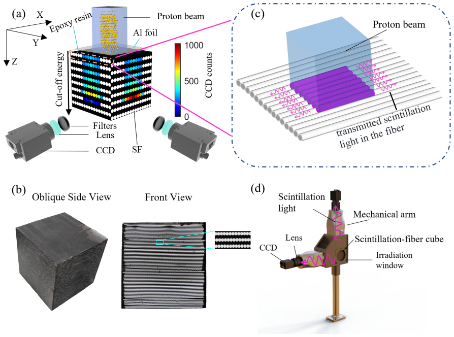
The scintillation-fiber cube spectrometer reconstructs the energy spectrum and transverse beam profile of protons by recording scintillation light along an array of fibers. Calibration with monoenergetic, spatially uniform synchrotron beams yields an energy range of 6-93 MeV, a relative energy uncertainty of 0.6 percent at 80 MeV, and a pixel size of 0.5 mm for profile reconstruction. When the same instrument is exposed to a broadband, spatially complex proton beam generated with a custom energy degrader, it successfully returns both the spectrum and the spatial distribution in a single shot.
What carries the argument
The scintillation-fiber cube, a three-dimensional array of scintillating optical fibers whose light signals encode proton penetration depth for energy and transverse location for beam profile.
If this is right
- The device enables online, single-shot diagnosis of energy spectrum and spatial distribution for high-peak-current proton beams.
- Calibration results support reconstruction over 6-93 MeV with 0.6 percent relative uncertainty at the upper end.
- A 0.5 mm pixel size permits detailed beam-profile mapping even for pulsed, non-uniform beams.
- The custom degrader plus fiber-cube combination provides a practical route to test and validate the spectrometer under realistic complex-beam conditions.
Where Pith is reading between the lines
- The approach could be extended to other ion species once fiber response curves are calibrated for their specific energy-loss characteristics.
- Integration with fast readout electronics would allow the spectrometer to serve as a real-time monitor in high-repetition-rate accelerator facilities.
- The demonstrated spatial resolution suggests the cube could also map beam emittance or divergence in a single exposure if paired with appropriate analysis.
Load-bearing premise
Calibration data from monoenergetic, spatially uniform synchrotron beams can be applied directly to reconstruct spectra and profiles of complex broadband beams without major errors from fiber cross-talk or non-linear light response.
What would settle it
Simultaneous measurement of the same complex degrader beam with the fiber cube and an independent magnetic spectrometer, followed by quantitative comparison of the reconstructed energy spectra and profiles.
Figures


read the original abstract
Advanced particle acceleration methods have produced high-peak-current ion beams with broad energy spread and complex spatial distribution. There is an urgent need to develop online spatial-resolved energy spectrometers for high-energy pulsed ions. This paper introduces a novel spectrometer based on a scintillation-fiber cube for online diagnosis of proton beams with broadband energy spread and complex spatial distribution. We present its working principles, experimental setup, and comprehensive calibration using monoenergetic and spatially uniform proton beams generated by a synchrotron accelerator. Calibration results confirm an energy measurement range of 6-93 MeV, a relative energy uncertainty of 0.6% at 80 MeV, and a pixel size of 0.5 mm for beam profile reconstruction. By exploiting a custom-designed energy degrader, we generated a complex proton beam and measured it with the scintillation-fiber cube spectrometer (SFICS). The results demonstrate the spectrometer's potential for online measurement of the energy spectrum and spatial distribution of complex proton beams.
Editorial analysis
A structured set of objections, weighed in public.
Referee Report
Summary. The manuscript introduces a scintillation-fiber cube spectrometer (SFICS) for online, spatially resolved energy spectrometry of proton beams with broad energy spreads and complex spatial distributions. It describes the working principles and setup, reports calibration on monoenergetic synchrotron beams yielding a 6-93 MeV range, 0.6% relative energy uncertainty at 80 MeV, and 0.5 mm pixel size for profiles, and demonstrates application to a complex beam produced by a custom energy degrader.
Significance. If the calibration transfer holds, the instrument offers a compact diagnostic for simultaneous energy spectrum and spatial profile measurements on high-peak-current ion beams from advanced accelerators, addressing a clear need in accelerator physics for online characterization of broadband, non-uniform beams.
major comments (1)
- Complex beam demonstration: the reconstruction of spectrum and profile for the degrader-generated beam is presented without an independent reference (magnetic spectrometer, time-of-flight, or Monte-Carlo simulation of the degrader output). This leaves untested the assumption that monoenergetic uniform-beam calibration transfers without artifacts from fiber cross-talk or non-linear quenching under mixed-energy, non-uniform illumination, which is load-bearing for the claim of successful demonstration on complex beams.
minor comments (1)
- The abstract and results section could explicitly note the unverified transfer of calibration to the complex-beam case as a limitation.
Simulated Author's Rebuttal
We thank the referee for the careful reading and constructive feedback. The single major comment is addressed point-by-point below. We agree that additional validation is warranted and will revise the manuscript accordingly.
read point-by-point responses
-
Referee: Complex beam demonstration: the reconstruction of spectrum and profile for the degrader-generated beam is presented without an independent reference (magnetic spectrometer, time-of-flight, or Monte-Carlo simulation of the degrader output). This leaves untested the assumption that monoenergetic uniform-beam calibration transfers without artifacts from fiber cross-talk or non-linear quenching under mixed-energy, non-uniform illumination, which is load-bearing for the claim of successful demonstration on complex beams.
Authors: We agree that an independent reference strengthens the claim. In the revised manuscript we will add Geant4 Monte-Carlo simulations of the custom energy degrader, providing the expected energy spectrum and spatial distribution at the SFICS location. Direct comparison with the measured SFICS reconstruction will quantify any discrepancies attributable to fiber cross-talk or quenching. We will also expand the discussion of the calibration transfer, including linearity checks across the 6–93 MeV range and estimates of quenching effects under mixed-energy illumination based on the Birks model. These additions directly test the load-bearing assumption. revision: yes
Circularity Check
No circularity: experimental calibration and demonstration are externally grounded
full rationale
The paper reports direct calibration of the scintillation-fiber cube against monoenergetic synchrotron proton beams (6-93 MeV) to establish energy response, uncertainty (0.6% at 80 MeV), and spatial resolution (0.5 mm). It then applies the same device to a degrader-generated broadband beam as a demonstration. No equations, fitted parameters, or self-citations reduce the reported performance metrics or reconstruction results to the inputs by construction; the calibration data and complex-beam measurement remain independent experimental outcomes. The transferability assumption to complex beams is a potential correctness limitation but does not constitute circularity under the defined patterns.
Axiom & Free-Parameter Ledger
axioms (2)
- standard math Proton energy loss follows established Bethe-Bloch-type relations in the fiber and degrader materials
- domain assumption Scintillation light output is proportional to deposited energy within the calibrated range
Reference graph
Works this paper leans on
-
[1]
R. Mohan, D. Grosshans et al., Proton therapy–present and future. Adv. Drug Delivery Rev.109, 26-44 (2017).https://doi.org/10.1016/j.addr.2016.11.006
-
[2]
R. Mohan et al., A review of proton therapy – Current status and future directions. Precis. Radiat. Oncol.6, 164-176 (2022).https://doi.org/10.1002/pro6.1149
-
[3]
Y. Q. Yang, W. C. Fang, X. X. Huang et al., Static superconducting gantry-based proton CT combined with X-ray CT as prior image for FLASH proton therapy. Nucl. Sci. Tech.34, 11 (2023). https://doi.org/10.1007/s41365-022-01163-2
-
[4]
Schmor et al., Review of Cyclotrons for the Production of Radioactive Isotopes for Medical and Industrial Applications
P. Schmor et al., Review of Cyclotrons for the Production of Radioactive Isotopes for Medical and Industrial Applications. Rev. Accel Sci. Technol.04, 103-116 (2011).https://doi.org/10.1142/ s1793626811000574
2011
-
[5]
J. R. Griswold, D. G. Medvedev, J. W. Engle et al., Large scale accelerator production of 225Ac: Effective cross sections for 78–192 MeV protons incident on 232Th targets. Appl. Radiat. Isot.118, 366-374 (2016).https://doi.org/10.1016/j.apradiso.2016.09.026
-
[6]
B. Jiang, B. B. Tian, H. T. Jing et al., Feasibility of medical radioisotope production based on the proton beams at China Spallation Neutron Source. Nucl. Sci. Tech.35, 102 (2024).https: //doi.org/10.1007/s41365-024-01438-w
-
[7]
K. Matsushita, T. Nishio, S. Tanaka et al., Measurement of proton-induced target fragmentation cross sections in carbon. Nucl. Phys. A946, 104-116 (2016).https://doi.org/10.1016/j.nuclphysa. 2015.11.007
-
[8]
W. P. Liu, B. Guo, Z. An et al., Recent progress in nuclear astrophysics research and its astrophysical implications at the China Institute of Atomic Energy. Nucl. Sci. Tech.35, 217 (2024).https: //doi.org/10.1007/s41365-024-01590-3
-
[9]
Jiang, B
B. Jiang, B. B. Tian, H. T. Jing et al., Measurement of232Th(p,x) 225Ac reaction cross section at CSNS APEP facility up to 80 MeV. Nucl. Sci. Tech.36, 54 (2025).https://doi.org/10.1007/ s41365-024-01616-w
2025
-
[10]
Unser et al., A Toroidal DC Beam Current Transformer with High Resolution
K. Unser et al., A Toroidal DC Beam Current Transformer with High Resolution. IEEE Trans. Nucl. Sci.28, 2344-2346 (1981).https://doi.org/10.1109/TNS.1981.4331686
-
[11]
M. Ghergherehchi, H. Afarideh, M. Ghannadi et al., Proton Beam Dosimetry: a Comparison between a Plastic Scintillator, Ionization Chamber and Faraday Cup. J. Radiat. Res.51, 423-430 (2010). https://doi.org/10.1269/jrr.09121
-
[12]
S. Giordanengo, L. F. Guarachi, S. Braccini et al., Fluence Beam Monitor for High-Intensity Particle Beams Based on a Multi-Gap Ionization Chamber and a Method for Ion Recombination Correction. Applied Sciences12, 12160 (2022).https://doi.org/10.3390/app122312160
-
[13]
Y. W. Zhang, G. Guo, S. Y. Xiao et al., Measurement of medium-energy proton flux. Acta Physica Sinica71, 012902-012901-012902-012908 (2022).https://doi.org/10.7498/aps.71.20211561
-
[14]
E. M. Donegani, A. Olsson, L. Page et al., Design and performance of the shielded DTL4 Faraday cup for the commissioning of the high-power ESS proton linac. Nucl. Instrum. Meth. A1057, 168727 (2023).https://doi.org/10.1016/j.nima.2023.168727
-
[15]
B. Q. Zhao, M. H. Zhao, M. Liu et al., The front-end electronics design of dose monitors for beam delivery system of Shanghai Advanced Proton Therapy Facility. Nucl. Sci. Tech.28, 83 (2017). https://doi.org/10.1007/s41365-017-0230-y
-
[16]
R. J. Leeper, J. R. Lee, L. Kissel et al., Direct measurement of the energy spectrum of an intense proton beam. J. Appl. Phys.60, 4059-4063 (1986).https://doi.org/10.1063/1.337536
-
[17]
K. P. Nesteruk, L. Ramseyer, T. S. Carzaniga et al., Measurement of the Beam Energy Distribution of a Medical Cyclotron with a Multi-Leaf Faraday Cup. Instruments3, 4 (2019).https://doi.org/ 10.3390/instruments3010004
-
[18]
https://doi.org/10.1002/mp.17018
D.S.Levin, P.S.Friedman, C.Ferrettietal., Aprototypescintillatorreal-timebeammonitorforultra- highdoserateradiotherapy.Med.Phys.51, 2905-2923(2024). https://doi.org/10.1002/mp.17018
-
[19]
J. Bosser, J. Mann, G. Ferioli et al., Optical transition radiation proton beam profile monitor. Nucl. Instrum. Meth. A238, 45-52 (1985).https://doi.org/10.1016/0168-9002(85)91025-3
-
[20]
H. Blümer, C. Ebersberger, A scintillating fiber profile monitor for intense proton beams. Nucl. Instrum. Meth. A365, 268-272 (1995).https://doi.org/10.1016/0168-9002(95)00513-7
-
[21]
M. Hori, K. Hanke, Spatial and temporal beam profile monitor with nanosecond resolution for 18 CERN’s Linac4 and Superconducting Proton Linac. Nucl. Instrum. Meth. A588, 359-374 (2008). https://doi.org/10.1016/j.nima.2008.01.078
-
[22]
S. Levasseur, B. Dehning, S. Gibson et al., Development of a rest gas ionisation profile monitor for the CERN Proton Synchrotron based on a Timepix3 pixel detector. J. Instrum.12, C02050 (2017). https://doi.org/10.1088/1748-0221/12/02/C02050
-
[23]
Q. Z. Xing, L. Du, X. L. Guan et al., Transverse profile tomography of a high current proton beam with a multi-wire scanner. Phys. Rev. Accel. Beams21, 072801 (2018).https://doi.org/10.1103/ PhysRevAccelBeams.21.072801
2018
-
[24]
T. Ziegler, I. Göthel, S. Assenbaum et al., Laser-driven high-energy proton beams from cascaded acceleration regimes. Nat. Phys. (2024).https://doi.org/10.1038/s41567-024-02505-0
-
[25]
M. Schollmeier, M. Geissel, A. B. Sefkow et al., Improved spectral data unfolding for radiochromic film imaging spectroscopy of laser-accelerated proton beams. Rev. Sci. Instrum.85, 043305 (2014). https://doi.org/10.1063/1.4870895
-
[26]
B. Dromey, M. Coughlan, L. Senje et al., Picosecond metrology of laser-driven proton bursts. Nat. Commun.7, 10642 (2016).https://doi.org/10.1038/ncomms10642
-
[27]
W.-J. Ma, Z.-P. Liu, P.-J. Wang et al., Experimental progress of laser-driven high-energy proton acceleration and new acceleration schemes. Acta Physica Sinica70, 084102-084101-084102-084115 (2021).https://doi.org/10.7498/aps.70.20202115
-
[28]
Margarone, L
D. Margarone, L. Torrisi, S. Cavallaro et al., Diamond detectors for characterization of laser- generated plasma. Radiat. Eff. Defects Solids163, 463-470 (2008).https://doi.org/10.1080/ 10420150701780540
2008
-
[29]
Milluzzo, V
G. Milluzzo, V. Scuderi, A. G. Amico et al., Laser-accelerated ion beam diagnostics with TOF detectors for the ELIMED beam line. J. Instrum.12, C02025 (2017).https://doi.org/10.1088/ 1748-0221/12/02/C02025
2017
-
[30]
G. Milluzzo, V. Scuderi, A. Alejo et al., A new energy spectrum reconstruction method for time- of-flight diagnostics of high-energy laser-driven protons. Rev. Sci. Instrum.90, 083303 (2019). https://doi.org/10.1063/1.5082746
-
[31]
V. Scuderi, G. Milluzzo, D. Doria et al., TOF diagnosis of laser accelerated, high-energy protons. Nucl. Instrum. Meth. A978, 164364 (2020).https://doi.org/10.1016/j.nima.2020.164364
-
[32]
A. Ngai, K. Dulitz, S. Hartweg et al., Method of kinetic energy reconstruction from time-of-flight mass spectra. Rev. Sci. Instrum.95, 033305 (2024).https://doi.org/10.1063/5.0201425
-
[33]
Prasad, D
R. Prasad, D. Doria, S. Ter-Avetisyan et al., Calibration of Thomson parabola—MCP assembly for multi-MeV ion spectroscopy. Nucl. Instrum. Meth. A623, 712-715 (2010).https://doi.org/10. 1016/j.nima.2010.02.078
2010
-
[34]
D. Jung, R. Hörlein, D. Kiefer et al., Development of a high resolution and high dispersion Thomson parabola. Rev. Sci. Instrum.82, 013306 (2011).https://doi.org/10.1063/1.3523428
-
[35]
R. Prasad, F. Abicht, M. Borghesi et al., Thomson spectrometer–microchannel plate assembly calibration for MeV-range positive and negative ions, and neutral atoms. Rev. Sci. Instrum.84, 053302 (2013).https://doi.org/10.1063/1.4803670
-
[36]
D. Gwynne, S. Kar, D. Doria et al., Modified Thomson spectrometer design for high energy, multi-species ion sources. Rev. Sci. Instrum.85(2014).https://doi.org/10.1063/1.4866021
-
[37]
Nürnberg, M
F. Nürnberg, M. Schollmeier, E. Brambrink et al., Radiochromic film imaging spectroscopy of laser-accelerated proton beams. Rev. Sci. Instrum.80, 033301 (2009).https://dx.doi.org/10. 1063/1.3086424
2009
-
[38]
J. Kaufman, D. Margarone, G. Candiano et al., Radiochromic film diagnostics for laser-driven ion beams. In: Research Using Extreme Light: Entering New Frontiers with Petawatt-Class Lasers II). SPIE (2015).https://doi.org/10.1117/12.2179354
-
[39]
X. H. Xu, Q. Liao, M. J. Wu et al., Detection and analysis of laser driven proton beams by calibrated Gafchromic HD-V2 and MD-V3 radiochromic films. Rev. Sci. Instrum.90, 033306 (2019). https://doi.org/10.1063/1.5049499
-
[40]
Zhang, H
Y. Zhang, H. W. Wang, Y. G. Ma et al., Energy calibration of a CR-39 nuclear-track detec- tor irradiated by charged particles. Nucl. Sci. Tech.30, 87 (2019).https://doi.org/10.1007/ s41365-019-0619-x
2019
-
[41]
Y. F. He, X. F. Xi, S. L. Guo et al., Calibration of CR-39 solid-state track detectors for study of laser-driven nuclear reactions. Nucl. Sci. Tech.31, 42 (2020). https://doi.org/10.1007/ 19 s41365-020-0749-1
2020
-
[42]
Y. Zhang, L.-X. Liu, H.-W. Wang et al., Primary yields of protons measured using CR-39 in laser-induced deuteron–deuteron fusion reactions. Nucl. Sci. Tech.31, 62 (2020).https://doi.org/ 10.1007/s41365-020-00769-8
-
[43]
M. M. Günther, A. Britz, R. J. Clarke et al., NAIS: Nuclear activation-based imaging spectroscopy. Rev. Sci. Instrum.84, 073305 (2013).https://doi.org/10.1063/1.4815826
-
[44]
Y. R. Shou, X. Z. Wu, G. E. Ahn et al., Spatial and spectral measurement of laser- driven protons through radioactivation. Nucl. Sci. Tech.34(2023). https://doi.org/10.1007/ s41365-023-01324-x
2023
-
[45]
J. Metzkes, L. Karsch, S. D. Kraft et al., A scintillator-based online detector for the angularly resolved measurement of laser-accelerated proton spectra. Rev. Sci. Instrum.83, 123301 (2012). https://doi.org/10.1063/1.4768672
-
[46]
J. Metzkes, K. Zeil, S. D. Kraft et al., An online, energy-resolving beam profile detector for laser- driven proton beams. Rev Sci Instrum87, 083310 (2016).https://doi.org/10.1063/1.4961576
-
[47]
N. P. Dover, M. Nishiuchi, H. Sakaki et al., Scintillator-based transverse proton beam profiler for laser- plasma ion sources. Rev. Sci. Instrum.88, 073304 (2017).https://doi.org/10.1063/1.4994732
-
[48]
M. Huault, D. De Luis, J. Apiñaniz et al., A 2D scintillator-based proton detector for high repetition rate experiments. High Power Laser Sci. Eng.7, e60 (2019).https://doi.org/10.1017/hpl.2019. 43
-
[49]
K. M. Schwind, E. Aktan, R. Prasad et al., An online beam profiler for laser-accelerated protons. Rev. Sci. Instrum.90, 053307 (2019).https://doi.org/10.1063/1.5086248
-
[50]
D. A. Mariscal, B. Z. Djordjevíc, E. S. Grace et al., Design of flexible proton beam imaging energy spectrometers (PROBIES). Plasma Phys. Controlled Fusion63, 114003 (2021). https: //doi.org/10.1088/1361-6587/ac234a
-
[51]
Hesse, OCTOPUS–A High Repetition Rate Detector for Laser-Accelerated Particles
M. Hesse, OCTOPUS–A High Repetition Rate Detector for Laser-Accelerated Particles. Technische Universität Darmstadt (2022).http://tuprints.ulb.tu-darmstadt.de/20248/
2022
-
[52]
M. Huault, M. Ehret, D. D. Luis et al., A Scintillator Detector for Spatiospectral Characterization of Proton Beams at High Repetition Rate. IEEE Trans. Instrum. Meas.73, 1-12 (2024).https: //doi.org/10.1109/TIM.2024.3398120
-
[53]
M. H. Xu, H. W. Li, F. Liu et al., Experimental studies of the characteristics of a real-time ion detector-plastic scintillator. Acta Physica Sinica61, 105202-105202 (2012).https://doi.org/10. 7498/aps.61.105202
2012
-
[54]
Torrisi, Plastic scintillator investigations for relative dosimetry in proton-therapy
L. Torrisi, Plastic scintillator investigations for relative dosimetry in proton-therapy. Nucl. Instrum. Meth. B170, 523-530 (2000).https://doi.org/10.1016/S0168-583X(00)00237-8
-
[55]
S. Agostinelli, J. Allison, K. a. Amako et al., GEANT4—a simulation toolkit. Nucl. Instrum. Meth. A506, 250-303 (2003).https://doi.org/10.1016/S0168-9002(03)01368-8
-
[56]
L. L. W. Wang, L. A. Perles, L. Archambault et al., Determination of the quenching correction factors for plastic scintillation detectors in therapeutic high-energy proton beams. Physics in Medicine & Biology57, 7767 (2012).https://doi.org/10.1088/0031-9155/57/23/7767
-
[57]
Z. V. P. Murthy, Nonlinear Regression: Levenberg-Marquardt Method. In: Encyclopedia of Membranes, ed. by Drioli E., Giorno L. (Springer, Berlin Heidelberg, 2015), pp. 1–3.https: //doi.org/10.1007/978-3-642-40872-4_1656-1
-
[58]
Z. M. Wang, W. Chen, M.-t. Qiu et al., Construction and beam commissioning of a compact proton synchrotron for space radiation environment simulation. Nucl. Instrum. Meth. A1027, 166283 (2022).https://doi.org/10.1016/j.nima.2021.166283
-
[59]
Reinhardt, M
S. Reinhardt, M. Hillbrand, J. J. Wilkens et al., Comparison of Gafchromic EBT2 and EBT3 films for clinical photon and proton beams. Med. Phys.39, 5257-5262 (2012).https://doi.org/10. 1118/1.4737890
2012
-
[60]
A. M. Gueli, N. Cavalli, R. De Vincolis et al., Background fog subtraction methods in Gafchromic® dosimetry. Radiat. Meas.72, 44-52 (2015).https://doi.org/10.1016/j.radmeas.2014.11.005
-
[61]
M. Almurayshid, Y. Helo, A. Kacperek et al., Quality assurance in proton beam therapy using a plastic scintillator and a commercially available digital camera. J. Appl. Clin. Med. Phys.18, 210-219 (2017).https://doi.org/10.1002/acm2.12143 20
discussion (0)
Sign in with ORCID, Apple, or X to comment. Anyone can read and Pith papers without signing in.