Recognition: unknown
Comparative Study of Weighted and Coupled Second- and Fourth-Order PDEs for Image Despeckling in Grayscale, Color, SAR, and Ultrasound
Pith reviewed 2026-05-08 06:31 UTC · model grok-4.3
The pith
Weighted and coupled second- and fourth-order PDEs suppress speckle noise in images more effectively than telegraph diffusion models.
A machine-rendered reading of the paper's core claim, the machinery that carries it, and where it could break.
Core claim
The paper claims that its weighted formulation of combined second- and fourth-order PDEs, along with its coupled iterative PDE framework, provides superior despeckling performance compared to the Telegraph Diffusion Model and Fourth-Order Telegraph Diffusion Model across multiple image types, as evidenced by improved quantitative metrics and visual preservation of structures.
What carries the argument
A weighting parameter that blends second-order diffusion coefficients based on grayscale and gradient with fourth-order terms based on Laplacian, and a coupled system solving second and fourth-order components separately in iterations.
Load-bearing premise
That the weighting parameter and the grayscale, gradient, and Laplacian indicators for the diffusion coefficients can be selected to remove noise without creating artifacts or losing details in all image types tested.
What would settle it
An experiment on a held-out set of SAR or ultrasound images where the proposed models show lower PSNR or SSIM values or visible new artifacts compared to the TDM and TDFM baselines.
Figures


read the original abstract
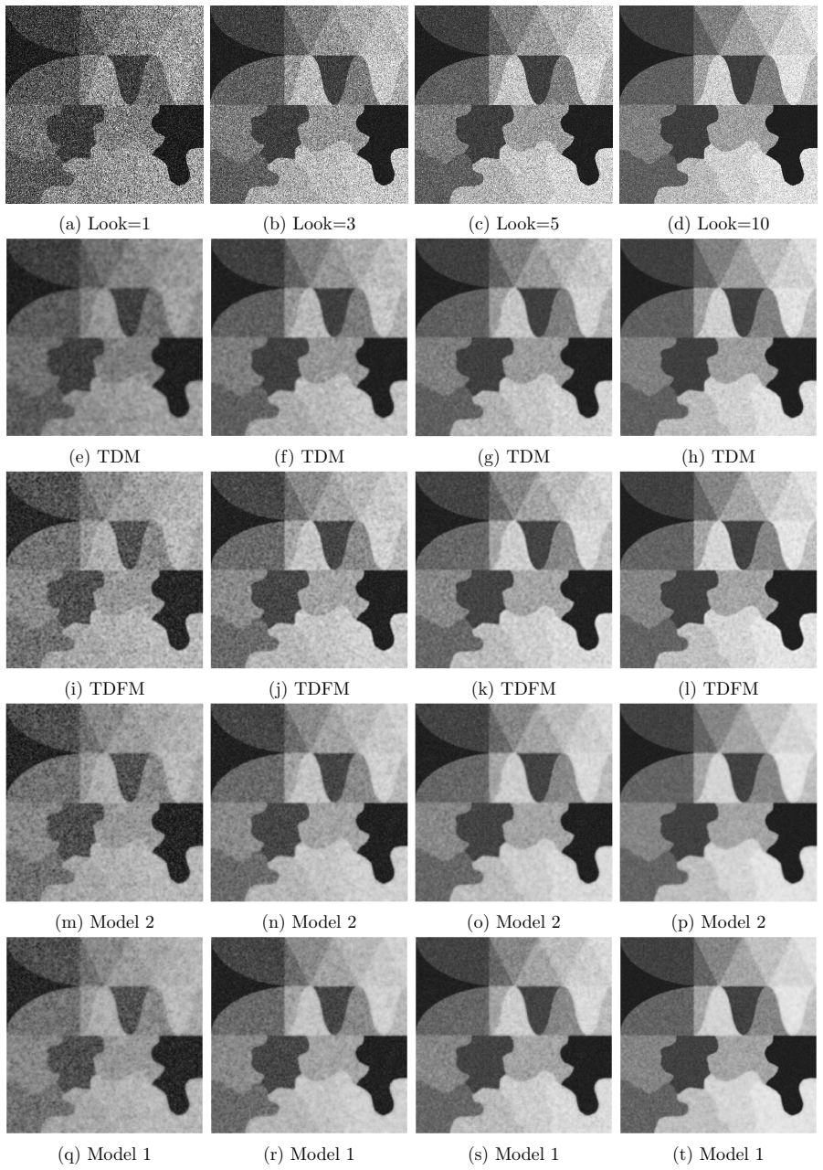
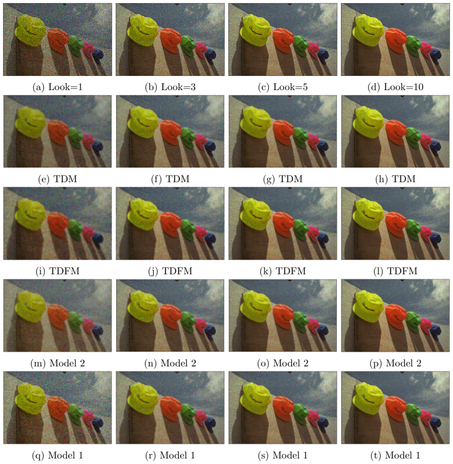
Partial Differential Equation (PDE)-based approaches have gained significant attention in image despeckling due to their strong capability to preserve structural details while suppressing noise. However, conventional second-order PDE models tend to generate blocky artifacts, whereas higher-order models often introduce speckle patterns. To resolve it, this paper proposes and comparatively analyzes two advanced PDE-based frameworks designed for speckle noise suppression while preserving the fine edges. The first model introduces a novel weighted formulation that combines second and fourth-order PDEs through a weighting parameter. The second-order diffusion coefficient employs grayscale and gradient-based indicators, while the fourth-order term is guided solely by a Laplacian-based indicator. The second model constructs a coupled PDE framework, where independent fourth and second-order components are explicitly solved in an iterative manner. In this coupled structure, each diffusion coefficient is defined separately to enhance adaptability in varying image regions. Both models are implemented using the explicit finite difference method. The proposed techniques are extensively evaluated on a variety of datasets, including standard grayscale, color, Synthetic Aperture Radar (SAR), and ultrasound images. Comparative experiments with the existing Telegraph Diffusion Model (TDM) and Fourth-Order Telegraph Diffusion Model (TDFM) demonstrate the superiority of the proposed approaches in reducing speckle noise while effectively preserving fine image structures and edges. Quantitative evaluations using PSNR, SSIM and Speckle Index metrics confirm that the proposed models produce higher image quality and enhanced visual perception. Overall, the presented PDE-based formulations provide a reliable and efficient framework for image despeckling in both natural and medical imaging.
Editorial analysis
A structured set of objections, weighed in public.
Referee Report
Summary. The paper proposes two PDE-based frameworks for speckle noise removal in images: (1) a weighted combination of second- and fourth-order PDEs controlled by a scalar weighting parameter, with the second-order diffusion coefficient using grayscale and gradient indicators and the fourth-order term using a Laplacian indicator; (2) a coupled iterative PDE system with separately defined diffusion coefficients for each order. Both are discretized via explicit finite differences and evaluated on grayscale, color, SAR, and ultrasound datasets against TDM and TDFM baselines, claiming superior PSNR, SSIM, and Speckle Index performance with better edge and structure preservation.
Significance. If the reported metric gains prove robust rather than the result of per-dataset tuning, the work would provide practical refinements to hybrid PDE despeckling methods, offering improved adaptability to the distinct noise statistics of natural versus medical/SAR imagery. The multi-modality experimental design is a constructive element that could support broader applicability in remote sensing and diagnostic imaging.
major comments (3)
- [Section 3] Section 3 (weighted model formulation): the scalar weighting parameter that combines the second- and fourth-order terms is introduced without any selection procedure, sensitivity analysis, or ablation; because the central claim of superiority rests entirely on the comparative PSNR/SSIM/Speckle Index results, the absence of these checks leaves open the possibility that the gains reflect manual per-image optimization rather than an intrinsic property of the PDE construction.
- [Section 5] Section 5 (experimental evaluation): no ablation studies are presented on the weighting parameter values or the choice of indicator functions (grayscale/gradient versus Laplacian); without such controls or perturbation tests, the ranking of the proposed models over TDM and TDFM cannot be shown to be stable across reasonable parameter ranges.
- [Section 5] Section 5 (quantitative tables): the reported metric improvements lack accompanying statistical significance tests (e.g., paired tests across images) or variability measures; this makes it difficult to determine whether the observed differences are reliable or could arise from random variation in the test sets.
minor comments (2)
- [Abstract] The abstract states that the models are 'extensively evaluated' yet supplies no information on the number of images per modality or the ranges explored for the weighting parameter.
- [Section 3] Notation for the diffusion coefficients and indicator functions could be made more uniform between the weighted and coupled models to aid readability.
Simulated Author's Rebuttal
We thank the referee for the constructive and detailed comments. We address each major comment point by point below. Revisions will be made to strengthen the manuscript where the concerns are valid.
read point-by-point responses
-
Referee: [Section 3] Section 3 (weighted model formulation): the scalar weighting parameter that combines the second- and fourth-order terms is introduced without any selection procedure, sensitivity analysis, or ablation; because the central claim of superiority rests entirely on the comparative PSNR/SSIM/Speckle Index results, the absence of these checks leaves open the possibility that the gains reflect manual per-image optimization rather than an intrinsic property of the PDE construction.
Authors: We acknowledge that the original manuscript does not describe the procedure used to select the weighting parameter. In practice, the parameter was determined via preliminary experiments on a small validation subset drawn from each modality (grayscale, color, SAR, ultrasound) and then held fixed for all test images within that modality. This fixed-value approach was intended to prevent per-image tuning. In the revised manuscript we will add an explicit subsection in Section 3 documenting this selection process together with a sensitivity plot showing metric variation for nearby parameter values, confirming that the reported ranking versus TDM and TDFM remains stable. revision: yes
-
Referee: [Section 5] Section 5 (experimental evaluation): no ablation studies are presented on the weighting parameter values or the choice of indicator functions (grayscale/gradient versus Laplacian); without such controls or perturbation tests, the ranking of the proposed models over TDM and TDFM cannot be shown to be stable across reasonable parameter ranges.
Authors: We agree that ablation studies would provide stronger evidence of robustness. The revised Section 5 will include (i) a table varying the weighting parameter over a reasonable interval while keeping all other settings constant, and (ii) a direct comparison of the proposed indicator functions against plausible alternatives (e.g., replacing the Laplacian indicator with a gradient-based one). These additions will demonstrate that the performance advantage over the baselines is not confined to a single narrow parameter choice. revision: yes
-
Referee: [Section 5] Section 5 (quantitative tables): the reported metric improvements lack accompanying statistical significance tests (e.g., paired tests across images) or variability measures; this makes it difficult to determine whether the observed differences are reliable or could arise from random variation in the test sets.
Authors: We recognize the importance of statistical validation. In the revised tables we will report standard deviations computed across the images of each dataset and will add the results of paired t-tests (or Wilcoxon signed-rank tests where normality assumptions are violated) on the per-image PSNR, SSIM, and Speckle Index differences between our models and the TDM/TDFM baselines. These tests will be performed separately for each modality to quantify the reliability of the observed gains. revision: yes
Circularity Check
No circularity: models are explicitly defined then evaluated empirically against external baselines.
full rationale
The paper defines two new PDE frameworks (weighted second+fourth-order with explicit grayscale/gradient/Laplacian indicators, and an iteratively coupled variant) using a scalar weighting parameter and modality-specific diffusion coefficients. These are discretized via explicit finite differences and tested on independent grayscale, color, SAR, and ultrasound datasets, with quantitative comparison to the pre-existing TDM and TDFM models using PSNR/SSIM/Speckle Index. No equation or result is shown to reduce to its own inputs by construction, no self-citation chain bears the central claim, and the reported superiority is an empirical outcome rather than a definitional or fitted renaming. Parameter selection is acknowledged as part of model construction, not presented as a derived prediction.
Axiom & Free-Parameter Ledger
free parameters (1)
- weighting parameter
axioms (1)
- domain assumption Explicit finite-difference discretization remains stable for the proposed weighted and coupled PDEs
Reference graph
Works this paper leans on
-
[1]
Speckle reduction in synthetic- aperture radars,
L. J. Porcello, N. G. Massey, R. B. Innes, and J. M. Marks, “Speckle reduction in synthetic- aperture radars,”J. Opt. Soc. Amer., vol. 66, no. 11, pp. 1305–1311, Nov. 1976
1976
-
[2]
A tutorial on speckle reduction in synthetic aperture radar images,
F. Argenti, A. Lapini, L. Alparone, and T. Bianchi, “A tutorial on speckle reduction in synthetic aperture radar images,”IEEE Geosci. Remote Sens. Mag., vol. 1, no. 3, pp. 6–35, Sep. 2013
2013
-
[3]
Some fundamental properties of speckle,
J. W. Goodman, “Some fundamental properties of speckle,”J. Opt. Soc. Amer., vol. 66, no. 11, pp. 1145–1150, Nov. 1976
1976
-
[4]
When is speckle noise multiplicative?
M. K. Tur, C. Chin, and J. W. Goodman, “When is speckle noise multiplicative?”Appl. Opt., vol. 21, no. 7, pp. 1157–1159, 1982
1982
-
[5]
A model for radar images and its application to adaptive digital filtering of multiplicative noise,
V. S. Frost, J. A. Stiles, K. S. Shanmugan, and J. C. Holtzman, “A model for radar images and its application to adaptive digital filtering of multiplicative noise,”IEEE Trans. Pattern Anal. Mach. Intell., vol. PAMI-4, no. 2, pp. 157–166, Mar. 1982
1982
-
[6]
A nonlocal SAR image denoising algorithm based on LLMMSE wavelet shrink- age,
S. Parrilli, M. Poderico, C. V. Angelino, and L. Verdoliva, “A nonlocal SAR image denoising algorithm based on LLMMSE wavelet shrink- age,” IEEE Trans. Geosci. Remote Sens., vol. 50, no. 2, pp. 606–616, Feb. 2012
2012
-
[7]
SAR image despeckling using Bayesian nonlocal means filter with sigma preselection,
H. Zhong, Y. Li, and L. Jiao, “SAR image despeckling using Bayesian nonlocal means filter with sigma preselection,” IEEE Geosci. Remote Sens. Lett., vol. 8, no. 4, pp. 809–813, Jul. 2011
2011
-
[8]
Iterative weighted maximum likelihood denoising with probabilistic patch-based weights,
C.-A. Deledalle, L. Denis, and F. Tupin, “Iterative weighted maximum likelihood denoising with probabilistic patch-based weights,” IEEE Trans. Image Process., vol. 18, no. 12, pp. 2661–2672, Dec. 2009
2009
-
[9]
SAR image denoising via Bayesian wavelet shrinkage based on heavy-tailed modeling,
A. Achim, P. Tsakalides, and A. Bezerianos, “SAR image denoising via Bayesian wavelet shrinkage based on heavy-tailed modeling,” IEEE Trans. Geosci. Remote Sens., vol. 41, no. 8, pp. 1773–1784, Aug. 2003
2003
-
[10]
Homomorphic wavelet-based statistical despeck- ling of SAR im- ages,
S. Solbo and T. Eltoft, “Homomorphic wavelet-based statistical despeck- ling of SAR im- ages,” IEEE Trans. Geosci. Remote Sens., vol. 42, no. 4, pp. 711–721, Apr. 2004
2004
-
[11]
Non-linear diffusion models for despeckling of images: Achieve- ments and future challenges,
S. K. Jain and R. K. Ray, “Non-linear diffusion models for despeckling of images: Achieve- ments and future challenges,” IETE Tech. Rev., vol. 37, no. 1, pp. 66–82, 2020
2020
-
[12]
A nonlinear coupled diffusion system for image despeckling and application to ultrasound images,
S. K. Jain, R. K. Ray, and A. Bhavsar, “A nonlinear coupled diffusion system for image despeckling and application to ultrasound images,” Circuits, Syst., Signal Process., vol. 38, no. 4, pp. 1654–1683, 2019
2019
-
[13]
A gray level indicator-based reg- ularized telegraph diffusion model: Application to image despeckling,
S. Majee, R. K. Ray, and A. K. Majee, “A gray level indicator-based reg- ularized telegraph diffusion model: Application to image despeckling,” SIAM J. Imag. Sci., vol. 13, no. 2, pp. 844–870, Jan. 2020
2020
-
[14]
Weickert, Anisotropic Diffusion in Image Processing, vol
J. Weickert, Anisotropic Diffusion in Image Processing, vol. 1. Stuttgart, Germany: Teub- ner, 1998. 20
1998
-
[15]
Speckle reducing anisotropic diffusion,
Y. Yu and S. T. Acton, “Speckle reducing anisotropic diffusion,” IEEE Trans. Image Pro- cess., vol. 11, no. 11, pp. 1260–1270, Nov. 2002
2002
-
[16]
A variational approach to removing mul- tiplicative noise,
G. Aubert and J.-F. Aujol, “A variational approach to removing mul- tiplicative noise,” SIAM J. Appl. Math., vol. 68, no. 4, pp. 925–946, 2008
2008
-
[17]
SAR image regulariza- tion with fast approximate discrete minimization,
L. Denis, F. Tupin, J. Darbon, and M. Sigelle, “SAR image regulariza- tion with fast approximate discrete minimization,” IEEE Trans. Image Process., vol. 18, no. 7, pp. 1588–1600, Jul. 2009
2009
-
[18]
Speckle reduction via higher order total variation approach,
W. Feng, H. Lei, and Y. Gao, “Speckle reduction via higher order total variation approach,” IEEE Trans. Image Process., vol. 23, no. 4, pp. 1831–1843, Apr. 2014
2014
-
[19]
A fuzzy edge de- tector driven telegraph total variation model for image despeckling,
S. Majee, S. K. Jain, R. K. Ray, and A. K. Majee, “A fuzzy edge de- tector driven telegraph total variation model for image despeckling,” In- verse Problems Imag., vol. 16, no. 2, pp. 367 396, 2022. [Online]. Available: https://www.aimsciences.org/article/doi/10.3934/ipi.2021054
-
[20]
Analysis of a new variational model for multiplica- tive noise removal,
Z. Jin and X. Yang, “Analysis of a new variational model for multiplica- tive noise removal,” J. Math. Anal. Appl., vol. 362, no. 2, pp. 415–426, 2010
2010
-
[21]
Multiplicative denoising and deblurring: Theory and algorithms,
L. Rudin, P.-L. Lions, and S. Osher, “Multiplicative denoising and deblurring: Theory and algorithms,” in Geometric Level Set Methods in Imaging, Vision, and Graphics. New York, NY, USA: Springer, 2003, pp. 103–119
2003
-
[22]
A nonlinear inverse scale space method for a convex multiplicative noise model,
J. Shi and S. Osher, “A nonlinear inverse scale space method for a convex multiplicative noise model,” SIAM J. Imag. Sci., vol. 1, no. 3, pp. 294–321, 2008
2008
-
[23]
SAR image despeckling through convolutional neural networks,
G. Chierchia, D. Cozzolino, G. Poggi, and L. Verdoliva, “SAR image despeckling through convolutional neural networks,” in Proc. IEEE Int. Geosci. Remote Sens. Symp. (IGARSS), Jul. 2017, pp. 5438–5441
2017
-
[24]
Speckle2Void: Deep self-supervised SAR despeckling with blind-spot convolutional neural networks,
A. B. Molini, D. Valsesia, G. Fracastoro, and E. Magli, “Speckle2Void: Deep self-supervised SAR despeckling with blind-spot convolutional neural networks,” IEEE Trans. Geosci. Remote Sens., vol. 60, pp. 1–17, 2022
2022
-
[25]
DeSpeckNet: Generalizing deep learning-based SAR image despeckling,
A. G. Mullissa, D. Marcos, D. Tuia, M. Herold, and J. Reiche, “DeSpeckNet: Generalizing deep learning-based SAR image despeckling,” IEEE Trans. Geosci. Remote Sens., vol. 60, 2020, Art. no. 5200315
2020
-
[26]
SAR image despeckling using a convolutional neural network,
P. Wang, H. Zhang, and V. M. Patel, “SAR image despeckling using a convolutional neural network,” IEEE Signal Process. Lett., vol. 24, no. 12, pp. 1763–1767, Dec. 2017
2017
-
[27]
Adaptive total variation regu- larization based SAR image despeckling and despeckling evaluation index,
Y. Zhao, J. G. Liu, B. Zhang, W. Hong, and Y. R. Wu, “Adaptive total variation regu- larization based SAR image despeckling and despeckling evaluation index,” IEEE Trans. Geosci. Remote Sens., vol. 53, no. 5, pp. 2765–2774, May 2015
2015
-
[28]
A review of deep-learning techniques for SAR image restoration,
L. Denis, E. Dalsasso, and F. Tupin, “A review of deep-learning techniques for SAR image restoration,” in Proc. IEEE Int. Geosci. Remote Sens. Symp., Jul. 2021, pp. 411–414
2021
-
[29]
How to compute a multi-look SAR image?
H. Cantalloube and C. Nahum, “How to compute a multi-look SAR image?” Eur. Space Agency Publications, vol. 450, pp. 635–640, Mar. 2000
2000
-
[30]
Image denoising by sparse 3-D transform-domain collaborative filtering,
K. Dabov, A. Foi, V. Katkovnik, and K. Egiazarian, “Image denoising by sparse 3-D transform-domain collaborative filtering,” IEEE Trans. Image Process., vol. 16, no. 8, pp. 2080–2095, Aug. 2007. 21
2080
-
[31]
(2023).Speckle noise removal via learned variational models
Cuomo, S., De Rosa, M., Izzo, S., Piccialli, F., Pragliola, M. (2023).Speckle noise removal via learned variational models
2023
-
[32]
Y.-L. You, M. Kaveh, Fourth-order partial differential equations for noise removal,IEEE Trans. Image Process., 9(10) (2000), pp. 1723–1730
2000
-
[33]
A new non-linear hyperbolic-parabolic coupled PDE model for image despeckling,
S. Majee, R. K. Ray, and A. K. Majee, “A new non-linear hyperbolic-parabolic coupled PDE model for image despeckling,”IEEE Trans. Image Process., vol. 31, pp. 1963–1976, Feb. 2022
1963
-
[34]
Perona, J
P. Perona, J. Malik, Scale-space and edge detection using anisotropic diffusion,IEEE Trans. Pattern Anal. Mach. Intell., 12(7) (1990), pp. 629–639
1990
-
[35]
Z. Zhou, Z. Guo, D. Zhang, B. Wu, A nonlinear diffusion equation-based model for ultra- sound speckle noise removal,J. Nonlinear Sci., 28 (2018), pp. 443–470
2018
-
[36]
Z. Zhou, Z. Guo, G. Dong, J. Sun, D. Zhang, B. Wu, A doubly degenerate diffusion model based on the gray level indicator for multiplicative noise removal,IEEE Trans. Image Process., 24(1) (2014), pp. 249–260
2014
-
[37]
X. Shan, J. Sun, Z. Guo, Multiplicative noise removal based on the smooth diffusion equa- tion,J. Math. Imaging Vision, 61(6) (2019), pp. 763–779
2019
-
[38]
New Fourth-Order Grayscale Indicator-Based Telegraph Diffusion Model for Image Despeckling
R. K. Ray and M. Kumar, “New fourth-order grayscale indicator-based telegraph diffusion model for image despeckling,”arXiv preprint arXiv:2509.26010, Sep. 2025. 22
work page internal anchor Pith review Pith/arXiv arXiv 2025
discussion (0)
Sign in with ORCID, Apple, or X to comment. Anyone can read and Pith papers without signing in.