Recognition: unknown
SPARSE -- Efficient High-Resolution SEM Imaging of Rare Microstructural Features Across Large Areas by Selective Rescanning
Pith reviewed 2026-05-10 12:11 UTC · model grok-4.3
The pith
A two-stage SEM framework identifies rare microstructural features with a fast scan then rescans only those regions at high resolution.
A machine-rendered reading of the paper's core claim, the machinery that carries it, and where it could break.
Core claim
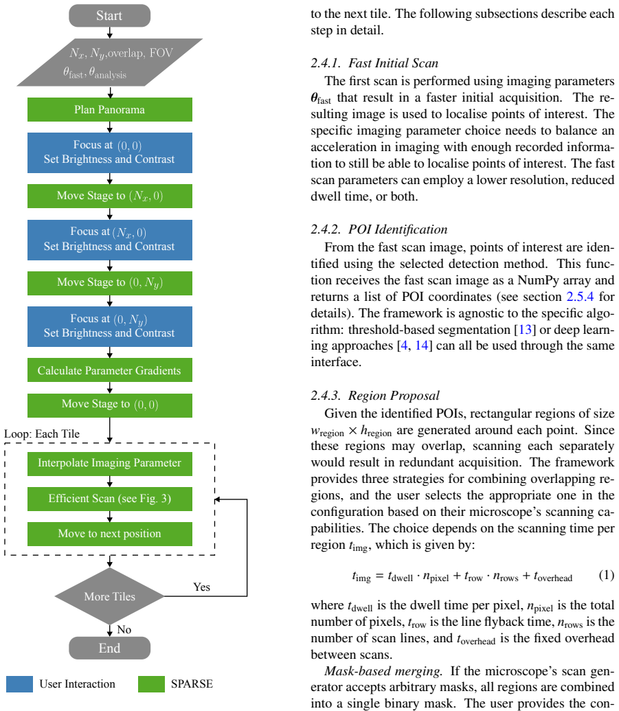
The framework defines a generic microscope interface and a modular detection interface to enable a two-stage workflow: a fast scan identifies regions of interest that are then selectively rescanned at high resolution. Parallel execution via separate processes and queue-based communication ensures detection overhead does not increase acquisition time. On a Tescan Clara SEM examining damage in DP800 steel, representative settings achieve 99 percent detection at approximately 58 percent of standard time and 95 percent detection at 19 percent, with time savings expressed as lower bounds from the ratio of scanned pixels.
What carries the argument
The two-stage selective rescanning workflow that uses a fast overview scan to locate regions of interest for targeted high-resolution follow-up imaging.
If this is right
- Large-area high-resolution characterization of rare features becomes practical because total acquisition time scales with the number of detected regions rather than the full area.
- The modular interfaces allow the same framework to work with different SEM platforms and alternative detection algorithms without rewriting core scanning logic.
- Parallel processing of detection keeps computational cost from extending the physical scan duration.
- Time savings scale directly with how sparse the features are, since fewer pixels require high-resolution acquisition.
- The lower-bound estimates based on pixel ratios indicate that actual savings could be even larger once minor overheads are reduced.
Where Pith is reading between the lines
- The same selective-rescan logic could be applied to other imaging modalities such as optical microscopy or atomic force microscopy when rare surface features must be examined over large areas.
- Reduced acquisition time would enable statistical sampling over sample sizes that are currently impractical, improving the reliability of microstructure statistics.
- Integration with real-time analysis could allow the detection step to adapt scan parameters on the fly for even higher efficiency.
- The open-source release would let researchers test the framework on their own instruments and feature types to quantify time savings for specific use cases.
Load-bearing premise
The initial fast scan together with the chosen detection method will locate essentially all rare microstructural features of interest without missing any that would need high-resolution imaging.
What would settle it
A controlled test that reveals one or more known rare features present in the sample but undetected by the fast scan and detection step, resulting in incomplete high-resolution coverage.
Figures


read the original abstract
Characterisation of rare microstructural features in scanning electron microscopy (SEM) requires imaging large areas at high resolution. This leads to prohibitively long acquisition times. We present an open-source Python framework that addresses this bottleneck through a two-stage approach: a fast scan identifies regions of interest, which are then selectively rescanned with imaging parameters suitable for quantitative analysis. The framework defines a generic microscope interface and a modular detection interface, allowing adaptation to different microscope platforms and detection methods. Scanning, detection, and rescanning are parallelized using separate processes, ensuring that computation time does not extend acquisition time. The two processes communicate exclusively through queues, avoiding shared mutable state and eliminating the need for explicit synchronization. We validate the framework on damage detection in dual-phase DP800 steel using a Tescan Clara SEM. For a representative configuration a detection rate of 99 % is achieved at approximately 58 % of the conventional acquisition time. At 95 % detection rate, acquisition time drops to 19 %. These time savings estimates represent lower bounds based on the ratio of scanned pixels. The complete implementation will be made available upon publication and upon request during peer-review.
Editorial analysis
A structured set of objections, weighed in public.
Referee Report
Summary. The manuscript presents SPARSE, an open-source Python framework for efficient high-resolution SEM imaging of rare microstructural features over large areas. It implements a two-stage workflow: a fast low-resolution scan identifies candidate regions of interest using a modular detection interface, followed by targeted high-resolution rescanning of those regions. The system uses separate processes for scanning, detection, and rescanning that communicate exclusively via queues to enable parallelization without shared mutable state or added acquisition time. Validation is reported on damage detection in dual-phase DP800 steel using a Tescan Clara SEM, with a representative configuration achieving 99% detection at ~58% of conventional acquisition time and 95% detection at 19% time; these savings are characterized as lower bounds based on the ratio of scanned pixels.
Significance. If the fast initial scan plus chosen detector reliably locates essentially all instances of the target rare features, the framework could meaningfully reduce acquisition times for large-area quantitative SEM characterization in materials science. Notable strengths include the generic microscope interface for platform adaptation, the modular detection interface, the queue-based parallelization that keeps computation off the critical path, and the commitment to open-source release. The concrete empirical results on a commercial instrument provide practical evidence of utility for one specific use case.
major comments (1)
- Validation section: the reported 99% detection rate at 58% acquisition time (and 95% at 19%) for DP800 steel damage is presented as the central performance result, yet the manuscript supplies no description of the detection algorithm, its parameters/thresholds, false-negative rates, or direct comparison against exhaustive high-resolution ground truth across the full scanned area. This assumption—that the fast scan locates every relevant rare feature—is load-bearing for the efficiency claims; without such characterization the time savings cannot be guaranteed to correspond to complete population coverage.
minor comments (2)
- Abstract: the statement that 'the complete implementation will be made available upon publication' would be strengthened by including a repository URL or DOI even if under embargo during review.
- The manuscript would benefit from a concise flowchart or pseudocode in the methods section illustrating the queue-based inter-process communication to clarify how parallelism is achieved without synchronization primitives.
Simulated Author's Rebuttal
We thank the referee for their constructive feedback on our manuscript. The single major comment raises an important point about the characterization of detection performance in the validation experiments. We address it directly below and will revise the manuscript accordingly.
read point-by-point responses
-
Referee: Validation section: the reported 99% detection rate at 58% acquisition time (and 95% at 19%) for DP800 steel damage is presented as the central performance result, yet the manuscript supplies no description of the detection algorithm, its parameters/thresholds, false-negative rates, or direct comparison against exhaustive high-resolution ground truth across the full scanned area. This assumption—that the fast scan locates every relevant rare feature—is load-bearing for the efficiency claims; without such characterization the time savings cannot be guaranteed to correspond to complete population coverage.
Authors: We agree that the validation section would benefit from greater detail on the detection component. In the revised manuscript we will add a dedicated subsection describing the specific detection algorithm employed for the DP800 damage features (including whether it is threshold-based, edge-detection, or a simple machine-learning classifier), all tunable parameters and thresholds used, and the procedure for estimating false-negative rates. These details were omitted from the original submission for brevity but are available from our experimental records. With respect to exhaustive high-resolution ground truth across the entire scanned area, we note that acquiring such a dataset would require a conventional full-area high-resolution scan—the very procedure whose time cost the framework is designed to reduce. Instead, detection performance was quantified by (i) manual expert review of all rescanned high-resolution images and (ii) comparison against a smaller set of independently acquired high-resolution reference images covering representative regions. We will make this evaluation protocol explicit, report the resulting false-negative statistics, and emphasize that the reported time savings are therefore lower bounds conditioned on the observed detection rate rather than an absolute guarantee of complete coverage. If the referee considers these additions insufficient, we are prepared to include additional supplementary figures showing example detections and missed features. revision: yes
Circularity Check
No circularity; empirical performance metrics from direct validation
full rationale
The paper introduces a two-stage SEM framework (fast scan + selective rescanning) and reports measured detection rates (99% at 58% time, 95% at 19%) from a single experimental configuration on DP800 steel. These are direct empirical outcomes, not predictions derived from equations, fitted parameters, or self-citations. No mathematical derivation chain exists, no parameters are fitted then renamed as predictions, and no load-bearing claims reduce to self-referential definitions or prior author work. The result is self-contained against external benchmarks via the reported pixel-ratio time savings and detection counts.
Axiom & Free-Parameter Ledger
axioms (1)
- domain assumption A fast low-resolution scan combined with the detection method will locate essentially all rare features of interest.
Reference graph
Works this paper leans on
-
[1]
C. C. Tasan, M. Diehl, D. Yan, M. Bech- told, F. Roters, L. Schemmann, C. Zheng, N. Peranio, D. Ponge, M. Koyama, K. Tsuzaki, D. Raabe, An Overview of Dual-Phase Steels: Advances in Microstructure-Oriented Process- ing and Micromechanically Guided Design, An- nual Review of Materials Research 45 (V olume 45, 2015) (2015) 391–431.doi:10.1146/ annurev-matsc...
2015
-
[2]
PLOS ONE9(12), 113490 (2014) https://doi.org/10.1371/journal
C. Kusche, T. Reclik, M. Freund, T. Al- Samman, U. Kerzel, S. Korte-Kerzel, Large- area, high-resolution characterisation and clas- sification of damage mechanisms in dual-phase steel using deep learning, PLOS ONE 14 (5) (2019) e0216493.doi:10.1371/journal. pone.0216493
-
[3]
H. V . Atkinson, G. Shi, Characterization of inclu- sions in clean steels: a review including the statis- tics of extremes methods, Progress in Materials Science 48 (5) (2003) 457–520.doi:10.1016/ S0079-6425(02)00014-2
2003
-
[4]
M. A. Wollenweber, C. F. Kusche, T. Al-Samman, S. Korte-Kerzel, On the automated characteri- sation of inclusion-induced damage in 16Mn- CrS5 case-hardening steel, Advances in Industrial and Manufacturing Engineering 7 (2023) 100123. doi:10.1016/j.aime.2023.100123
-
[5]
T. Dahmen, M. Engstler, C. Pauly, P. Trampert, N. de Jonge, F. Mücklich, P. Slusallek, Feature Adaptive Sampling for Scanning Electron Mi- croscopy, Scientific Reports 6 (1) (2016) 25350. doi:10.1038/srep25350
-
[6]
Ultramicroscopy273, 114138 (2025) https://doi.org/10.1016/j.ultramic
S. Clusiau, N. Piché, N. Brodusch, M. Strauss, R. Gauvin, Workflow automation of SEM acqui- sitions and feature tracking, Ultramicroscopy 269 (2025) 114093.doi:10.1016/j.ultramic. 2024.114093
-
[7]
A. E. Tekkaya, N. Ben Khalifa, O. Hering, R. Meya, S. Myslicki, F. Walther, Forming- induced damage and its effects on product prop- erties, CIRP Annals 66 (1) (2017) 281–284.doi: 10.1016/j.cirp.2017.04.113
-
[8]
O. Hering, A. E. Tekkaya, Damage-induced per- formance variations of cold forged parts, Journal of Materials Processing Technology 279 (2020) 116556.doi:10.1016/j.jmatprotec.2019. 116556
-
[9]
Medghalchi, C
S. Medghalchi, C. F. Kusche, E. Karimi, U. Kerzel, S. Korte-Kerzel, Damage Analysis in Dual-Phase Steel Using Deep Learning: Transfer from Uniaxial to Biaxial Straining Conditions by Image Data Augmentation, JOM 72 (12) (2020) 4420–4430, tLDR: Overall, the network perfor- mance could be greatly improved and an anal- ysis of changes in damage behavior, her...
2020
-
[10]
F. Hannard, T. Pardoen, E. Maire, C. Le Bour- lot, R. Mokso, A. Simar, Characterization and mi- cromechanical modelling of microstructural het- erogeneity effects on ductile fracture of 6xxx alu- minium alloys, Acta Materialia 103 (2016) 558– 572.doi:10.1016/j.actamat.2015.10.008
-
[11]
G. Avramovic-Cingara, Y . Ososkov, M. K. Jain, D. S. Wilkinson, Effect of martensite distribution on damage behaviour in DP600 dual phase steels, Materials Science and Engineering: A 516 (1) (2009) 7–16.doi:10.1016/j.msea.2009.03. 055
-
[12]
E. E. A¸ sık, E. S. Perdahcıo ˘glu, A. H. van den Boogaard, Microscopic investigation of damage mechanisms and anisotropic evolution of damage in DP600, Materials Science and Engineering: A 739 (2019) 348–356.doi:10.1016/j.msea. 2018.10.018
-
[13]
L. Zhou, H. Wen, I. C. Kuschnerus, S. L. Y . Chang, Efficientand Robust Automated Segmentation of Nanoparticles and Aggregates from Transmission Electron Microscopy Images with Highly Com- plex Backgrounds, Nanomaterials 14 (14) (2024) 1169.doi:10.3390/nano14141169
-
[14]
A. Shah, J. A. Schiller, I. Ramos, J. Serrano, D. K. Adams, S. Tawfick, E. Ertekin, Auto- mated image segmentation of scanning electron microscopy images of graphene using U-Net Neu- ral Network, Materials Today Communications 35 (2023) 106127.doi:10.1016/j.mtcomm.2023. 106127
-
[15]
M. Rocklin, Dask: Parallel Computa- tion with Blocked algorithms and Task Scheduling, SciPy 2015 (Jun. 2015). doi:10.25080/Majora-7b98e3ed-013. 15
-
[16]
F. Kolpak, H. Traphöner, O. Hering, A. E. Tekkaya, Large strain flow curves of sheet met- als by sheet extrusion, CIRP Annals 70 (1) (2021) 247–250.doi:10.1016/j.cirp.2021.03.023
-
[17]
Ester, H.-P
M. Ester, H.-P. Kriegel, J. Sander, X. Xu, A density-based algorithm for discovering clusters in large spatial databases with noise, in: Proceedings of the Second International Conference on Knowl- edge Discovery and Data Mining, KDD’96, AAAI Press, Portland, Oregon, 1996, pp. 226–231
1996
-
[18]
C. E. Duchon, Lanczos Filtering in One and Two Dimensions, Journal of Applied Meteorology and Climatology 18 (8) (1979) 1016–1022. doi:10.1175/1520-0450(1979)018<1016: LFIOAT>2.0.CO;2
-
[19]
L. R. Dice, Measures of the Amount of Ecologic Association Between Species, Ecology 26 (3) (1945) 297–302, _eprint: https://esajournals.onlinelibrary.wiley.com/doi/pdf/10.2307/1932409. doi:10.2307/1932409
-
[20]
T. Reclik, S. Medghalchi, P. Schumacher, M. A. Wollenweber, T. Al-Samman, S. Korte-Kerzel, U. Kerzel, Resolution enhancement of scanning electron micrographs using artificial intelligence, Materials & Design 253 (2025) 113955.doi: 10.1016/j.matdes.2025.113955
-
[21]
S. Medghalchi, E. Karimi, S.-H. Lee, B. Berkels, U. Kerzel, S. Korte-Kerzel, Three-dimensional characterisation of deformation-induced damage in dual phase steel using deep learning, Materials & Design 232 (2023) 112108.doi:10.1016/j. matdes.2023.112108
work page doi:10.1016/j 2023
-
[22]
S. Medghalchi, J. Kortmann, S.-H. Lee, E. Karimi, U. Kerzel, S. Korte-Kerzel, Automated segmen- tation of large image datasets using artificial intelligence for microstructure characterisation and damage analysis, Materials & Design 243 (2024) 113031.doi:10.1016/j.matdes.2024. 113031. 16 Appendix A. 0.6 0.8 1.0 0.4 0.5 0.6 0.7 0.8 0.9 1.0Detection Rate = ...
discussion (0)
Sign in with ORCID, Apple, or X to comment. Anyone can read and Pith papers without signing in.