Recognition: unknown
A geometry aware framework enhances noninvasive mapping of whole human brain dynamics
Pith reviewed 2026-05-07 13:40 UTC · model grok-4.3
The pith
Participant-specific eigenmodes from each person's cortical surface geometry resolve the EEG/MEG inverse problem and reconstruct whole-brain dynamics as linear combinations of a few hundred modes.
A machine-rendered reading of the paper's core claim, the machinery that carries it, and where it could break.
Core claim
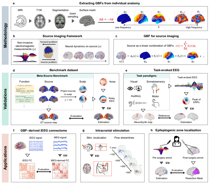
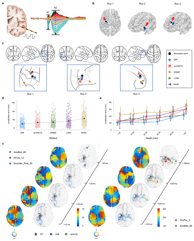
Neural sources can be reconstructed as linear combinations of participant-specific Geometric Basis Functions obtained as eigenmodes of each individual's cortical surface; this anatomic constraint resolves the inverse problem, produces high localization accuracy, recovers fast spatiotemporal dynamics consistent with anatomical pathways, and shows that both spontaneous and evoked whole-brain activity can be described by hundreds of geometric modes.
What carries the argument
Geometric Basis Functions (GBFs): eigenmodes derived from each participant's own cortical surface geometry, used as the spanning set for representing and estimating neural sources.
If this is right
- GBF achieves high localization accuracy on the Meta-Source Benchmark and across task, resting-state, and clinical datasets.
- Reconstructed dynamics align with anatomical pathways and capture fast spatiotemporal features.
- Both spontaneous and evoked whole-brain activity admit compact representation by hundreds of geometric modes.
- The same framework applies to epilepsy data and intracranial stimulation validation without modality-specific changes.
Where Pith is reading between the lines
- The method could be combined with individual connectome data to test whether geometric modes also predict propagation delays along white-matter tracts.
- If the number of required modes stays low across subjects, the approach may enable subject-specific priors that reduce the need for heavy regularization in clinical source imaging.
- Extending the basis construction to include subcortical surfaces would test whether the same geometric principle applies beyond cortex.
Load-bearing premise
Eigenmodes computed from cortical surface geometry form a sufficient and accurate basis for neural sources, so that linear combinations of them solve the inverse problem without introducing new biases.
What would settle it
A case in which GBF source estimates systematically mismatch simultaneous intracranial electrode recordings in both location and timing for a known focal activation.
Figures

read the original abstract
Non-invasive electrophysiology lacks methods that accurately reconstruct whole-brain spatiotemporal dynamics while incorporating individual cortical geometry, leaving current electroencephalography and magnetoencephalography source imaging limited by simplistic or biologically implausible priors. Here, we show that embedding participant-specific Geometric Basis Functions (GBFs), eigenmodes derived from each individual's cortical surface, provides a powerful anatomic constraint that resolves the inverse problem and improves reconstruction fidelity. The method reconstructs neural sources as linear combinations of geometric basis functions, thereby aligning source estimates with the geometric organization of neural dynamics. We validate GBF across the Meta-Source Benchmark, task-evoked data, resting-state networks, intracranial stimulation, and epilepsy data. The results demonstrate that GBF yields high localization accuracy and captures fast spatiotemporal dynamics consistent with anatomical pathways. These findings suggest that both spontaneous and evoked whole-brain activity can be described by hundreds of geometric modes, providing a compact yet accurate representation of neural sources. By linking cortical geometry to electrophysiological dynamics, GBF offers a versatile source imaging tool for both scientific and clinical applications.
Editorial analysis
A structured set of objections, weighed in public.
Referee Report
Summary. The manuscript introduces a geometry-aware framework for EEG/MEG source imaging that embeds participant-specific Geometric Basis Functions (GBFs), defined as eigenmodes derived from each individual's cortical surface. Neural sources are reconstructed as linear combinations of these GBFs to impose an anatomic constraint and address the ill-posed inverse problem. The authors validate the method on the Meta-Source Benchmark, task-evoked data, resting-state networks, intracranial stimulation, and epilepsy datasets, claiming high localization accuracy, capture of fast spatiotemporal dynamics consistent with anatomical pathways, and that both spontaneous and evoked whole-brain activity can be compactly described by hundreds of geometric modes.
Significance. If the central claims hold, the framework would advance noninvasive electrophysiology by supplying a biologically grounded, participant-specific prior based on cortical geometry rather than generic or implausible assumptions. The compact representation using hundreds of modes and validation across multiple data types could improve reconstruction fidelity for both scientific and clinical applications in mapping whole-brain dynamics.
major comments (2)
- [Abstract] Abstract: The abstract asserts validation across multiple datasets and improved accuracy but supplies no quantitative metrics, error bars, statistical tests, or description of how the inverse problem is actually solved or regularized. This omission is load-bearing for assessing whether the GBF approach genuinely resolves the ill-posedness.
- [Methods/Results] GBF construction and inverse solution (Methods/Results): The claim that surface-derived eigenmodes suffice as a basis for whole-brain volumetric reconstruction rests on the untested assumption that the 2-D manifold span adequately covers 3-D sources including subcortical generators. The lead-field operator maps volume currents, yet without explicit volume terms or depth-weighted regularization the linear combinations may attenuate or mislocalize non-cortical activity, directly affecting the whole-brain fidelity claim.
Simulated Author's Rebuttal
We thank the referee for their constructive and insightful comments. We have addressed each point below and believe the revisions will strengthen the manuscript's clarity and rigor.
read point-by-point responses
-
Referee: [Abstract] Abstract: The abstract asserts validation across multiple datasets and improved accuracy but supplies no quantitative metrics, error bars, statistical tests, or description of how the inverse problem is actually solved or regularized. This omission is load-bearing for assessing whether the GBF approach genuinely resolves the ill-posedness.
Authors: We agree that the abstract would benefit from greater specificity. In the revised manuscript, we will incorporate key quantitative metrics from the Meta-Source Benchmark and other validations (including localization errors, reconstruction correlations, and statistical comparisons), along with a concise description of the regularization strategy employed in the GBF inverse solution. This will allow readers to immediately evaluate the method's performance while respecting abstract length constraints. revision: yes
-
Referee: [Methods/Results] GBF construction and inverse solution (Methods/Results): The claim that surface-derived eigenmodes suffice as a basis for whole-brain volumetric reconstruction rests on the untested assumption that the 2-D manifold span adequately covers 3-D sources including subcortical generators. The lead-field operator maps volume currents, yet without explicit volume terms or depth-weighted regularization the linear combinations may attenuate or mislocalize non-cortical activity, directly affecting the whole-brain fidelity claim.
Authors: We thank the referee for highlighting this important consideration. Our GBFs are explicitly cortical, but the forward model (lead-field) incorporates volume conduction effects from the entire brain, allowing cortical basis functions to approximate projected activity from deeper sources. We acknowledge that this is an approximation and that subcortical signals may be attenuated without dedicated depth weighting. In the revision, we will add a dedicated limitations paragraph clarifying the cortical focus, note that validations (e.g., epilepsy and intracranial datasets) primarily involve cortical generators, and outline plans to incorporate depth-weighted regularization in future extensions. No new empirical subcortical tests are added at this stage as they would require substantial additional simulations beyond the current scope. revision: partial
Circularity Check
No significant circularity; GBF basis and inverse reconstruction remain independent of fitted outputs
full rationale
The derivation begins with participant-specific eigenmodes computed directly from each individual's cortical surface geometry (GBFs), then uses linear combinations of these modes to constrain the EEG/MEG inverse problem. No equation or step equates a reported performance metric to a quantity defined by the same fitted parameters that generated the basis. Validation proceeds on held-out benchmarks (Meta-Source), task-evoked recordings, resting-state networks, intracranial stimulation, and epilepsy data, none of which are used to define the GBFs themselves. The surface-to-volume coverage concern is a modeling assumption that can be tested externally and does not reduce the reported reconstruction fidelity to a definitional identity.
Axiom & Free-Parameter Ledger
Reference graph
Works this paper leans on
-
[1]
Nature neuroscience20(3), 327–339 (2017)
Baillet, S.: Magnetoencephalography for brain electrophysiology and imaging. Nature neuroscience20(3), 327–339 (2017)
2017
-
[2]
Nature com- munications9(1), 2987 (2018)
Vidaurre, D., Hunt, L.T., Quinn, A.J., Hunt, B.A., Brookes, M.J., Nobre, A.C., Woolrich, M.W.: Spontaneous cortical activity transiently organises into frequency specific phase-coupling networks. Nature com- munications9(1), 2987 (2018)
2018
-
[3]
Nature Reviews Neuroscience20(12), 746–762 (2019)
Adamantidis, A.R., Gutierrez Herrera, C., Gent, T.C.: Oscillating cir- cuitries in the sleeping brain. Nature Reviews Neuroscience20(12), 746–762 (2019)
2019
-
[4]
Neuron92(2), 544–554 (2016)
Shine, J.M., Bissett, P.G., Bell, P.T., Koyejo, O., Balsters, J.H., Gor- golewski, K.J., Moodie, C.A., Poldrack, R.A.: The dynamics of func- tional brain networks: integrated network states during cognitive task performance. Neuron92(2), 544–554 (2016)
2016
-
[5]
Vogels, T.P., Rajan, K., Abbott, L.F.: Neural network dynamics. Annu. Rev. Neurosci.28(1), 357–376 (2005)
2005
-
[6]
European Journal of Neuroscience55(11-12), 3209– 3223 (2022)
Gaillard, C., Ben Hamed, S.: The neural bases of spatial attention and perceptual rhythms. European Journal of Neuroscience55(11-12), 3209– 3223 (2022)
2022
-
[7]
Trends in Cognitive Sciences (2024)
Staresina, B.P.: Coupled sleep rhythms for memory consolidation. Trends in Cognitive Sciences (2024)
2024
-
[8]
Nature human behaviour7(7), 1196–1215 (2023)
Xu, Y., Long, X., Feng, J., Gong, P.: Interacting spiral wave patterns underlie complex brain dynamics and are related to cognitive processing. Nature human behaviour7(7), 1196–1215 (2023)
2023
-
[9]
Science advances7(30), 2709 (2021)
Raut, R.V., Snyder, A.Z., Mitra, A., Yellin, D., Fujii, N., Malach, R., Raichle, M.E.: Global waves synchronize the brain’s functional systems with fluctuating arousal. Science advances7(30), 2709 (2021)
2021
-
[10]
Current Opinion in Physiology15, 172–182 31 (2020)
Timofeev, I., Schoch, S.F., LeBourgeois, M.K., Huber, R., Riedner, B.A., Kurth, S.: Spatio-temporal properties of sleep slow waves and impli- cations for development. Current Opinion in Physiology15, 172–182 31 (2020)
2020
-
[11]
Journal of Neuroscience41(16), 3665–3678 (2021)
Liang, Y., Song, C., Liu, M., Gong, P., Zhou, C., Kn¨ opfel, T.: Cortex- wide dynamics of intrinsic electrical activities: propagating waves and their interactions. Journal of Neuroscience41(16), 3665–3678 (2021)
2021
-
[12]
IEEE Transactions on Biomedical Engineering63(1), 4–14 (2015)
Edelman, B.J., Baxter, B., He, B.: Eeg source imaging enhances the decoding of complex right-hand motor imagery tasks. IEEE Transactions on Biomedical Engineering63(1), 4–14 (2015)
2015
-
[13]
Pro- ceedings of the National Academy of Sciences120(11), 2207831120 (2023)
Weiner, V.S., Zhou, D.W., Kahali, P., Stephen, E.P., Peterfreund, R.A., Aglio, L.S., Szabo, M.D., Eskandar, E.N., Salazar-Gomez, A.F., Samp- son, A.L.,et al.: Propofol disrupts alpha dynamics in functionally distinct thalamocortical networks during loss of consciousness. Pro- ceedings of the National Academy of Sciences120(11), 2207831120 (2023)
2023
-
[14]
IEEE Transac- tions on Biomedical Engineering63(12), 2619–2628 (2016)
Coito, A., Michel, C.M., Van Mierlo, P., Vulliemoz, S., Plomp, G.: Directed functional brain connectivity based on eeg source imaging: methodology and application to temporal lobe epilepsy. IEEE Transac- tions on Biomedical Engineering63(12), 2619–2628 (2016)
2016
-
[15]
Proceedings of the National Academy of Sciences119(31), 2201128119 (2022)
Sun, R., Sohrabpour, A., Worrell, G.A., He, B.: Deep neural networks constrained by neural mass models improve electrophysiological source imaging of spatiotemporal brain dynamics. Proceedings of the National Academy of Sciences119(31), 2201128119 (2022)
2022
-
[16]
Nature communications13(1), 994 (2022)
Cao, M., Galvis, D., Vogrin, S.J., Woods, W.P., Vogrin, S., Wang, F., Woldman, W., Terry, J.R., Peterson, A., Plummer, C.,et al.: Vir- tual intracranial eeg signals reconstructed from meg with potential for epilepsy surgery. Nature communications13(1), 994 (2022)
2022
-
[17]
IEEE reviews in biomedical engineering1, 23–40 (2008)
He, B., Liu, Z.: Multimodal functional neuroimaging: integrating func- tional mri and eeg/meg. IEEE reviews in biomedical engineering1, 23–40 (2008)
2008
-
[18]
Neuroimage66, 436–448 (2013) 32
Katwal, S.B., Gore, J.C., Gatenby, J.C., Rogers, B.P.: Measuring relative timings of brain activities using fmri. Neuroimage66, 436–448 (2013) 32
2013
-
[19]
Neuroimage102, 80–91 (2014)
Hall, E.L., Robson, S.E., Morris, P.G., Brookes, M.J.: The relationship between meg and fmri. Neuroimage102, 80–91 (2014)
2014
-
[20]
Nature reviews neuroscience13(6), 407–420 (2012)
Buzs´ aki, G., Anastassiou, C.A., Koch, C.: The origin of extracel- lular fields and currents—eeg, ecog, lfp and spikes. Nature reviews neuroscience13(6), 407–420 (2012)
2012
-
[21]
Nature Communications15(1), 5253 (2024)
Jaber, K., Avigdor, T., Mansilla, D., Ho, A., Thomas, J., Abdallah, C., Chabardes, S., Hall, J., Minotti, L., Kahane, P.,et al.: A spatial perturbation framework to validate implantation of the epileptogenic zone. Nature Communications15(1), 5253 (2024)
2024
-
[22]
NeuroImage270, 119953 (2023)
Wens, V.: Exploring the limits of meg spatial resolution with multipolar expansions. NeuroImage270, 119953 (2023)
2023
-
[23]
Frontiers in neuroinformatics12, 4 (2018)
Liu, Q., Ganzetti, M., Wenderoth, N., Mantini, D.: Detecting large-scale brain networks using eeg: impact of electrode density, head modeling and source localization. Frontiers in neuroinformatics12, 4 (2018)
2018
-
[24]
Nature communications11(1), 1946 (2020) https://doi.org/10.1038/s41467-020-15781-0
Sohrabpour, A., Cai, Z., Ye, S., Brinkmann, B., Worrell, G., He, B.: Non- invasive electromagnetic source imaging of spatiotemporally distributed epileptogenic brain sources. Nature communications11(1), 1946 (2020) https://doi.org/10.1038/s41467-020-15781-0
-
[25]
Annual review of biomedical engineering20, 171–196 (2018)
He, B., Sohrabpour, A., Brown, E., Liu, Z.: Electrophysiological source imaging: a noninvasive window to brain dynamics. Annual review of biomedical engineering20, 171–196 (2018)
2018
-
[26]
National Science Review, 457 (2025)
Zhang, Y., Liu, D., Liang, Z., Cheng, J., Lou, K., Duan, J., Gao, T., Hu, B., Liu, Q.: Artificial intelligence as a surrogate brain: Bridging neural dynamical models and data. National Science Review, 457 (2025)
2025
-
[27]
NeuroImage54(2), 851–859 (2011)
Haufe, S., Tomioka, R., Dickhaus, T., Sannelli, C., Blankertz, B., Nolte, G., M¨ uller, K.-R.: Large-scale eeg/meg source localization with spatial flexibility. NeuroImage54(2), 851–859 (2011)
2011
-
[28]
Plos one7(10), 44439–44439 (2012) 33
Petrov, Y.: Harmony: Eeg/meg linear inverse source reconstruction in the anatomical basis of spherical harmonics. Plos one7(10), 44439–44439 (2012) 33
2012
-
[29]
PloS one8(2), 55969 (2013)
Chowdhury, R.A., Lina, J.M., Kobayashi, E., Grova, C.: Meg source localization of spatially extended generators of epileptic activity: com- paring entropic and hierarchical bayesian approaches. PloS one8(2), 55969 (2013)
2013
-
[30]
Nature, 1–9 (2023)
Pang, J.C., Aquino, K.M., Oldehinkel, M., Robinson, P.A., Fulcher, B.D., Breakspear, M., Fornito, A.: Geometric constraints on human brain function. Nature, 1–9 (2023)
2023
-
[31]
Trends in neurosciences36(5), 275–284 (2013)
Zilles, K., Palomero-Gallagher, N., Amunts, K.: Development of cortical folding during evolution and ontogeny. Trends in neurosciences36(5), 275–284 (2013)
2013
-
[32]
Nature Communications15(1), 3570 (2024)
Koller, D.P., Schirner, M., Ritter, P.: Human connectome topology directs cortical traveling waves and shapes frequency gradients. Nature Communications15(1), 3570 (2024)
2024
-
[33]
Nature Methods19(11), 1472–1479 (2022)
Markello, R.D., Hansen, J.Y., Liu, Z.-Q., Bazinet, V., Shafiei, G., Su´ arez, L.E., Blostein, N., Seidlitz, J., Baillet, S., Satterthwaite, T.D.,et al.: Neuromaps: structural and functional interpretation of brain maps. Nature Methods19(11), 1472–1479 (2022)
2022
-
[34]
Physical Review E96(3), 032413 (2017)
Gabay, N.C., Robinson, P.: Cortical geometry as a determinant of brain activity eigenmodes: Neural field analysis. Physical Review E96(3), 032413 (2017)
2017
-
[35]
Nature Communications14(1), 375 (2023)
Cabral, J., Fernandes, F.F., Shemesh, N.: Intrinsic macroscale oscillatory modes driving long range functional connectivity in female rat brains detected by ultrafast fmri. Nature Communications14(1), 375 (2023)
2023
-
[36]
The Neuroscientist24(3), 277–293 (2018)
Atasoy, S., Deco, G., Kringelbach, M.L., Pearson, J.: Harmonic brain modes: a unifying framework for linking space and time in brain dynamics. The Neuroscientist24(3), 277–293 (2018)
2018
-
[37]
Neuron104(2), 189–204 (2019)
Gross, J.: Magnetoencephalography in cognitive neuroscience: a primer. Neuron104(2), 189–204 (2019)
2019
-
[38]
NeuroImage275, 120179 (2023) 34
Thio, B.J., Grill, W.M.: Relative contributions of different neural sources to the eeg. NeuroImage275, 120179 (2023) 34
2023
-
[39]
IEEE Transactions on Biomedical Engineering70(7), 2080–2090 (2023)
Qin, X., Du, L., Jiao, X., Wang, J., Tong, S., Yuan, T., Sun, J.: Evaluation of brain source localization methods based on test-retest reli- ability with multiple session eeg data. IEEE Transactions on Biomedical Engineering70(7), 2080–2090 (2023)
2080
-
[40]
Frontiers in Neuroscience18, 1444935 (2024)
Reynaud, S., Merlini, A., Ben Salem, D., Rousseau, F.: Comprehensive analysis of supervised learning methods for electrical source imaging. Frontiers in Neuroscience18, 1444935 (2024)
2024
-
[41]
In: 2024 46th Annual International Conference of the IEEE Engineering in Medicine and Biology Society (EMBC), pp
Wang, S., Wei, C., Lou, K., Gu, D., Liu, Q.: Advancing eeg/meg source imaging with geometric-informed basis functions. In: 2024 46th Annual International Conference of the IEEE Engineering in Medicine and Biology Society (EMBC), pp. 1–4 (2024). IEEE
2024
-
[42]
NeuroImage271, 120006 (2023)
Allouch, S., Kabbara, A., Duprez, J., Khalil, M., Modolo, J., Hassan, M.: Effect of channel density, inverse solutions and connectivity mea- sures on eeg resting-state networks reconstruction: A simulation study. NeuroImage271, 120006 (2023)
2023
-
[43]
NeuroImage303, 120896 (2024)
Leone, F., Caporali, A., Pascarella, A., Perciballi, C., Maddaluno, O., Basti, A., Belardinelli, P., Marzetti, L., Di Lorenzo, G., Betti, V.: Investigating the impact of the regularization parameter on eeg resting- state source reconstruction and functional connectivity using real and simulated data. NeuroImage303, 120896 (2024)
2024
-
[44]
Bore, J.C., Li, P., Jiang, L., Ayedh, W.M.A., Chen, C., Harmah, D.J., Yao, D., Cao, Z., Xu, P.: A long short-term memory network for sparse spatiotemporal eeg source imaging. IEEE Transactions on Medical Imag- ing40(12), 3787–3800 (2021) https://doi.org/10.1109/TMI.2021.30977 58
-
[45]
Liang, J., Yu, Z.L., Gu, Z., Li, Y.: Electromagnetic source imaging with a combination of sparse bayesian learning and deep neural network. IEEE Transactions on Neural Systems and Rehabilitation Engineering 31, 2338–2348 (2023) https://doi.org/10.1109/TNSRE.2023.3251420
-
[46]
Huang, G., Liu, K., Liang, J., Cai, C., Gu, Z.H., Qi, F., Li, Y., Yu, Z.L., Wu, W.: Electromagnetic source imaging via a data-synthesis- based convolutional encoder–decoder network. IEEE Transactions on 35 Neural Networks and Learning Systems35(5), 6423–6437 (2024) https: //doi.org/10.1109/TNNLS.2022.3209925
-
[47]
Nature neuroscience 24(12), 1733–1744 (2021)
Beam, E., Potts, C., Poldrack, R.A., Etkin, A.: A data-driven frame- work for mapping domains of human neurobiology. Nature neuroscience 24(12), 1733–1744 (2021)
2021
-
[48]
Nature Communications15(1), 8452 (2024)
Pacella, V., Nozais, V., Talozzi, L., Abdallah, M., Wassermann, D., Forkel, S.J., Schotten, M.: The morphospace of the brain-cognition organisation. Nature Communications15(1), 8452 (2024)
2024
-
[49]
Nature communications11(1), 5363 (2020)
Arnulfo, G., Wang, S.H., Myrov, V., Toselli, B., Hirvonen, J., Fato, M., Nobili, L., Cardinale, F., Rubino, A., Zhigalov, A.,et al.: Long-range phase synchronization of high-frequency oscillations in human cortex. Nature communications11(1), 5363 (2020)
2020
-
[50]
Nature neuroscience 20(3), 353–364 (2017)
Bassett, D.S., Sporns, O.: Network neuroscience. Nature neuroscience 20(3), 353–364 (2017)
2017
-
[51]
Proceedings of the National Academy of Sciences113(44), 12574–12579 (2016)
Margulies, D.S., Ghosh, S.S., Goulas, A., Falkiewicz, M., Huntenburg, J.M., Langs, G., Bezgin, G., Eickhoff, S.B., Castellanos, F.X., Petrides, M.,et al.: Situating the default-mode network along a principal gra- dient of macroscale cortical organization. Proceedings of the National Academy of Sciences113(44), 12574–12579 (2016)
2016
-
[52]
NeuroImage292, 120604 (2024)
Ma, L., Braun, S.E., Steinberg, J.L., Bjork, J.M., Martin, C.E., Keen II, L.D., Moeller, F.G.: Effect of scanning duration and sample size on reliability in resting state fmri dynamic causal modeling analysis. NeuroImage292, 120604 (2024)
2024
-
[53]
Nature biomedical engineering3(11), 902–916 (2019)
Betzel, R.F., Medaglia, J.D., Kahn, A.E., Soffer, J., Schonhaut, D.R., Bassett, D.S.: Structural, geometric and genetic factors predict inter- regional brain connectivity patterns probed by electrocorticography. Nature biomedical engineering3(11), 902–916 (2019)
2019
-
[54]
Network Neuroscience9(1), 421–446 (2025) 36
Afnan, J., Cai, Z., Lina, J.-M., Abdallah, C., Pellegrino, G., Arcara, G., Khajehpour, H., Frauscher, B., Gotman, J., Grova, C.: Validating meg estimated resting-state connectome with intracranial eeg. Network Neuroscience9(1), 421–446 (2025) 36
2025
-
[55]
Brain141(4), 1130–1144 (2018)
Frauscher, B., Von Ellenrieder, N., Zelmann, R., Doleˇ zalov´ a, I., Minotti, L., Olivier, A., Hall, J., Hoffmann, D., Nguyen, D.K., Kahane, P.,et al.: Atlas of the normal intracranial electroencephalogram: neurophysiolog- ical awake activity in different cortical areas. Brain141(4), 1130–1144 (2018)
2018
-
[56]
Scientific data7(1), 127 (2020)
Mikulan, E., Russo, S., Parmigiani, S., Sarasso, S., Zauli, F.M., Rubino, A., Avanzini, P., Cattani, A., Sorrentino, A., Gibbs, S.,et al.: Simul- taneous human intracerebral stimulation and hd-eeg, ground-truth for source localization methods. Scientific data7(1), 127 (2020)
2020
-
[57]
Brain Communications5(1), 023 (2023)
Unnwongse, K., Rampp, S., Wehner, T., Kowoll, A., Parpaley, Y., Lehe, M., Lanfer, B., Rusiniak, M., Wolters, C., Wellmer, J.: Validat- ing eeg source imaging using intracranial electrical stimulation. Brain Communications5(1), 023 (2023)
2023
-
[58]
Nature communications12(1), 5259 (2021)
Alberto, G.E., Stapleton-Kotloski, J.R., Klorig, D.C., Rogers, E.R., Constantinidis, C., Daunais, J.B., Godwin, D.W.: Meg source imag- ing detects optogenetically-induced activity in cortical and subcortical networks. Nature communications12(1), 5259 (2021)
2021
-
[59]
Nature communications10(1), 1056 (2019)
Roberts, J.A., Gollo, L.L., Abeysuriya, R.G., Roberts, G., Mitchell, P.B., Woolrich, M.W., Breakspear, M.: Metastable brain waves. Nature communications10(1), 1056 (2019)
2019
-
[60]
Proceedings of the National Academy of Sciences118(48), 2105031118 (2021)
Veit, M.J., Kucyi, A., Hu, W., Zhang, C., Zhao, B., Guo, Z., Yang, B., Sava-Segal, C., Perry, C., Zhang, J.,et al.: Temporal order of signal propagation within and across intrinsic brain networks. Proceedings of the National Academy of Sciences118(48), 2105031118 (2021)
2021
-
[61]
Brain145(5), 1653–1667 (2022)
Lemar´ echal, J.-D., Jedynak, M., Trebaul, L., Boyer, A., Tadel, F., Bhat- tacharjee, M., Deman, P., Tuyisenge, V., Ayoubian, L., Hugues, E.,et al.: A brain atlas of axonal and synaptic delays based on modelling of cortico-cortical evoked potentials. Brain145(5), 1653–1667 (2022)
2022
-
[62]
Brain 124(9), 1683–1700 (2001)
Rosenow, F., L¨ uders, H.: Presurgical evaluation of epilepsy. Brain 124(9), 1683–1700 (2001)
2001
-
[63]
Nature Reviews Neurology 15(10), 594–606 (2019)
Zijlmans, M., Zweiphenning, W., Van Klink, N.: Changing concepts in 37 presurgical assessment for epilepsy surgery. Nature Reviews Neurology 15(10), 594–606 (2019)
2019
-
[64]
Clinical Neurophysiology131(8), 1815–1823 (2020)
Sebastiano, D.R., Tassi, L., Duran, D., Visani, E., Gozzo, F., Cardinale, F., Nobili, L., Del Sole, A., Rubino, A., Dotta, S.,et al.: Identifying the epileptogenic zone by four non-invasive imaging techniques ver- sus stereo-eeg in mri-negative pre-surgery epilepsy patients. Clinical Neurophysiology131(8), 1815–1823 (2020)
2020
-
[65]
Network Neuroscience2(02), 218–240 (2018)
Li, A., Chennuri, B., Subramanian, S., Yaffe, R., Gliske, S., Stacey, W., Norton, R., Jordan, A., Zaghloul, K.A., Inati, S.K.,et al.: Using network analysis to localize the epileptogenic zone from invasive eeg recordings in intractable focal epilepsy. Network Neuroscience2(02), 218–240 (2018)
2018
-
[66]
Frontiers in neurology8, 14 (2017)
Tamilia, E., Madsen, J.R., Grant, P.E., Pearl, P.L., Papadelis, C.: Cur- rent and emerging potential of magnetoencephalography in the detection and localization of high-frequency oscillations in epilepsy. Frontiers in neurology8, 14 (2017)
2017
-
[67]
Scientific Data12(1), 1441 (2025)
Vorderw¨ ulbecke, B.J., Carboni, M., Tourbier, S., Spinelli, L., Brunet, D., Seeber, M., Korff, C.M., Momjian, S., Vargas, M., Seeck, M.,et al.: High- density eeg source localisation of averaged interictal epileptic discharges validated by surgical outcome. Scientific Data12(1), 1441 (2025)
2025
-
[68]
Imaging Neuroscience3, (2025)
Simpson, C., Hall, G., Duncan, J.S., Wang, Y., Taylor, P.N.: Automated generation of epilepsy surgery resection masks: The ramps pipeline. Imaging Neuroscience3, (2025)
2025
-
[69]
Neuroinformatics8, 285–299 (2010)
Zwoli´ nski, P., Roszkowski, M., ˙Zygierewicz, J., Haufe, S., Nolte, G., Durka, P.J.: Open database of epileptic eeg with mri and postopera- tional assessment of foci—a real world verification for the eeg inverse solutions. Neuroinformatics8, 285–299 (2010)
2010
-
[70]
Neuron80(5), 1112– 1128 (2013)
Silva, F.L.: Eeg and meg: relevance to neuroscience. Neuron80(5), 1112– 1128 (2013)
2013
-
[71]
NeuroImage26(2), 38 356–373 (2005)
Mattout, J., P´ el´ egrini-Issac, M., Garnero, L., Benali, H.: Multivariate source prelocalization (msp): use of functionally informed basis functions for better conditioning the meg inverse problem. NeuroImage26(2), 38 356–373 (2005)
2005
-
[72]
Nature communications7(1), 10340 (2016)
Atasoy, S., Donnelly, I., Pearson, J.: Human brain networks function in connectome-specific harmonic waves. Nature communications7(1), 10340 (2016)
2016
-
[73]
Journal of Magnetic Resonance Imaging53(6), 1666–1682 (2021)
Yeh, C.-H., Jones, D.K., Liang, X., Descoteaux, M., Connelly, A.: Mapping structural connectivity using diffusion mri: challenges and opportunities. Journal of Magnetic Resonance Imaging53(6), 1666–1682 (2021)
2021
-
[74]
Nature Communications15(1), 2289 (2024)
Xia, J., Liu, C., Li, J., Meng, Y., Yang, S., Chen, H., Liao, W.: Decomposing cortical activity through neuronal tracing connectome- eigenmodes in marmosets. Nature Communications15(1), 2289 (2024)
2024
-
[75]
Communica- tions Biology6(1), 1128 (2023)
Yang, Y., Tang, S., Wang, X., Zhen, Y., Zheng, Y., Zheng, H., Liu, L., Zheng, Z.: Eigenmode-based approach reveals a decline in brain structure–function liberality across the human lifespan. Communica- tions Biology6(1), 1128 (2023)
2023
-
[76]
Neuroimage61(2), 371–385 (2012)
Michel, C.M., Murray, M.M.: Towards the utilization of eeg as a brain imaging tool. Neuroimage61(2), 371–385 (2012)
2012
-
[77]
In: 2021 International Joint Conference on Neural Networks (IJCNN), pp
Wei, C., Lou, K., Wang, Z., Zhao, M., Mantini, D., Liu, Q.: Edge sparse basis network: a deep learning framework for eeg source localization. In: 2021 International Joint Conference on Neural Networks (IJCNN), pp. 1–8 (2021). IEEE
2021
-
[78]
Nature communications11(1), 325 (2020)
Kucyi, A., Daitch, A., Raccah, O., Zhao, B., Zhang, C., Esterman, M., Zeineh, M., Halpern, C.H., Zhang, K., Zhang, J.,et al.: Electro- physiological dynamics of antagonistic brain networks reflect attentional fluctuations. Nature communications11(1), 325 (2020)
2020
-
[79]
Imaging Neuroscience3, (2025) 39
Xavier, M., Esteves, I., Jorge, J., Abreu, R., Giraud, A.-L., Sadaghiani, S., Wirsich, J., Figueiredo, P.: Consistency of resting-state correlations between fmri networks and eeg band power. Imaging Neuroscience3, (2025) 39
2025
-
[80]
Brain topography26(1), 98–109 (2013)
Meyer, M.C., Oort, E.S., Barth, M.: Electrophysiological correlation pat- terns of resting state networks in single subjects: a combined eeg–fmri study. Brain topography26(1), 98–109 (2013)
2013
discussion (0)
Sign in with ORCID, Apple, or X to comment. Anyone can read and Pith papers without signing in.