Recognition: 2 theorem links
· Lean TheoremCharacterising Water Exchange in Gliomas Using Diffusion MRI with Free Gradient Waveforms
Pith reviewed 2026-05-08 19:26 UTC · model grok-4.3
The pith
Diffusion MRI with free gradient waveforms maps water exchange rates across cell membranes in gliomas.
A machine-rendered reading of the paper's core claim, the machinery that carries it, and where it could break.
Core claim
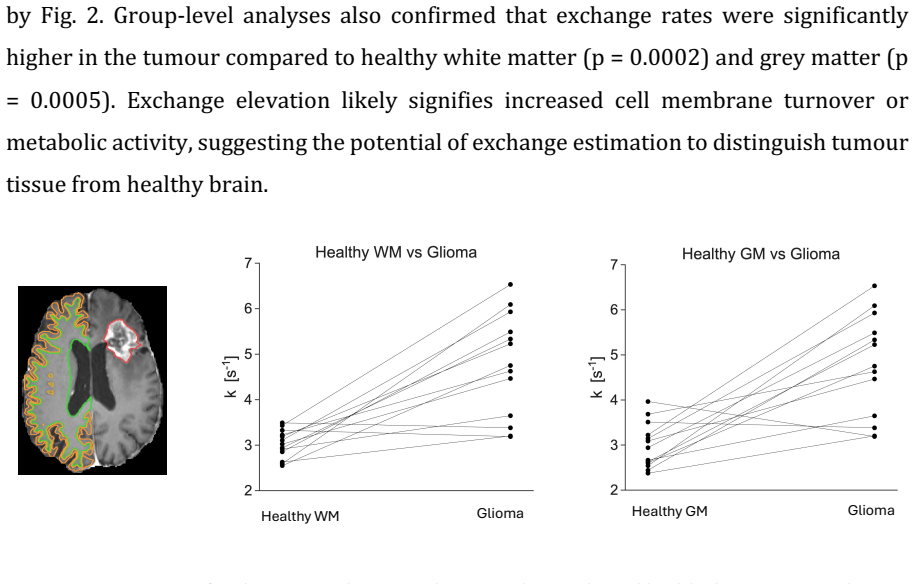
The Restriction-Exchange (ResEx) approach with free gradient waveforms permits voxel-wise estimation of water exchange rates in gliomas that is independent of restricted diffusion effects. In thirteen patients the derived maps showed heterogeneous exchange, commonly elevated in enhancing tumor margins and sometimes extending into non-enhancing peritumoural tissue, while oedema values were modestly above those in healthy brain.
What carries the argument
The Restriction-Exchange (ResEx) protocol, which acquires multiple diffusion-weighted images with free gradient waveforms tuned for selective exchange sensitivity to isolate transmembrane water exchange from intracellular restriction.
Load-bearing premise
The ResEx protocol isolates and quantifies water exchange rate without being confounded by restricted diffusion or other tissue properties in the heterogeneous glioma environment.
What would settle it
Direct comparison of ResEx exchange-rate maps with histological measurements of cell density, membrane permeability, and aquaporin-4 expression in the same resected tissue samples showing no systematic correspondence.
Figures

read the original abstract
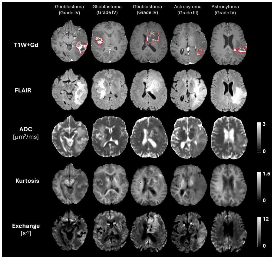
Transmembrane water permeability, which regulates cellular water exchange and is influenced by water channels such as aquaporin-4 (AQP4), has been implicated in glioma progression and may affect tumour infiltration and treatment response. Non-invasive mapping of water exchange may therefore provide biomarkers of glioma pathology. This study investigates the feasibility of characterizing water exchange in gliomas using diffusion MRI with free gradient waveforms, known as the Restriction-Exchange (ResEx) approach, which enables exchange quantification independent of restricted diffusion effects. Thirteen patients with histologically confirmed gliomas (ten glioblastomas, three astrocytomas) underwent preoperative MRI at 3T using a custom ResEx protocol. Multiple diffusion-weighted acquisitions with selective exchange sensitivity were performed to estimate voxel-wise maps of the apparent diffusion coefficient (ADC), diffusion kurtosis, and water exchange rate. ResEx-derived maps revealed heterogeneous spatial patterns across and within tumours. Elevated exchange rates were commonly observed in enhancing tumour margins, potentially reflecting smaller cells, increased membrane permeability or AQP4 upregulation. In some cases, elevated exchange extended into non-enhancing peritumoural regions. Exchange values in oedema were slightly higher than in healthy tissue, suggesting potential infiltration or membrane disruption. Diffusion MRI with free gradient waveforms permits non-invasive mapping of water exchange in gliomas and reveals physiological information not captured by standard imaging. Exchange rate mapping may offer novel biomarkers of tumour aggressiveness, infiltration, and treatment response, and holds promise for surgical and radiotherapy planning.
Editorial analysis
A structured set of objections, weighed in public.
Referee Report
Summary. The manuscript reports a feasibility study applying the Restriction-Exchange (ResEx) diffusion MRI protocol with free gradient waveforms to non-invasively map water exchange rates in 13 patients with histologically confirmed gliomas (10 GBM, 3 astrocytomas). Voxel-wise maps of ADC, diffusion kurtosis, and exchange rate are generated from selective-exchange-sensitive acquisitions at 3T; the maps show heterogeneous intra- and inter-tumoral patterns, with elevated exchange commonly observed at enhancing margins and extending into some non-enhancing peritumoral regions, interpreted as possible signatures of smaller cells, increased permeability, AQP4 upregulation, or infiltration.
Significance. If the ResEx protocol demonstrably decouples transmembrane exchange from intra-compartment restriction across the range of cell sizes and packing densities present in gliomas, the work would supply a new physiological biomarker orthogonal to standard DWI metrics, with potential relevance to tumor aggressiveness, infiltration assessment, and treatment planning. The reported spatial patterns are physiologically plausible and extend beyond conventional imaging contrast.
major comments (2)
- [Abstract] Abstract: the central claim that ResEx 'enables exchange quantification independent of restricted diffusion effects' is presented as a property of the acquisition scheme, yet the manuscript provides no forward simulations with known ground-truth exchange in multi-compartment geometries matching GBM histology, nor any paired comparison against an independent exchange method (e.g., FEXI) to verify that recovered exchange rates remain unbiased when restriction parameters vary across tumor subregions.
- [Results] Results (voxel-wise maps): no quantitative assessment of fit quality, parameter uncertainty, or residual restriction leakage is reported for the exchange-rate maps; without these, it is impossible to determine whether the observed elevation at enhancing margins reflects true exchange differences or residual confounding from the heterogeneous restriction environment.
minor comments (2)
- [Abstract] The abstract would be strengthened by stating the exact number of diffusion-weighted acquisitions, b-values, and waveform parameters used in the custom ResEx protocol.
- Clarify whether the reported exchange values are apparent or model-derived parameters and how they are normalized across patients.
Simulated Author's Rebuttal
We thank the referee for their thoughtful and constructive review of our manuscript. We value the recognition of the potential of the ResEx protocol as a new biomarker in gliomas. We address the major comments below and have prepared revisions to the manuscript where the concerns are valid.
read point-by-point responses
-
Referee: [Abstract] Abstract: the central claim that ResEx 'enables exchange quantification independent of restricted diffusion effects' is presented as a property of the acquisition scheme, yet the manuscript provides no forward simulations with known ground-truth exchange in multi-compartment geometries matching GBM histology, nor any paired comparison against an independent exchange method (e.g., FEXI) to verify that recovered exchange rates remain unbiased when restriction parameters vary across tumor subregions.
Authors: The independence from restricted diffusion is a core design feature of the ResEx method using free gradient waveforms, as described in the foundational methodological papers. This clinical feasibility study does not include new ground-truth simulations or head-to-head comparisons with FEXI. We will revise the abstract to state that the ResEx protocol is designed to enable exchange quantification independent of restriction effects (with appropriate citations), rather than presenting the current patient data as a new demonstration of that independence. revision: partial
-
Referee: [Results] Results (voxel-wise maps): no quantitative assessment of fit quality, parameter uncertainty, or residual restriction leakage is reported for the exchange-rate maps; without these, it is impossible to determine whether the observed elevation at enhancing margins reflects true exchange differences or residual confounding from the heterogeneous restriction environment.
Authors: We agree that quantitative measures of fit quality and potential residual effects are needed to support interpretation of the exchange maps. In the revised manuscript we will add voxel-wise goodness-of-fit metrics (e.g., R²), parameter uncertainty estimates, and residual analysis to assess whether restriction leakage could contribute to the observed spatial patterns. revision: yes
Circularity Check
No significant circularity; exchange-rate maps are data-driven estimates from ResEx acquisitions.
full rationale
The paper presents voxel-wise estimation of water exchange rate directly from custom ResEx diffusion acquisitions in glioma patients. No equations are shown that define the target quantity in terms of itself or that rename a fitted parameter as an independent prediction. The claim that ResEx isolates exchange independent of restriction is stated as a property of the protocol rather than re-derived here; any supporting reference to prior method development is not load-bearing for the present feasibility results in gliomas. The reported spatial patterns and elevated exchange in tumour margins are therefore outputs of the fitting procedure applied to new data, not tautological restatements of the inputs.
Axiom & Free-Parameter Ledger
axioms (1)
- domain assumption ResEx approach enables exchange quantification independent of restricted diffusion effects
Lean theorems connected to this paper
-
IndisputableMonolith.Cost.FunctionalEquationwashburn_uniqueness_aczel unclearln(S/S0) = −b[E_D + V_ω E_R] + (1/2) b² (3 V_D + 2 V_ω C_{D,R} + V_ω² V_R)·(1 − k Γ)
Reference graph
Works this paper leans on
-
[1]
Centre for Medical Imaging and Function, Skane University Hospital, Lund, Sweden Corresponding author: Arthur Chakwizira Department of Radiology, Brigham and Women's Hospital, Harvard Medical School, Boston, MA, United States Email address: achakwizira@bwh.harvard.edu Sponsors/Grant numbers: VR (Swedish Research Council) o 2024-04968 o 2023-02412 eSSENCE ...
2024
-
[2]
which has previously demonstrated ability to differentiate between gliomas and meningiomas (Lampinen et al., 2017). However, FEXI is susceptible to biased exchange estimation due to effects of restricted diffusion and imaging gradients (Lasič et al., 2018; Ohene et al., 2023; Ulloa et al., 2017). A more recent diffusion MRI approach using free gradient w...
2017
-
[3]
(Fig. 1A-B). The waveforms were differentially sensitive to exchange but similarly sensitive to restricted diffusion, that is, they had different Γ (10 and 40 ms) but equal 𝑉# (3500 s-2). Due to scan time constraints, it was not feasible to also include waveforms with variable sensitivity to restrictions. Waveforms were one-dimensional (linear tensor enco...
2023
-
[4]
+16𝑏%𝐸"%𝐾'⋅(1−𝑘Γ) (5) where 𝐾'=3𝑉
with extrapolated references (Nilsson et al, 2016). The images were then powder-averaged for further analysis. To allow comparison between exchange measurements and contrast enhancement, diffusion-weighted images for each 8 participant were rigidly coregistered with the corresponding T1-weighted volume. FLAIR images were also coregistered to the T1-weight...
2016
-
[5]
Contralateral healthy grey and white matter were also segmented using SynthSeg (Billot et al., 2023)
with the glioma pre- and post-treatment segmentor (Jain et al., 2025), which delineated three mutually exclusive regions of interest: oedema, tumour core, and contrast-enhancing parts of the tumour. Contralateral healthy grey and white matter were also segmented using SynthSeg (Billot et al., 2023). Quantitative comparisons of exchange estimates in the tu...
2025
-
[6]
(Fig. 2-3). The variability in exchange effects was not consistently predicted by gadolinium enhancement, suggesting that exchange imaging provides complementary information to conventional contrast-enhanced MRI (Fig. 2). Oedema generally showed low exchange rates relative to enhancing tumour, but slightly elevated rates relative to healthy white matter (...
-
[7]
https://doi.org/10.1186/2045-8118-10-18 19 Chakwizira, A., Westin, C.-F., Brabec, J., Lasič, S., Knutsson, L., Szczepankiewicz, F., & Nilsson, M. (2022). Diffusion MRI with pulsed and free gradient waveforms: Effects of restricted diffusion and exchange. NMR in Biomedicine, n/a(n/a), e4827. https://doi.org/10.1002/nbm.4827 Chakwizira, A., Zhu, A., Foo, T....
-
[8]
https://doi.org/10.1186/s12987-023-00422-7 Omuro, A., & DeAngelis, L. M. (2013). Glioblastoma and other malignant gliomas: A clinical review. JAMA, 310(17), 1842–1850. https://doi.org/10.1001/jama.2013.280319 Ostrom, Q. T., Bauchet, L., Davis, F. G., Deltour, I., Fisher, J. L., Langer, C. E., Pekmezci, M., Schwartzbaum, J. A., Turner, M. C., Walsh, K. M.,...
-
[9]
https://doi.org/10.3390/cancers12030667 Szczepankiewicz, F., Sjölund, J., Ståhlberg, F., Lätt, J., & Nilsson, M. (2019). Tensor-valued diffusion encoding for diffusional variance decomposition (DIVIDE): Technical feasibility in clinical MRI systems. PLOS ONE, 14(3), e0214238. https://doi.org/10.1371/journal.pone.0214238 Tan, Y., Zhang, H., Zhao, R.-F., Wa...
-
[10]
Denoising of diffusion MRI using random matrix theory
https://doi.org/10.1016/j.neuroimage.2016.08.016 Verkman, A. S., Hara-Chikuma, M., & Papadopoulos, M. C. (2008). Aquaporins—New players in cancer biology. Journal of Molecular Medicine (Berlin, Germany), 86(5), 523–529. https://doi.org/10.1007/s00109-008-0303-9 Wang, Z., Wang, B., Li, Z., Han, G., Meng, C., Jiao, B., Guo, K., Hsu, Y.-C., Sun, Y., Liu, Y.,...
discussion (0)
Sign in with ORCID, Apple, or X to comment. Anyone can read and Pith papers without signing in.